Study on anatomy and clinical application of Kambin’s triangle and modified quadrangle
2022-10-10QiuYuPengHuanXiongChenZhenHaoZhongXueJianYangGuoJunLiZhiBinMeng
Qiu-Yu Peng, Huan-Xiong Chen, Zhen-Hao Zhong, Xue-Jian Yang, Guo-Jun Li, Zhi-Bin Meng
Department of Spinal Surgery, The First Affiliated Hospital of Hainan Medical University, Haikou 570102, China
Keywords:Kambin’s triangle Transforaminal lumbar interbody fusion Lumbar vertebra Transforaminal percutaneous endoscopic
ABSTRACT Objective: We through the anatomy of cadavers to study the " Kambin’s triangle " in the safe working area of lumbar intervertebral foramen and to provide anatomical reference for clinical lumbar fusion through Kambin’s triangle approach. Methods: five complete cadaveric specimens were taken, the soft tissue of the lumbar back was removed, the transverse process,upper and lower articular processes and part of the vertebral lamina were bitten, the Kambin’s triangle area of the lumbar spine was completely exposed, the bottom edge and height of the Kambin’s triangle were measured, and the area of the Kambin’s triangle was calculated;Using Kirschner wire, pull and fix the traveling nerve root to make the Kambin’s triangle into a rectangle, measure the length of the bottom edge and height again, calculate the area,and compare the two groups of data. Results: the average height of the Kambin’s triangle was 11.20mm ± 2.10mm, and the average height of the improved four corners was 11.19mm± 1.93mm. The height of the improved four corners was slightly shorter than that of the Kambin’s triangle. There was a significant correlation between the two, but the difference was not statistically significant. The average bottom of Kambin’s triangle is 10.78mm ± 1.95mm,and the average bottom of improved four corners is 12.14mm ± 1.78mm. The length of the bottom edge of improved four corners is greater than that of Kambin’s triangle. There is a significant correlation between them, and the difference is statistically significant; The average area of Kambin’s triangle is 61.79mm2 ± 20.71mm2, and the area of improved four corners is 137.71mm2 ± 38.20mm2. The area of improved four corners is significantly larger than that of Kambin’s triangle. There is a significant correlation between the two, and the difference is statistically significant. Conclusion: there is a narrow right angle triangle area surrounded by traveling nerve root, dural sac and superior endplate of lower vertebral body in the lumbar intervertebral foramen. If the traveling nerve root is pulled and fixed to turn the traditional Kambin’s triangle into a quadrilateral, the bottom edge of the Kambin’s triangle area can be significantly longer and the area can be significantly expanded, which can be operated more safely.✉Corresponding author: MENG Zhi-bin, Chief Physician.
1. Introduction
Kambin's triangle is a right-angled triangular region, which is located on the posterolateral side of the vertebral body and is composed of the traversing nerve root or dural sac,the exiting nerve root and superior border of the caudal vertebra. Due to the lack of nerves and blood vessels in this area, it can be operated safely, and it is one of the most popular surgical approaches in clinic[1].Lumbar fusion via Kambin's triangle approach such as minimally invasive transforaminal lumbar fusion (MIS-TLIF) and percutaneous endoscopic transforaminal lumbar interbody fusion (PE-TLIF) are the main methods for the treatment of lumbar degenerative diseases.Compared with anterior or posterior lumbar approach, Kambin's triangle approach has obvious advantages: there is no need to worry about the injury of important organs and large vessels, and there is no need to destroy the posterior column structure of the spine.It does not affect the stability of the spine, and with the use of PETLIF, the operation is more minimally invasive, less traumatic, can completely retain the function of paraspinal muscles, significantly reduce intraoperative bleeding, reduce the risk of anesthesia,and ensure the safety of the operation[2, 3].However, because the space between the nerve root and the dural sac is very narrow, it is difficult to thoroughly decompress, prepare the endplate and insert a sufficient fusion cage during the kambin triangle approach, and there is a risk of nerve root injury.[4]。In the report of Villavicencio[5],the incidence of nerve root related complications in the MISTLIF group was as high as 10.5%, while in the Jacquot[6] report on percutaneous endoscopic lumbar interbody fusion, the incidence of complications was 36%,The main complications were the cage migration and nerve root injury. Among the 57 patients, 15 patients had cage migration. Among them, 13 cases needed reoperation,and 8 patients had corresponding symptoms of nerve root injury after operation. For this reason, more and more scholars at home and abroad have devoted themselves to the study of Kambin's triangle, using cadaveric specimens or imaging techniques to study the anatomical characteristics of Kambin's triangle and measure the relevant parameters[7-14]. However, there are few reports on Kambin's triangle in China, especially in the study of cadaveric specimens. Lertudomphonwanit[15] points out that the bottom edge of kambin's triangle can be widened and the area can be enlarged by nerve root traction during the operation. Therefore, in this study, the length of each side of lumbar Kambin's triangle was measured, the area was calculated, and the nerve root was fixed by Kirschner wire and traction, so as to make the neurogenic Kambin's triangle into a rectangle. at the same time, the length of each side was measured,the area was calculated, and the two groups of data were compared,so as to provide anatomical reference for avoiding nerve root injury in clinical lumbar fusion via Kambin's triangle approach.
2. Materials and methods
2.1 Materials
Five embalmed human (all specimens were provided by the Department of Anatomy, Hainan Medical College). All cadavers had no history of lumbar surgery and no obvious lumbar deformities.
2.2 Method of operation
After complete excision of paraspinal muscles and exposure of lumbar osseous elements, soft tissues such as intervertebral foramen ligaments were removed, transverse process, superior and inferior articular process and part of vertebral lamina were removed by rongeur, pedicle and isthmus were preserved, dural sac, the traversing nerve root and the exiting nerve root were fully exposed, Kambin's triangle was exposed (figure.1), and electronic Vernier calipers were used(Accuracy 0.01mm) measure the base and height of Kambin's triangle of each segment of lumbar vertebrae in turn, and the assistant is responsible for recording the measurement data and taking pictures. After measuring the relevant parameters of Kambin's triangle, three Kirschner wires and one Kirschner wire were used to pull the travelling nerve root, which was fixed under the lateral edge of the upper vertebral pedicle, and the latter two were fixed at the intersection of the dural sac, the exiting nerve root and the superior border of the caudal vertebra respectively (figure.2) to prevent the dural sac from protruding laterally due to the traction of the exiting nerve root, so that the kambin triangle became a quadrangle, and the base edge and height of the improved quadrangle were measured again.
2.3 Measuring parameters
(1)The height of Kambin's triangle:The height of kambin triangle is defined as the intersection of dural sac and the exiting nerve root and the distance between dural sac and the superior endplate of the caudal vertebra.
(2)The base of Kambin's triangle:The distance between the superior endplate of the caudal vertebra and the intersection of the nerve root and dural sac is the length of the base.
(3)The base and height of improved quadrangle: Same as Kambin's triangle.
2.4 Area calculation
(1)The area of the Kambin's triangle:area = 0.5 (base x height)(2)The area of the improved quadrangle:area = base x height
2.5 Statistical analysis
The measured data are processed by SPSS Version 26.0, and the measured data are expressed as±s(mm). The triangular area of Kambin's triangle and the improved quadrangle are calculated,and the 95% confidence interval of each data is calculated. Using the paired design t-test of the Kambin's triangle and the improved quadrangle, the difference was statistically significant.
3. Results
A total of 50 lumbar intervertebral foramen Kambin's triangle were measured in 5 cadavers.
3.1 Kambin's triangle related parameters(Table 1)
①The height of Kambin's triangle:The height of the Kambin's triangle becomes longer with the decrease of the segment, the mean height for the kambin's triangle is 11.20mm ±2.10mm, with the L1 having the minimum height is 8.95mm ±1.38mm, and the L5 having the largest height is 11.20mm ±2.10mm.
②The base of Kambin's triangle:the base of the Kambin's triangle becomes longer with the decrease of the segment, the mean length of the base for the kambin's triangle is 10.78mm±1.95mm, with the L1 having the minimum length is 8.48mm±0.81mm, and the L5 having the largest length is 10.78mm±1.95mm.
③The area of the Kambin's triangle:in the L1~L5 segment, the average area of Kambin's triangle increased gradually, the mean surface area for the kambin triangle is 61.79mm2±20.71mm2,with L1 having the minimum area is 37.90mm2±6.52mm2, and L5 having the largest area is 88.46mm2±11.85mm2.
3.2 Improved quadrangle related parameters(Table 2)
①The height of improved quadrangle:The height of the Improved quadrangle gradually increases with the segment decline, the mean height for the improved quadrangle is 11.19mm ±1.93 mm, with the L1 having the minimum height is 9.38mm ±0.70mm, and the L5 having the largest height is 13.40mm ±1.46mm.
②The base of improved quadrangle:The bottom of the improved corner: the length of the bottom edge of the improved corner increases gradually with the decrease of the segment, the mean length of the base for the improved quadrangle is 12.14mm±1.78mm,with the L1 having the minimum length is 9.55mm ±0.90mm, and the L5 having the largest length is 13.80mm ±0.94mm.
③The area of the Improved quadrangle:The area of the improved four corners gradually increased with the decrease of the segment,the mean surface area for the kambin triangle is 137.71mm2±38.20mm2,with L1 having the minimum area is 89.69mm2±12.11mm2, and L5 having the largest area is 184.70mm2±21.31mm2.
Table 1 The base、height、area and limit 95% confidence interval of the kambin's triangle(±s)

Table 1 The base、height、area and limit 95% confidence interval of the kambin's triangle(±s)
Segments Height(mm) 95%CI Bottom(mm) 95%CI Area(mm2) 95%CI L1 8.95±1.38 7.96-9.94 8.48±0.81 7.90-9.06 37.90±6.52 33.24-42.56 L2 10.31±1.38 9.32-11.30 9.22±0.57 8.81-9.63 47.72±8.13 41.91-53.53 L3 11.05±1.54 9.95-12.15 11.32±1.08 10.54-12.09 62.49±10.03 55.32-69.67 L4 12.12±1.47 11.07-13.17 11.85±1.18 11.00-12.69 72.36±14.74 61.82-82.90 L5 13.56±1.41 12.55-14.57 13.05±1.11 12.25-13.84 88.46±11.85 79.98-90.51 Average 11.20±2.10 10.60±11.80 10.78±1.95 10.23-11.33 61.79±20.71 55.90-2.93
Table 2 The base、height、area and limit 95% confidence interval of the Improved quadrangle(±s)

Table 2 The base、height、area and limit 95% confidence interval of the Improved quadrangle(±s)
Segments Height(mm) 95%CI Bottom(mm) 95%CI Area(mm2) 95%CI L1 9.38±0.70 8.88-9.87 9.55±0.90 8.90-10.19 89.69±12.11 81.03-98.36 L2 9.93±1.13 9.12-10.74 11.10±0.53 10.72-11.48 109.00±11.52 101.76-118.24 L3 11.44±1.83 10.13-12.75 12.77±0.83 12.17-13.36 145.37±20.79 130.50-160.25 L4 11.79±1.29 10.87-12.71 13.46±0.62 13.02-13.90 158.81±18.74 145.41-172.21 L5 13.40±1.46 12.35-14.45 13.80±0.94 13.13-14.48 184.70±21.31 169.45-199.94 Average 11.19±1.93 10.64-11.74 12.14±1.78 11.63-12.64 137.71±38.20 126.86-148.57
3.3 Correlation comparison between the Kambin's triangle and the Improved quadrangle parameters and paired sample t-Test.
The high average value of Kambin's triangle was 11.20mm±2.10mm, the average height of Improved quadrangle was 11.19mm ±1.93 mm,t=0.050, P=0.961, P>0.05.There was significant correlation between them, but there was no significant difference between them. The average base of the Kambin's triangle is 10.78mm ±1.95mm, and the average base of the Improved quadrangle is 12.14mm ±1.78 mm,t=-8.270,P=0.000,P < 0.05.There is a significant correlation between them, and the bottom edge of the improved corner is obviously larger than that of the Kambin's triangle. The average area of Kambin's triangle was 61.79mm2±20.71mm2, and the area of the Improved quadrangle was 137.71mm2±38.20mm2. The area of the Improved quadrangle was significantly larger than that of Kambin's triangle (Table 3).
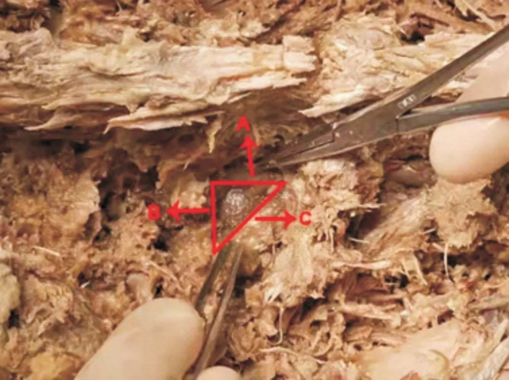
Figure1 The Kambin's triangle:A,the dural sac, is the height of Kambin's triangle;B, superior border of the caudal vertebra, is the base of Kambin's triangle;C, the exiting nerve root,is the hypotenuse of Kambin's triangle.
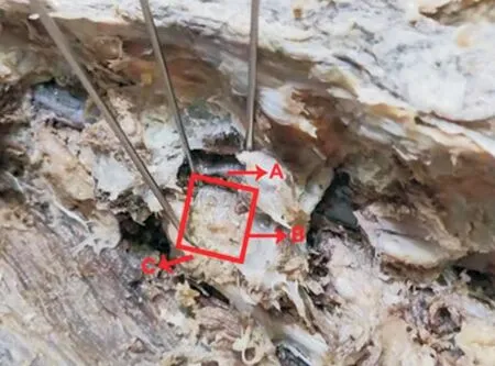
Figure2 Improved quadrangle:A, the dural sac, is the height of Improved quadrangle;B, superior border of the caudal vertebra, is the base of Improved quadrangle;C, the exiting nerve root,is the hypotenuse of Improved quadrangle.
Table 3 The paired design t-test and correlation of measurements for the kambin's triangle and the improved quadrangle(±s)

Table 3 The paired design t-test and correlation of measurements for the kambin's triangle and the improved quadrangle(±s)
Kambin Triangle Modified quadrangle t P Height(mm) 11.20±2.10 11.19±1.93 0.050 0.961 Bottom(mm) 10.78±1.95 12.14±1.78 -8.270 0.000 Area(mm2) 61.79±20.71 137.71±38.20 -25.262 0.000
4. Discussion
Kambin proposed in 1973 that there is a safe working area in the posterolateral side of the vertebral body, which is composed of the traversing nerve root or dural sac, the exiting nerve root, and the superior border of the caudal vertebra. This is a right-angled triangular area, in which there are no important blood vessels and nerves, so it can be operated safely. In order to commemorate kambin, later generations called this area Kambin's triangle. The concept of Kambin's triangle provides a solid theoretical basis for lumbar fusion via intervertebral foramen approach. Minimally invasive transforaminal lumbar fusion (MIS-TLIF) and percutaneous endoscopic transforaminal lumbar interbody fusion
(PE-TLIF) are the two most representative minimally invasive lumbar fusion procedures at present. Foley [16] proposed Minimally invasive transforaminal lumbar fusion (MIS-TLIF) in 2002. By establishing an expandable working channel in the Wiltse space between the longest muscle and the multifida muscle, the operation is performed in the "Kambin's triangle" to avoid the peeling of the paraspinal muscles, and can significantly reduce surgical trauma and intraoperative bleeding. In theory, MIS-TLIF can significantly reduce iatrogenic damage to paraspinal soft tissue by using expandable working channels for surgical operation. [17],However, during the operation, the long-term traction of the expandable channel affected the blood supply of the paraspinal muscle, and even damaged the posterior branch of the spinal nerve, resulting in the denervation of the paraspinal muscle and further leading to the ischemic necrosis of the paraspinal muscle after operation[18]. We can judge the effect of operation on paraspinal muscle by observing the cross-sectional area of multifida muscle after operation. [19].Su Kai [20] and others measured the cross-sectional area of the multifidus muscle after MIS-TLIF and PLIF surgery by MRI, and found that the crosssectional area of the multifidus muscle was significantly reduced regardless of whether it was MIS-TLIF or PLIF surgery, and both. There was no statistical difference between the two groups,indicating that MIS-TLIF surgery did not reduce the damage to the paraspinal muscles compared with traditional open surgery. The safety of MIS-TLIF surgery in elderly patients is also questioned.A study shows that after MIS-TLIF surgery for patients older than 80 years old, the incidence of postoperative complications is as high as 66.7%, 9.5% of patients died of postoperative septicemia and pulmonary embolism within 30 days after operation [21].Therefore,in order to further reduce surgical trauma and reduce the risk of anesthesia in elderly patients, Osman [22] and others began to try to use intervertebral foramen lens for lumbar fusion, which can completely perform nerve root decompression, endplate preparation and bone graft fusion, thus realizing the endoscopy of lumbar fusion.Compared with MIS-TLIF, it has the advantages of smaller incision,less trauma to patients, complete preservation of paraspinal muscle function, short postoperative recovery time, patients can get out of bed on the day of operation, and can be operated without general anesthesia. it is more suitable for patients with advanced age, poor general condition and high risk of anesthesia[23-25]. Regardless of whether it is MIS-TLIF or PE-TLIF, the operation is completed in the "Kambin's triangle", but because the space between the outgoing nerve root and the dural sac is very narrow, it is difficult to perform a thorough operation through the kambin triangle approach.Decompression, preparation of endplates, and placement of a cage of adequate size can easily cause damage to the traveling nerve root and displacement of the cage after surgery [4, 6]。How to reduce the incidence of complications is an urgent problem for spine surgeons The main problems of minimally invasive trans-kambin triangle lumbar fusion are that it is easy to damage the exiting nerve root and the displacement rate of the cage is high. Decompression,preparation of endplates, and placement of a cage of sufficient size can easily lead to the displacement of the cage after surgery.Once the cage is moved backward into the spinal canal, it is easy to compress the nerves and cause the patient to produce corresponding symptoms, and even size The possibility of fecal incontinence; due to the limitation of the narrow operating area, a little carelessness during the operation may damage the exiting nerve root [4, 6, 25-27]. If a special device is designed to pull the outgoing nerve root during the operation, the base of the Kambin's triangle can be made longer, the operating area can be larger, a larger interbody cage can be placed, and the risk of nerve injury can be reduced. Lee [28] If a special device is designed to pull the outgoing nerve root during the operation, the base of the Kambin's triangle can be made longer, the operating area can be larger, a larger interbody cage can be placed,and the risk of nerve injury can be reduced. Lee[29] tried to use an expandable cage to avoid damage to the exiting nerve root, but follow-up studies showed that the expandable cage not only failed to reduce the chance of nerve root injury, but also increased the risk of intraoperative subsidence. and expensive.
To master the anatomical structure of Kambin's triangle is the premise to improve the efficacy of surgery and ensure the safety of surgery. For this reason, more and more scholars at home and abroad have devoted themselves to the study of the kambin triangle, using cadaver specimens or imaging techniques to study the anatomical structure characteristics of kambin and measure related parameters.There are some differences in the data depending on the method.Ozer[9] divided the kambin triangle into three types through autopsy studies: type I is a closed triangle without any space in the middle,type II is a narrowed triangle, and type III is a normal kambin triangle. It was found that 82.4% of the patients' bone kambin belonged to type II and type III,and there is little space in the kambin triangle. Hardenbrook[4] measured the average surface area of this area to be 1.83cm2, and the area gradually became larger as the segment descended, and the area of L5~S1 was the largest at 2.19cm2. They also found that in the kambin triangle there is a trapezoidal safety zone without any nerve tissue limited by the width of the lower pedicle. The average area of this area is 1.19cm2, of which L5~S1 has the largest area of 1.26cm2. There is no nerve tissue in the safety zone, which can be safely of surgical operations.Ding Yi [12] used imaging technology to calculate the area of Kambin's triangle. The L1~L4 segment is between(153.73±37.34)mm2~(193.19±36.15)mm2. With the downward movement of the lumbar segment, The area gradually increased, but the angle between the exiting nerve root and the dural sac gradually became smaller, and the Kambin's triangle gradually became narrower and longer. Through autopsy, Liu Delong [14]measured the area of the Kambin's triangle of the L4 segment to be 104.65mm2±23.66mm2,the angle between the exiting nerve root and the dural sac was(44.40±2.21)°; the area of the Kambin's triangle of the L5 segment was(91.81±16.78)mm2, the angle between the exiting nerve root and the dural sac was(35.40±2.37)°.
Previous studies have done a lot of research on the anatomy of the intervertebral foramen and the Kambin's triangle, and also measured a large number of parameters, but they have not considered the possibility of expanding the Kambin's triangle.Lertudomphonwanit[15] pointed out that if the nerve root is used for traction pulling makes the base of the kambin triangle wider and the area larger.Nakamura[30] designed a new type of L-shaped nerve retractor. During the operation, the L-shaped nerve retractor was used to open the exiting nerve root, and the cage was safely placed under the protection of the L-shaped nerve retractor. Based on previous experience, KenNagahama et al [26] improved the surgical technique and equipment, and designed a new type of oval sleeve and J-shaped nerve retractor. For the first time, the patients with lumbar spondylolisthesis were treated with the aid of transforaminal mirror for the first time. Intervertebral fusion has achieved good surgical results. The incidence of postoperative nerve root-related complications is 8%, and the fusion rate reaches 88%.Yin Peng et al. used a self-designed bilingual bone graft sleeve for PE-TLIF surgery. During the operation, the bilingual flap outside the bone graft channel was used to protect the exiting nerve root,traversing nerve root and dural sac in the implant. Outside the bone channel, a bone graft channel with an outer diameter of 13 mm can be safely placed for surgical operations, which greatly improves the safety of surgery[31]. In the above studies, the surgical operation was performed by designing a special instrument to pull out the nerve root, and good surgical results were achieved, but there was no corresponding theoretical support. Therefore, in this study, cadaveric specimens were used to measure the length of each side of the lumbar kambain triangle, calculate the area, and use Kirschner wire to pull and fix the traveling nerve root to make the neurogenic kambin triangle into a rectangle. area, and compared the two groups of data to provide a certain theoretical reference for clinical practice. In this study, through the measurement of 5 cadaveric specimens, it was found that the height of the kambin triangle of the lumbar vertebrae ranged from(8.95±1.38)mm~(13.56±1.41)mm, and the length of the base ranged from(8.48±0.81)mm~Between(13.05±1.11)mm, the area is between(37.90±6.52)mm2~(88.46±11.85)mm2, in this narrow area, it is difficult to insert a conventional size interbody cage, and the exiting nerve root is pulled After fixing, the kambin triangle becomes a quadrangle, the height range is between(9.38±0.70)mm~(13.40±1.46)mm, the base edge is significantly longer, and the area is significantly enlarged between(9.55±0.90)mm~(13.80±0.94)mm, It is(89.69±12.11)mm2~(184.70±21.31)mm2. Through our research, we can find that the traditional kambin triangle is a very narrow area. If the surgical operation is performed in this area, it is difficult to perform thorough decompression, endplate preparation and placement of a cage of sufficient size, it is easy to cause damage to the exiting nerve root and the displacement of the cage after surgery. If the exiting nerve root is pulled and fixed during the operation, the kambin triangle can be turned into a quadrilateral, and the base edge of the operation area can be lengthened. Significantly expands the area of the kambin triangle.
This study shows that in the lumbar intervertebral foramen, there is a narrow surgical operation area surrounded by the exiting nerve roots, the dural sac and the superior border of the caudal vertebra.Intra-canal decompression, adequate endplate preparation and bone graft fusion, and it is very easy to damage the exiting nerve root during the operation. If a special device is designed, the exiting nerve root will be pulled and fixed, so that the traditional kambin triangle becomes a quadrilateral. Then, the base edge of the operation area can be significantly lengthened, the area can be significantly enlarged, and the operation can be performed more safely.
With the clinical application of percutaneous endoscopic transforaminal lumbar interbody fusion (PE-TLIF), it marks that lumbar fusion has completely entered the era of endoscopic surgery.Compared with traditional open surgery, its trauma is more severe.It is small, the risk of anesthesia is lower, and the postoperative bed rest time is shorter. However, this operation also has the problems of the displacement rate of the cage and the high rate of nerve root injury. Therefore, mastering the anatomy of Kambin's triangle is a necessary condition to improve the surgical efficacy and reduce the surgical risk. The current study shows that it is feasible to expand the kambin triangle area with special instruments to insert a larger cage and reduce the risk of nerve root injury.
Author’ Contribution
The first author: Peng Qiuyu, completed the research content,processed the data and wrote the article; the second author:Chen Huanxiong, responsible for the revision of the paper; the corresponding author: Meng Zhibin; proposed the research content and experimental design; Yu was responsible for assisting in the completion of the research content and data processing.
Conflict of Interest Statement:
The author declares that the content and opinions written in the paper are neutral and objective, and there is no conflict of interest.
杂志排行
Journal of Hainan Medical College的其它文章
- Research progress of autophagy in acute lung injury induced by multiple factors
- Research progress and comparison of the establishment of animal models of radiotherapy or chemotherapy-induced oral mucositis
- Based on the national patent database to explore the rule of traditional Chinese medicine in the treatment of hyperlipidemia
- Nonsurgical intervention for neuroclaudication due to lumbar spinal stenosis: Interpretation of the 2021 American Association for the Study of Pain Guidelines
- A mechanism to improve early diabetic nephropathy by modulating autophagy-related mechanisms with Yuye Decotion
- Study on the mechanism and active components of Radix et Rhizoma Rhei in the treatment of Alzheimer's disease based on molecular docking