Various configuration types of the foveal avascular zone with related factors in normal Chinese adults with or without myopia assessed by swept-source OCT angiography
2022-09-14YanMinDongHaiYanZhuZhenHuiLiuShuYingFuKeWangLiPingDuXueMinJin
INTRODUCTION
The foveal avascular zone (FAZ) is a ring-like vascular free zone located in the center of the macula, which is surrounded by a network of continuous capillary plexus from retina. As a highly refined area of the human retina, FAZ possesses unparalleled significance for maintaining normal visual function. The FAZ area is highly sensitive to ischemia for its high oxygen demand. It is found that FAZ area increased in some disease states such as retinal vascular diseasesand glaucoma. Moreover, the area of the FAZ is significantly correlated with visual acuity in diabetic retinopathy (DR) and retinal vein occlusion (RVO). Quantitative evaluation of FAZ can be used as a potential biomarker for macular ischemia in retinal vascular diseases. In order to investigate the variation of FAZ in different disease states, it is necessary to first explore the morphological characteristics of normal FAZ and the factors that may influence it. Although multiple studies have published data of FAZ parameter in normal individuals, less data was available using swept-source optical coherence tomography angiography (SS-OCTA).
But to this day I do not know what strange impulse made me take George to see her and to tell her, before I had confided13 in another living soul, of our engagement
SS-OCT is the latest development in optical coherence tomography (OCT) technology, which employs a swept-source light and optical detector. With a longer wavelength and faster scanning speed, SS-OCT can present fundus structures in a broad, high-resolution and non-invasive manner. Building on the principles of OCT, SS-OCT has also been applied to SS-OCTA, thus enabling a non-invasive and depth-resolved imaging of the retinal and choroidal microvasculature.Despite obvious advantages over traditional OCT/OCTA, the widespread of SS-OCT/OCTA remains relatively limited in clinical practice.
36. Is your name Conrad?: Note that the Queen, now sure of her victory, plays with the tiny man by guessing common names instead of the unusual ones she tried the previous two days. Critics often consider the queen s actions to be reprehensible. Critic Roger Sale, for example, condemns the queen for her cruelty to the only character who has shown her any sympathy and offered her any assistance (Sale 1978). In my view, this is the first time she has the upper hand in any situation and she is savoring it. She has been a victim of the three men in the story, her father, her king/husband, and her helper. Finally she has triumphed and gained some control, the control she needs to protect her child. Return to place in story.
In modern times, the most common romantic instance is perhaps the hiding of an engagement ring in a food item or drink as part of a marriage proposal.Return to place in story.
The present study aims to identify the morphological characteristics and potential influence factors of FAZ in normal individuals in order to provide normative data from a cohort of normal Chinese adults with or without myopia.
SUBJECTS AND METHODS
This study was conducted according to the principles of the World Medical Association Declaration of Helsinki and was approved by the Ethics Committee of the First Affiliated Hospital of Zhengzhou University. All participants in the study signed the informed consent prior to participation.
Normal Chinese adults with or without myopia were consecutively recruited from August to December 2021. The following inclusion criteria were adopted: age range between 18 and 60y; best-corrected visual acuity (BCVA) ≥1.0 and intraocular pressure (IOP) range 10-21 mm Hg; refractive error ≤-10 D; axial length (AL) ≤28 mm; and slit lamp microscope and fundus examination with no abnormalities.Subjects with any kind of systemic disease (such as hypertension and diabetes mellitus), current pregnancy, any ocular disease (except for ametropia), any ocular trauma or any history of eye surgery were excluded. One eye per person was then randomly selected for examination.
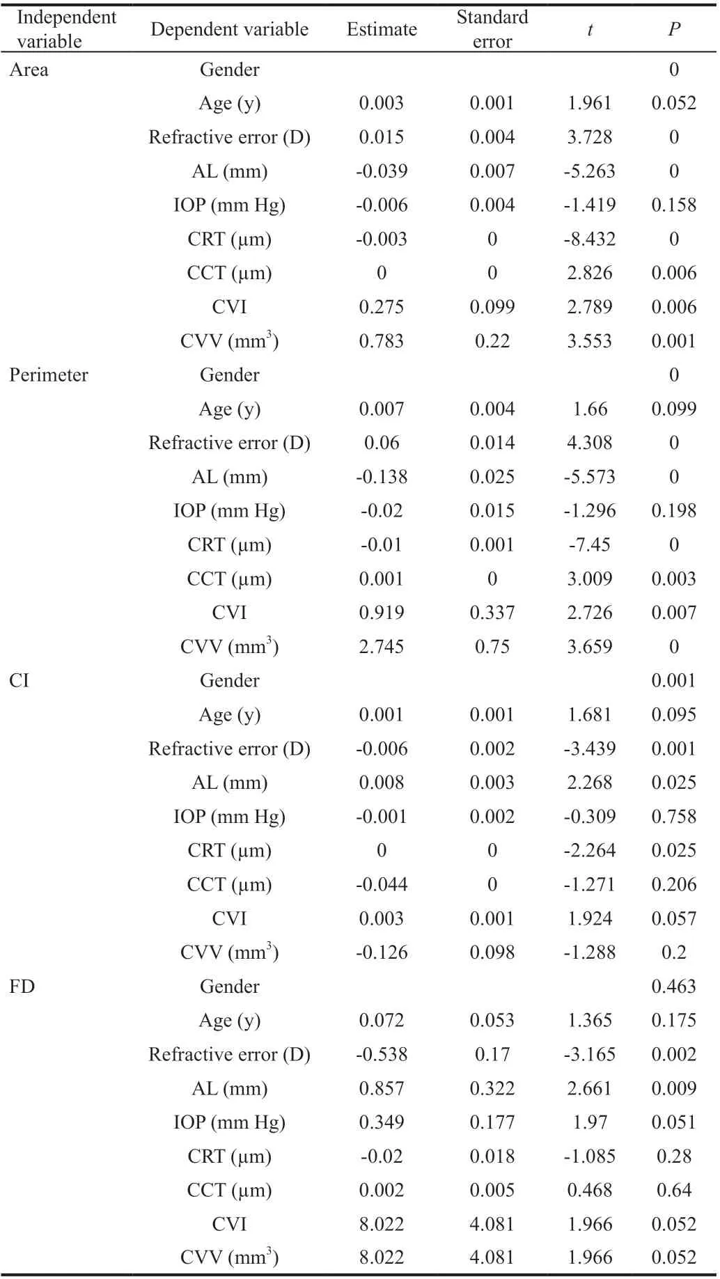
In this study, females possessed a significantly larger FAZ area compared with that of males. This result was found to be consistent with previous studies, which may be due to thinner fovea retinal thickness in females than males.There was no correlation between FAZ parameters and age,as reported by other researchers. In fact, the association between age and FAZ area is always a controversial topic.Fujiwaradescribed a positive correlation between age and FAZ area among subjects aged 40 years or older.Moreover, Yureported that the FAZ area increased annually by 1.48%, and they speculated the main reason for the trend was relative reductions in oxygen and nutrient demand.Various studies have shown that the FAZ area is related to retinal thickness under the macular fovea, but not to choroid thicknesswhich was supported in this study.Furthermore, the multivariate regression analysis demonstrated that CRT was significantly correlated with FAZ. A linear relationship was also present between CRT and FAZ area,perimeter, CI, and FD. AL is another important factor that may influence FAZ measurement. Previous studies have shown that AL was negatively correlated with FAZ area.According to univariate regression analysis, AL was found to be correlated with the area and perimeter of FAZ; however, in the multivariate regression analysis, there was no significant correlation between AL and FAZ area. Fujiwaraalso suggested AL was not significantly correlated with FAZ area.Therefore, this issue remains ambiguous and requires further study.
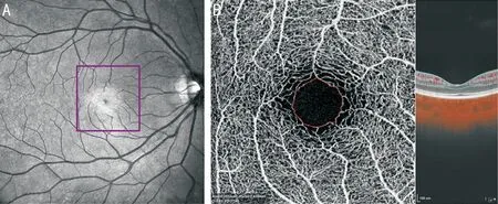
The SS-OCT/OCTA system (VG200D; SVision Imaging, Ltd., China) has been previously described. The core technology of the system contained: an SS laser with 1050 nm central wavelength and A-scans with a scanning rate of 200 K per second. The device had a longitudinal resolution of 5 μm and transverse resolution of 13 μm with a scan depth of 3 mm. The instrument was embedded with an eye-tracking utility and had a combining follow-up mode to eliminate eye-motion artifacts. All examinations were performed by equally skilled photographers and adhered to consistent parameters. Macular structure images were performed by ‘Star 18Line R32’ mode to observe retina and choroid. In terms of angiography, the FAZ images were obtained by scanning mode ‘Angio 3×3 521×512 R4’,centered on the fovea. The A-scan density was 512 lines(horizontal) ×512 lines (vertical), which was repeated four times, and the average value was taken to improve the SNR.Then images with signal intensity ≥8 were included in the data analysis. The area (mm), perimeter (mm), circularity index(CI) and fractal dimension (FD) of FAZ were automatically analyzed using van Gogh v1.36.10 instrument software(Figure 1).
The trees grew so thick and near together that it was almost impossible to see through them, only straight in front of him lay a little patch of meadowland
The macular area was divided into three regions: central fovea,parafovea, and perifovea according to the Early Treatment Diabetic Retinopathy Study (ETDRS) grid. Average central retinal thickness (CRT) and central choroidal thickness(CCT) in the central fovea were then analyzed. Choroidal vascularity index (CVI) and choroidal vessel volume (CVV) of central fovea were obtained from the research function of the instrument software.
Statistic software SPSS (version 25.0; IBM,Chicago, USA) was used for data analysis. All continuous variables were expressed as mean±standard deviation, and classification variables were expressed as frequency (%).Comparisons of factors between groups were performed using one-way ANOVA for variables with normal distributions,while Kruskal-Wallis test was adopted for variables with skewed distributions. Univariate and multivariate regression analyses were then performed in order to determine factors related to FAZ. The regression analysis of the FAZ values included: age, gender, refractive error, AL, IOP, CRT, CCT,CVI, and CVV. For all of the analyses,<0.05 was considered to be statistically significant.
RESULTS
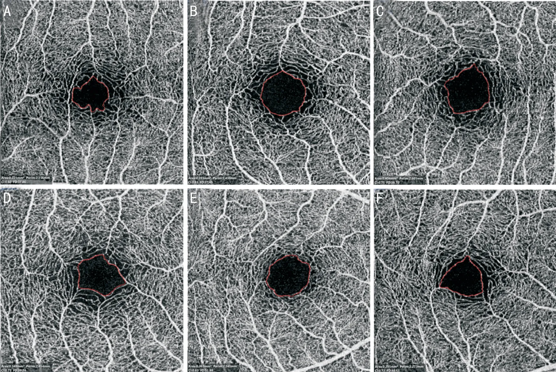
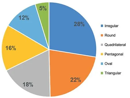
Eldalydetected five different FAZ patterns: horizontally oval, rounded, pentagon, vertically oval and nonspecific configuration in an Egyptian population study, in which horizontally oval superficial FAZ was shown to be the most common pattern. In this study, six FAZ forms were observed in normal Chinese adults, with most having specific patterns,including round, oval, triangle, quadrilateral and pentagonal that reached up to 72%, while a few exhibited an irregular configuration accounted for 28%. To our knowledge, this is the first study using SS-OCTA to describe FAZ morphology in a normal Chinese population. The results showed that the morphology of FAZ was variable in normal adults.

The shape of FAZ exhibited by SS-OCTA was found to be variable, in which most of the fundus images showed specific patterns.The round configuration was noted in 28 eyes (22%), while the quadrilateral configuration was present in 23 eyes (18%),pentagonal configuration in 20 eyes (16%), oval configuration in 15 eyes (12%), and triangular configuration in 6 eyes (5%).In addition, the irregular configuration was found in 35 eyes(28%). The characteristics and distribution of FAZ patterns are shown in Figures 2 and 3.
This study provided a detailed quantitative analysis of FAZ.Accordingly, the six patterns of FAZ in normal Chinese adults with or without myopia were initially described, and the obtained results support previous studies pertaining to the association between gender, CRT and FAZ area. This study is also the first to report on the variation of FAZ according to different degree of myopia.
DISCUSSION
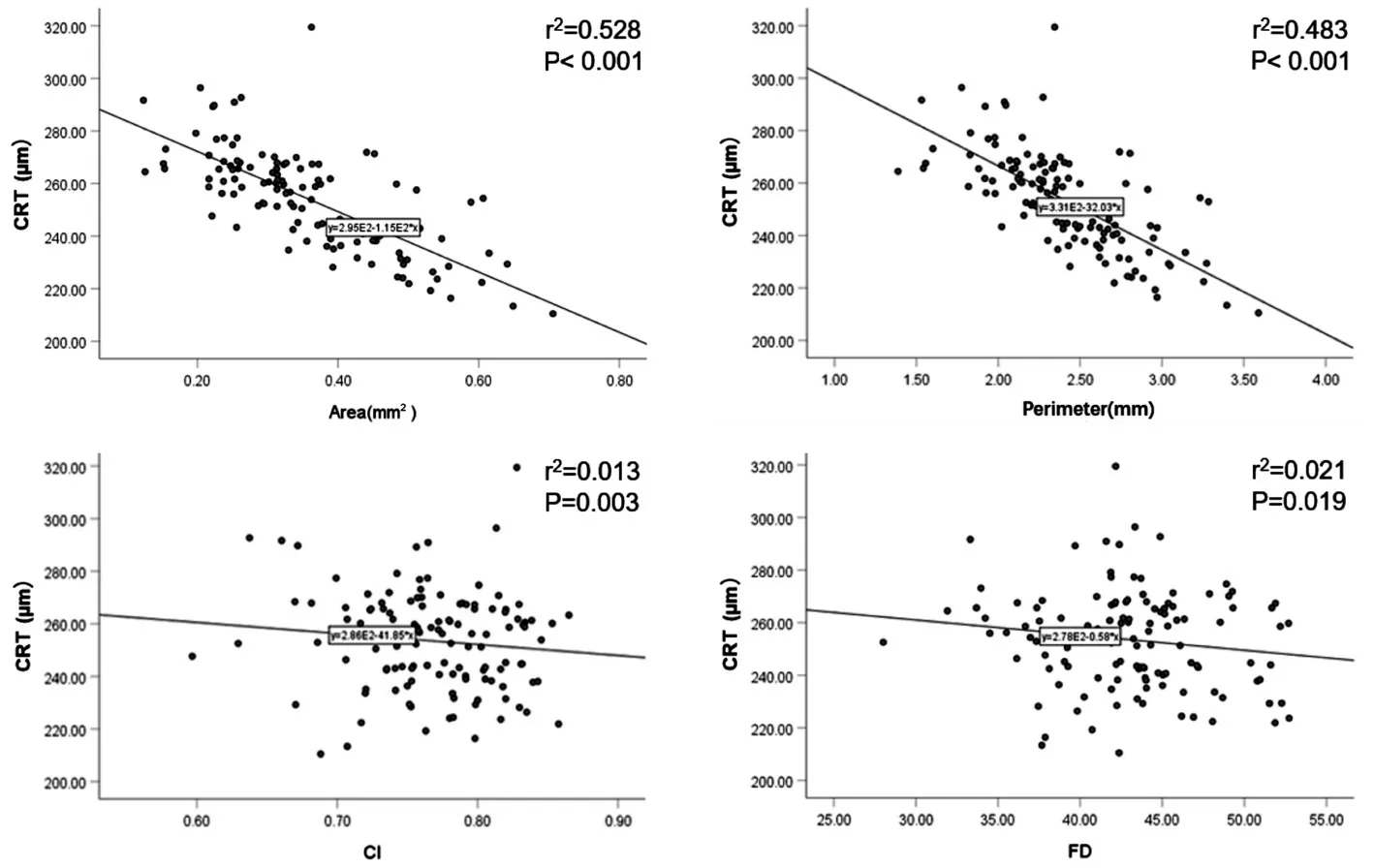
The results of the univariate regression analyses for FAZ parameters are shown in Table 3. According to the univariate regression analysis, multiple factors were noted to be correlated with FAZ. Specifically, gender, refractive error, AL, CRT, CCT,CVI, and CVV were significantly related to FAZ area and perimeter in the study. Refractive error, AL and CRT were also significantly related to the CI of FAZ. Moreover, refractive error and AL were significantly related to the FD of FAZ.CRT showed significant correlation with FAZ in multivariate regression analysis. In addition, a linear relationship was observed between CRT and FAZ area, perimeter, CI and FD.With a thinner CRT, FAZ area and perimeter exhibited an increasing trend (<0.01). The linear relationship between CRT and FAZ is shown in Figure 6. No obvious correlation between age and FAZ parameters was present in the univariate or multivariate regression analysis.
This observational, cross-sectional study enrolled a total of 127 normal volunteers, including 61 males (48%) and 66 females (52%), in which 127 eyes were examined. The mean age was noted to be 29.5±8.22y (18-58y).Meanwhile, the mean area and perimeter of FAZ was found to be 0.37±0.12 mmand 2.42±0.41 mm, respectively. The mean FAZ area of the female subjects was 0.41±0.11 mm,while that of male subjects was 0.32±0.11 mm. The average FAZ area of males was significantly smaller compared with that of females (<0.01). Moreover, males had a smaller FAZ perimeter and CI compared with those of females (P<0.01).The FAZ parameters of the subjects are shown in Table 1.
The main limitations of the present study are variability of refractive error and small sample size for subgroup. Therefore,it is necessary to further expand the sample size across different degrees of myopia so as to verify FAZ related variation.Furthermore, continuous monitoring of these indicators in the progression of myopia may provide more reference value for evaluation and prognosis of the disease.
CI and FD are another two quantitative indexes to evaluate FAZ morphology. Bigger CI and FD indicate more regular FAZ morphology and capillary distribution. Previous studies found that FD was significantly reduced in patients with DR,and FD was significantly associated with disease severity and visual acuity. Therefore, accurate measurement of the shape and size of FAZ can be used as an important quantitative indicator for monitoring early morphological variation of the disease.
All subjects underwent a comprehensive ophthalmic examination, including BCVA and refractive error assessmentautorefractor (CV-5000; Topcon Co, Japan), IOP measurement (CT-800; Topcon Co, Japan),slit-lamp microscope assessment of the anterior segment,fundus photography using Scanning Laser Ophthalmoscop(Daytona P200T; OPTOS, England), and AL measurement(Meditec IOL Master 500; Carl Zeiss, Germany).
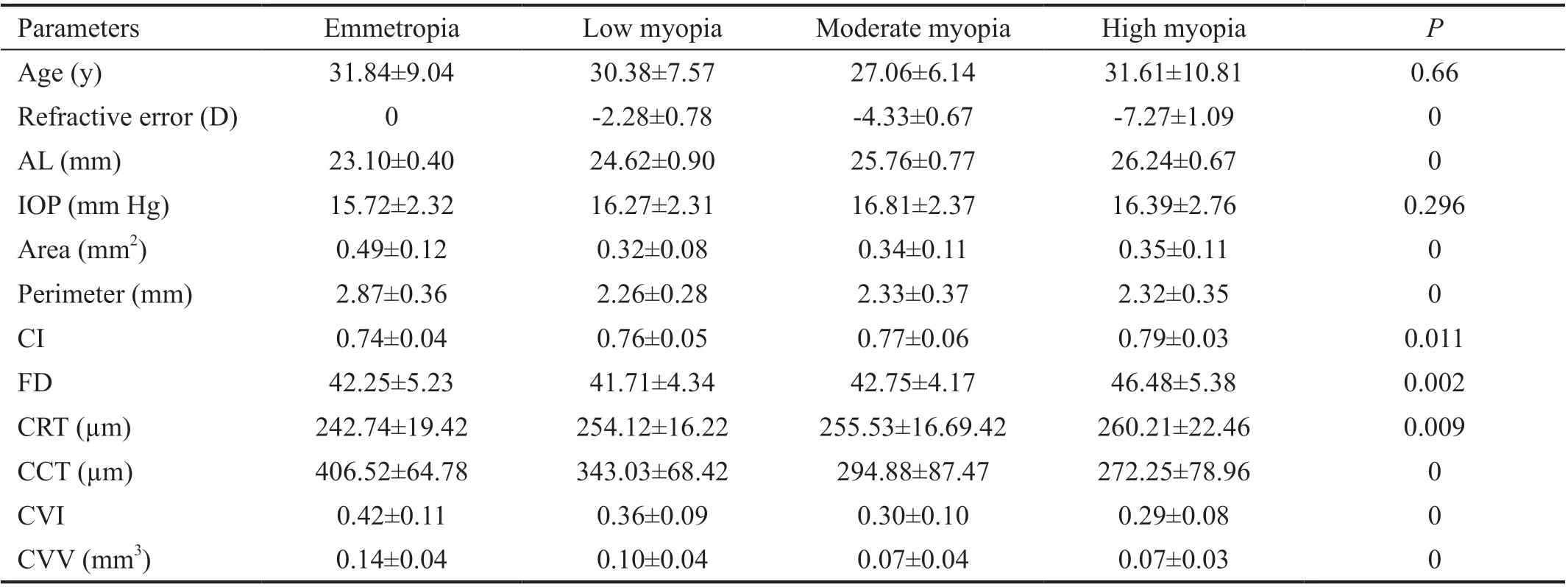
In this study, the subjects were detailed and grouped according to refractive errors. The variation of FAZ parameters in myopic eyes was also preliminarily discussed. All myopic eyes showed a smaller FAZ area and perimeter compared with those of the control (<0.01). Additionally, FAZ area had an increasing trend with myopia severity. Previous studies have also shown that FAZ area was significantly enlarged in highly myopic eyes than in those with non-high myopia. All myopic eyes exhibited thicker CRT and thinner CCT compared with that of emmetropic eyes. The CVI and CVV were also shown to be statistically decreased in the myopia groups. These results werealso consistent with previous studies. However, though a variation of FAZ in myopia group was noted, there was no significant correlation between the parameters and refractive error or AL in the regression analysis.
Several previous studies have described FAZ area in Chinese subjects using different OCTA instruments. Zhouevaluated young Chinese subjects with average age of 25.95±1.79y and found that the mean FAZ area was 0.30±0.11 mmusing the Optovue Fourier-domain OCTA system. In a study of 262 normal Chinese volunteers whose age ranged between 5 and 89y, Zhoureported that the mean FAZ area was 0.30±0.03 mmin all eyes using Zeiss HD-OCT with Angioplex. Moreover, by employing the Optovue RTVue XR OCTA, Wangfound that the mean FAZ area was 0.35±0.12 mmin 105 normal Chinese volunteers who had an average age of 35.9±13.8y. Notably, the mean FAZ area in the present study was found to be 0.37±0.12 mm, which was slightly greater than the above results. We believe that the main reason for the difference is the use of different instruments.Corvicompared FAZ values using a 3×3 mmvolume scan pattern centered on the fovea with seven different OCTA devices and suggested that making comparisons between instruments is nearly impossible and the set of measurements from various instruments are not interchangeable. Therefore,it may be necessary to use a single device in order to reduce variability in clinical practice. Other reasons may involve differences regarding age and gender distribution.
This study described the morphological characteristics of FAZ and quantified the parameters of FAZ in normal Chinese adults with or without myopia using SS-OCTA. The patterns of FAZ exhibited various forms, including: round, oval, triangle,quadrilateral, pentagonal, and irregular configurations. The results suggest that the morphology of normal FAZ can be variable. Additionally, all myopic eyes showed a smaller FAZ area and perimeter compared with those of emmetropic eyes.To determine the exact correlation between the parameters and myopia, it is necessary to conduct longitudinal studies to explore the continuous variation of FAZ. However, detailed quantitative evaluation of FAZ parameters, such as FAZ area, will help us to identify early morphological changes of macula diseases. Specifically, establishing quantitative parameters of FAZ would not only provide details of macular pathophysiology but also could possibly contribute as a biomarker in disease staging.
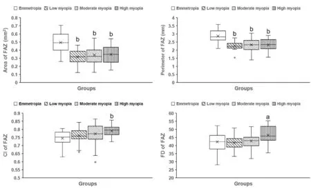
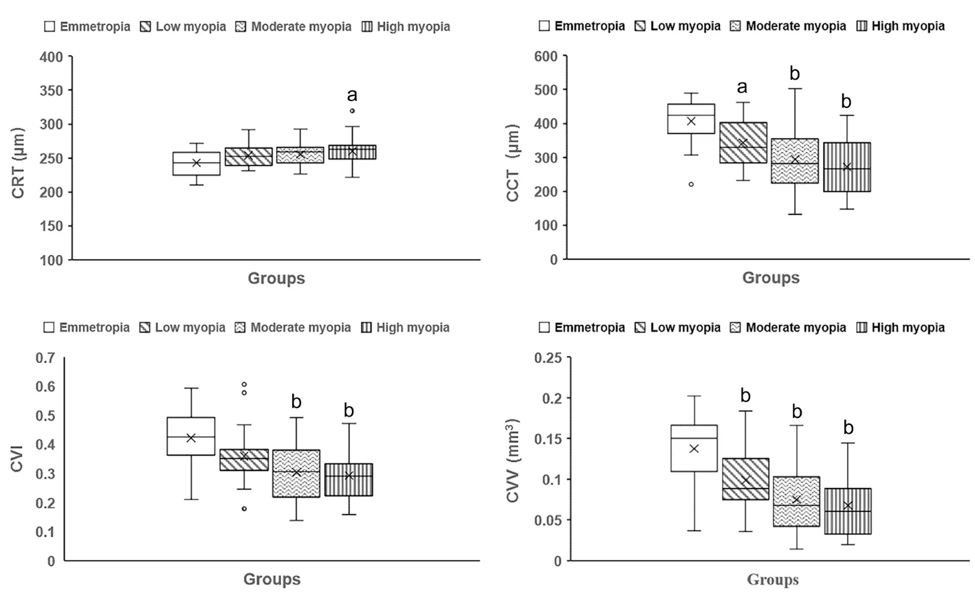
According to refractive error, subjects were divided into the emmetropia group (=25) and myopia group (=102). Next, the myopia group was divided into three subgroups: low degree myopia (=26)with diopter (D) range from -0.75 to -3.00 D, medium myopia(=53) with diopter range from -3.00 to -6.00 D and high myopia (=23) with diopter range from -6.00 to -10.00 D. The mean AL in the three groups was 24.62±0.90 mm (23.06-25.85 mm), 25.76±0.77 mm (23.68-27.18 mm), 26.24±0.67 mm(24.24-27.42 mm), respectively. All myopic eyes showed a smaller FAZ area and perimeter (<0.01). Only the high myopia group exhibited a statistically significant lager CI and FD when compared with the emmetropia group (Figure 4).All myopic eyes possessed a thicker CRT than the emmetropic eyes, which changed according to myopia severity. In contrast,the myopia group was shown to have a statistically thinner CCT than normal. Additionally, the CVI and CVV were found to be statistically decreased in both the moderate and high myopia group compared with those of the normal group(<0.01; Figure 5). The variables of different groups are shown in Table 2.
But the old man shook his head sadly, for he knew that the villain35 was only crushed for the moment, and that he would shortly be revenging himself upon them
Dong YM and Zhu HY designed and conducted the study, and drafted the article; Liu ZH, analyzed the data; Fu SY, Wang K, and Du LP collected the samples; Jin XM designed the study, and revised the article.
Supported by the National Natural Science Foundation of China (No.81970792; No.82171040); Medical Science and Technology Project of Health Commission of Henan Province (No.YXKC2020026).
You shall, however, only lend them to him on condition that you may accompany him when he goes to make the payment, and that you then have permission to run before him as a fool
None;None;
The stress and the loneliness began to destroy Marianne. She sucked down alcohol like it was water. She fed and clothed her sons, put them to bed, but refused to leave her home. She thought about slashing16 her wrists.
None;None;None;None;None.
杂志排行
International Journal of Ophthalmology的其它文章
- What can we learn from negative results in clinical trials for proliferative vitreoretinopathy?
- Suggestions on gut-eye cross-talk: about the chalazion
- A novel mutation of RPGR in a Chinese family with X-linked retinitis pigmentosa
- Novel technique of penetrating keratoplasty in high-risk grafts with significant corneal neovascularization
- COVlD-19 infection with keratitis as the first clinical manifestation
- Corneal histomorphology and electron microscopic observation of R124L mutated corneal dystrophy in a relapsed pedigree
