Dual histologic variants of schwannomas in orbital schwannomatosis
2022-09-14GaryJohnMercadoLeandroVictorArcenaElaineYatcoOmaaElizabethArcellanaNuquiRolandJosephTan
Dear Editor,
We present an extremely rare case of orbital schwannomatosis with dual histologic variants.Schwannomas are tumors resulting from the slow proliferation of Schwann cells located in peripheral nerve sheaths
. They are commonly seen in middle-aged individuals but had been reported in patients 20-70 years of age. The presence of multiple schwannomas in the body has often been associated with neurofibromatosis (NF). However, schwannomatosis is considered in the absence of coexisting criteria diagnostic of NF. Schwannomatosis is a rare genetic disorder with an annual estimated incidence of 0.58 cases per 1 000 000 persons and usually manifests in the 2
-3
decade of life
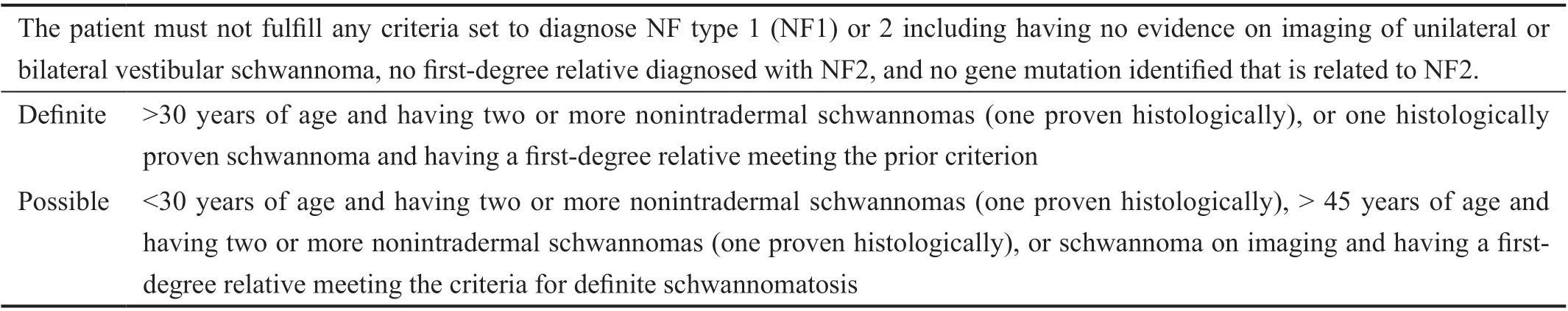
. A set of criteria to diagnose schwannomatosis proposed by Baser
is summarized in Table 1.
改革开放40年来中国的扶贫开发一直在占全国1/3左右的县(区)展开,不计其数的各级干部尤其是基层干部参与其中。仅精准扶贫开展以来,每年有近百万的干部参加帮扶。大量的干部工作到扶贫第一线,直接与贫困户、贫困村打交道,了解贫困户的致贫原因、帮助参谋和设计扶贫项目和脱贫方式,使参与其中的干部能够比较深入地了解和认识国情,培养踏实的工作作风,在工作过程中也锻炼了干部的能力。这些经历和锻炼成为参与帮扶干部的宝贵财富,也将成为提高政府部门尤其是基层组织的决策水平和管理绩效的重要推手。
Schwannomas found within or adjacent to the orbit are rare and represent only 1% of all orbital neoplasms
. A lone schwannoma in the orbit without association to NF is called primary orbital schwannoma
. Having multiple orbital ones is often NF-associated. However, it is extremely rare to have multiple orbital schwannomas without fulfilling any criteria diagnostic of NF, or orbital schwannomatosis.
This is a case of a 25-year-old Filipino female with a 3y history of gradually progressive proptosis of the right eye and enlarging mass at the superomedial anterior orbit of the same side. There were associated blurring of vision of the right eye,7/10 pain in right orbit, and diplopia on all gazes. She had no pre-existing medical conditions and there was no similar condition indicative of NF1 or 2 in the family. On examination,the best-corrected visual acuity (BCVA) were 20/50 on the right eye and 20/20 on the left eye. A proptosis of 4 millimeters(mm) was measured by exophthalmometry. A 10×5 mm
nodular, solitary, non-movable, and non-tender mass was palpated at the superomedial anterior orbit with an inferolateral displacement of the right globe. There was resistance to retropulsion. There was no relative afferent pupillary defect.Limitations in extraocular muscle movement in the right eye were graded at -1 on all gazes. Intraocular pressures were at 14 mm Hg for both eyes. Anterior and posterior segments were unremarkable. The fundoscopy was unremarkable. There was no Lisch nodule in the iris or cataract seen. The principles outlined in the Declaration of Helsinki were followed. Written informed consent was obtained from the patient for this case presentation.
On systemic examination, she had no clinical evidence indicative of NF1 or 2 (
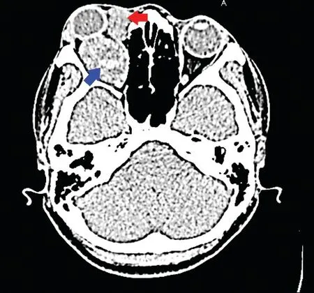
, café-au-lait macules, axillary or inguinal freckles, and palpable mass in any part of her body aside from the orbit). Cranial and orbital computed tomography(CT) revealed multiple well-circumscribed intraconal and extraconal isointense masses in the right orbit (Figure 1). The intraconal mass measured 15×23×15 mm
and was indenting the right globe posteriorly and displacing the intraorbital optic nerve segment medially. It had a central cystic component with fluid level. The extraconal mass measured 18×11×8 mm
,dumbbell-shaped, well-circumscribed, isointense, and was in the superomedial extraconal space. There was no other mass suggestive of meningioma or optic nerve glioma. There was also no mass appreciated in the vestibulocochlear nerve.
She underwent excision of the tumors through a lateral orbitotomy.Two orbital masses were removed from the superomedial anterior orbit measuring 6×8 mm
and 8×16 mm
, lightpink, and with smooth pseudocapsules. The larger intraconal mass measured 20×25 mm
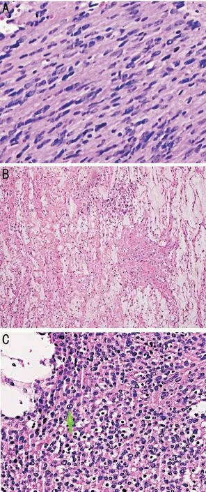
and was similarly encased in a pseudocapsule. The three excised lesions appeared to be distinct from each other. Histopathology read the tumors into two different types: schwannoma with myxoid degeneration(two superomedial anterior orbital masses), and schwannoma with degenerative atypia/ancient schwannoma (intraconal mass; Figure 2). On 6
week post-operative follow-up, her BCVA in the right eye improved to 20/25 and proptosis was resolved. She was advised to follow-up annually.
However, none of the cases reported having different types of histologic variants. Schwannoma has four major histologic variants: the classical/conventional type which is characterized by encapsulation and the presence of Antoni A composed of hypercellular spindle cells and Antoni B showing hypocellular myxomatous pattern; the relatively uncommon cellular type found in the vertebrae and exhibits Antoni A; the plexiform type commonly found in the skin and also exhibits Antoni A; and the melanotic type found in spinal roots,contains epithelioid cells and melanin and has potential to be malignant
. Additional variants include ancient schwannoma characterized by unusual hyperchromatic nuclei in the absence of mitoses. This is a rare variant and has been reported in the orbit only once
. Our patient presented both with classical and ancient schwannomas.
Specific identification of the nerve origin is difficult due to the anatomic complexity of the orbital soft tissues. However,the location of the extraconal masses in all the cases can be suggestive that the schwannomas developed from the supratrochlear, infratrochlear, or the supraorbital nerve
. CT and magnetic resonance imaging (MRI) are helpful for tumor localization. MRI can also show capsular enhancement and circumferential enhancement surrounding regions of cystic degeneration in the ancient subtype of schwannomas
. MRI is also necessary to ensure that there is no vestibular mass diagnostic of NF2. If present, ophthalmologists, who can be the first to see the patient, have to also screen for cataract and glaucoma. The patient also has to be assessed for other NF2 manifestations such as hearing loss, numbness, and weakness of the extremities and appropriate referrals shall be made.
This is the 4
, youngest, and 1
case of orbital schwannomatosis with two histologic types of schwannomas. Review of the literature showed only three other patients with orbital schwannomatosis
. Koktekir
reported a case of a 59-year-old male with two orbital masses on imaging with no associated NF findings whom they diagnosed as a case of orbital schwannomatosis. However, on excision, only one mass was removed with the smaller mass on the imaging as just a possible part of it making the case a probable primary orbital schwannoma. Although two more masses were removed 5y after, these could have been recurrences from the initial mass as both were excised from the same area
. Similar to the other reported cases, our patient presented with gradual proptosis,enlarging mass in the superior to medial anterior orbit with resulting hypoglobus, had multiple extra- and intraconal masses on imaging, had no findings associated with NF, and responded well to surgical excision
.
一杭想在楼下一个早餐点前喝碗豆浆,却见两个戴墨镜的男子一边狼吞虎咽地吃着包子,一边偷偷地看他,见一杭有所察觉,立即低下头吃东西。一杭的心提了起来,范坚强不会在路上对我下手吧?他迅速走过去,到前面一家早餐店买了两根油条,边走边回头,那两个人似乎并没有再注意他。一杭这才放下心来,看来是自己太多疑了,弄得草木皆兵。
无节幼体阶段依靠自身体内卵黄做营养,无须投喂饵料。在这一阶段,水温控制在20~22℃,PH保持在7.8~8.6,无节幼体期水质比较清净,幼体耗氧量低,微充气即可。无节幼体期历时约4d,经过6次蜕皮变态为蚤状幼体。无节幼体培育期间每天加水15~20cm。
Even though orbital schwannomas are usually asymptomatic,surgery is a mainstay in its management
. Schwannomas are commonly well-circumscribed, encapsulated solitary masses that adhere to the nerve, but can be separated from the nerve origin with careful dissection to avoid nerve transection.Early surgical excision is desired to avoid the sequelae of tissue compression. Schwannomas become symptomatic by compressing adjacent structures
. Compression of the globe leads to proptosis and the optic nerve to the blurring of vision. Its space-occupying effect can result in diplopia from limiting extraocular movement and to eye pain
. Complete surgical excision of the mass is desired, as incomplete excision may lead to tumor recurrence similar to the other patient reported by Koktekir
. For our patient, despite complete excision of masses, regular surveillance of tumor recurrence is necessary since recurrence was reported to be common in orbital schwannomatosis.
None;
None;
None;
None;
None.
1 Sheikh MM, De Jesus O. Schwannoma. Updated 2021 Aug 30. In:
. Treasure Island (FL): StatPearls Publishing; 2021.
2 Lee SH, Kim SH, Kim BJ, Lim DJ. Multiple schwannomas of the spine:review of the schwannomatosis or congenital neurilemmomatosis: a case report.
2015;12(2):91-94.
3 Baser ME, Friedman JM, Evans DG. Increasing the specificity of diagnostic criteria for schwannomatosis.
2006;66(5):730-732.
4 Kim KS, Jung JW, Yoon KC, Kwon YJ, Hwang JH, Lee SY.Schwannoma of the orbit.
2015;16(2):67-72.
5 Leung KCP, Lam NKY, Chan E, Ko TCS. Primary recurrent orbital schwannoma treated with surgical excision and mitomycin-C.
2020;19:100784.
6 Koktekir BE, Kim HJ, Geske M, Bloomer M, Vagefi R, Kersten RC.Orbital schwannomatosis in the absence of neurofibromatosis.
2014;25(6):2109-2111.
7 Golblum J. Soft Tissues. In: Golblum J, Lamps LW, Mckenney JK,Myers JL, editors.
. 11
ed.Cambridge: Elsevier; 2018.
8 Young SM, Kim YD, Jeon GS, Woo KI. Orbital frontal nerve schwannoma—distinctive radiological features.
2018;186:41-46.
9 Sun XL, Wen K, Xu ZZ, Wang XP. Magnetic resonance imaging findings for differential diagnosis of perianal plexiform schwannoma:case report and review of the literature.
2018;6(5):88-93.
猜你喜欢
杂志排行
International Journal of Ophthalmology的其它文章
- What can we learn from negative results in clinical trials for proliferative vitreoretinopathy?
- Suggestions on gut-eye cross-talk: about the chalazion
- A novel mutation of RPGR in a Chinese family with X-linked retinitis pigmentosa
- Novel technique of penetrating keratoplasty in high-risk grafts with significant corneal neovascularization
- COVlD-19 infection with keratitis as the first clinical manifestation
- Corneal histomorphology and electron microscopic observation of R124L mutated corneal dystrophy in a relapsed pedigree