Combination of spark discharge and nanoparticle-enhanced laser-induced plasma spectroscopy
2022-08-31QingXueLi李庆雪DanZhang张丹YuanFeiJiang姜远飞SuYuLi李苏宇AnMinChen陈安民andMingXingJin金明星
Qing-Xue Li(李庆雪), Dan Zhang(张丹), Yuan-Fei Jiang(姜远飞),Su-Yu Li(李苏宇), An-Min Chen(陈安民), and Ming-Xing Jin(金明星)
Institute of Atomic and Molecular Physics,Jilin University,Changchun 130012,China
Keywords: laser-induced plasma spectroscopy,spark discharge,nanoparticle,spectral enhancement
1. Introduction
Laser-induced breakdown spectroscopy (LIBS) plays a vital role in elemental analysis. It has developed rapidly in the past many years and has been used in many fields, including industrial detection,environmental monitoring,and biological research. The LIBS technology has low requirements for samples,so the pretreatment of samples is simple,or no pretreatment is required, and the damage to the sample is minimal.This advantage is significant in analyzing and detecting valuables and biological samples. And the LIBS can implement in-situ detection and multi-element real-time rapid detection of samples.[1–7]
In analyzing and detecting trace elements by the LIBS,sensitivity is crucial.[8–11]For traditional LIBS technology,the sensitivity mainly depends on the laser pulse energy,laser beam incident angle, beam focusing conditions, and the type of detection devices. Many researchers have optimized the parameters mentioned above. On this basis, the scheme is to enhance the spectral intensity by improving the sample and adding auxiliary enhancement devices in the generation and expansion stage of plasma to enhance the LIBS signal. As is well known, the emission signal of LIBS depends on the light absorption of the sample surface.[12]Therefore,improving the sample surface can improve the LIBS sensitivity. It should be noted that the chemical properties of the sample surface cannot be changed in the process of improving the sample surface. And the preparation method cannot be too complex; otherwise, the distinct advantages of LIBS technology will be lost. Considering the cases above, the nanoparticles(NPs)were chosen and used to improve the sample surface.
Gold NPs were deposited on the surface of brass samples by dropping NPs solution on the surface samples and drying. When the laser irradiated the sample surface, the deposited NPs were removed in the process of laser ablation,and there was almost no pollution to the sample. For solids, NPs could adjust the electromagnetic field to produce local surface plasma resonance. If they had good geometric optical properties, they could create a very high optical response.[13]By observing the images of ablation craters from scanning electron microscope,De Giacomoet al.found that the sample surface with NPs became irregular after having been ablated by laser,[14]and there existed many burst cavities with the sizes ranging from hundreds of nanometers to several microns. The conclusion was drawn that the NPs deposited on the sample surface were similar to multiple ignition points: each ignition point is the source of seed electrons through field emission.Owing to electron injection and multiphoton ionization, subsequent breakdown happened to the sample to realize the enhancement effect. Also,they investigated the dynamics of the plasma-induced during LIBS and nanoparticle enhanced LIBS(NE-LIBS) by acquiring spectrally resolved images, finding the increased quantity of material ejected during NE-LIBS.[15]Therefore, in this work we choose NPs as our method to improve the sample surface, and increase the spectral intensity without changing the chemical properties of the sample.
In recent years, there have been many mature methods of additionally enhancing the LIBS, such as spatial confinement LIBS,[16–18]sample pre-heating LIBS,[19–21]magnetic confinement LIBS,[22–24]and double pulse or multipulse LIBS.[25]Most of these methods require additional laser equipment or optical auxiliary equipment,thereby usually increasing the complexity and cost of the system. A spark discharge(SD)system coupled to the LIBS is considered to be a low-cost and straightforward method to overcome the low energy values of lasers by reheating the plasma,increasing emission intensity to improve the signal intensity of plasma. Liet al.compared the ablation craters and found that the SD will not increase the ablation mass of the sample.[26]Zhouet al.proved that SD-LIBS can obtain a stronger emission through lower discharge voltage. In addition, the SD-LIBS has made progress of reducing relative standard deviation and improving the signal-to-noise ratio,indicating the great advantage of using a nanosecond discharge circuit to increase the spectral emission of plasma[27]In general, the air resistance between the two electrodes is high, and SD will not occur. With the plasma generated,the SD size gradually becomes bigger with the expansion of the plasma.The electric field between the two electrodes re-excites the plasma. The atoms and ions within the plasma obtain more energy, and are excited to a higher level to achieve the enhancement effect.[28]
In addition, some previously published papers show that the enhancement effect after the two enhancement methods, such as a combination of electrical spark and pre-heated sample,[29]combining pre-heated sample and spatial confinement,[30]combining spatial and magnetic confinement,[31,32]combining spatial confinement and dualpulse excitation,[33,34]combining magnetic confinement dualpulse excitation,[24]and combining NPs and dual-pulse,[35]have been combined, is better than using only a specific enhancement method. In view with these studies, in this paper the NPs are combined with the SD to assist LIBS,which can improve LIBS signal.When the laser irradiates the sample surface,more atoms are excited due to the action of NPs. In the process of plasma expansion,the plasma is reheated by SD to absorb energy further. Also,plasma temperature and electron density are calculated. The plasma temperature and electron density are two characteristic parameters of plasma,which are helpful in gaining a more in depth understanding of the behavior of plasma and improving the application in elemental analysis.
2. Sample preparation and experimental devices
The sample preparation process is very simple as shown in Fig. 1. The sample Au NPs were provided by Shanghai Chao Wei Nanotechnology Company in the form of suspension. Its concentration was 1000 ppm. The Au NPs were diluted into a solution of target concentration with bottled purified water, and mixed with the solution thoroughly through ultrasonic vibration. In the ultrasonic cleaning instrument,the brass samples had been cleaned carefully with alcohol and pure water. A pipette gun was used to drop the resulting solution onto the surface of the brass sample quantitatively, the sample was transferred onto a constant temperature heating table,and then dried at 60◦C.The preparation method is similar to the surface enhancement method of converting liquid samples into solid samples.[36]
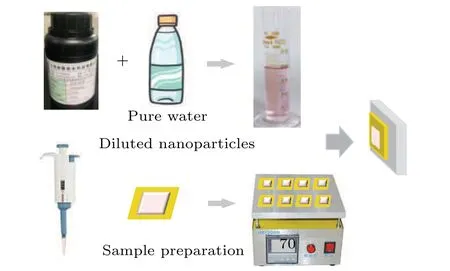
Fig.1. Nanoparticle solution preparation and sample drying process.
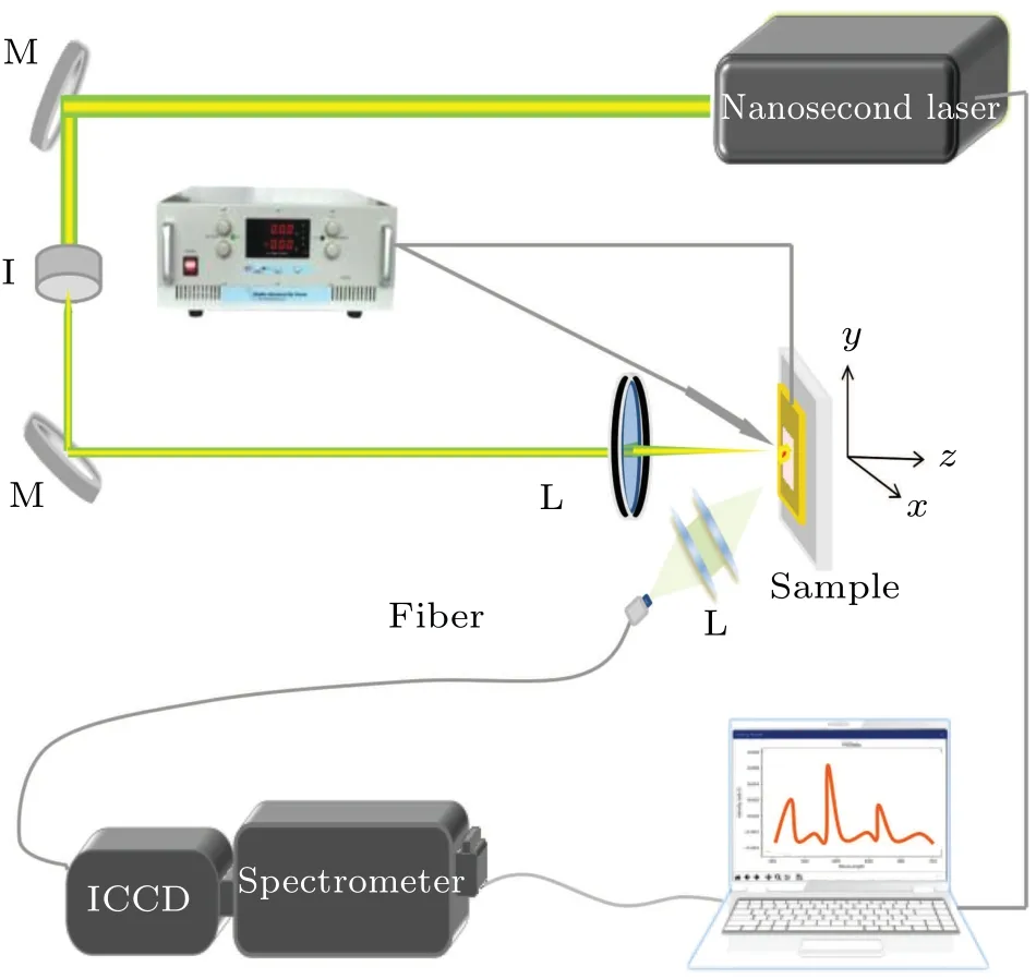
Fig.2. SD-LIBS experimental devices(I:iris;M and L:lens;ICCD:intensified charge-coupled device).
The experimental device is shown in Fig. 2. The whole discharge experiment was carried out under normal temperature and pressure.The laser source used in the experiment was an Nd:YAG nanosecond laser;the laser energy was 20 mJ and kept unchanged;the laser repetition rate was set to be 1 Hz. A direct-current power supply provided the voltage for the discharge circuit. The sample was used as one electrode, and another pure tungsten electrode was added to the edge of the plasma. The distance between the two electrodes was about 3 mm. This could improve the stability of the optical path and avoid influencing experimental results by the electrode angle.[37]The laser was focused on the sample by using a 100-mm focusing lens. The sample was fixed on a movable three-dimensional platform to ensure that each laser pulse and SD are at different sample positions. The plasma signal was collected by two lenses and coupled to a spectrometer(Spectra Pro 500, PI Acton, Princeton instruments) through an optical fiber. The spectrometer was equipped with an intensified charge-coupled device(ICCD,PI-MAX4). An external diode trigger was used to synchronize the time between emission and laser. The delay time used in this experiment was 1.0 µs for avoiding continuous emission, and the gate width was 50 µs for collecting more line emission.
3. Results and discussion
3.1. Spectral intensity
Figure 3 shows the comparison of spectral intensities for a selected nanoparticle concentration(6 ppm)and different discharge voltages. The voltages are 0, 2, and 4 kV. The above three panels show the spectra of brass samples without NPs,and the below three panels display the spectra obtained by depositing NPs with a concentration of 6 ppm on the surfaces of brass samples. It can be seen from the figure that the combination of SD and NPs has a noticeable enhancement effect on the spectral emission. After combining the two enhancement methods,the enhancement effect is more than ten times that of the traditional LIBS technology. For the case with NPs and 4 kV, the spectral intensities for 510.0 nm and 521.8 nm reach saturation,are close to those with NPs and 2 kV.So,the spectrum at 515.3 nm is selected as the curve with the concentration of NPs.
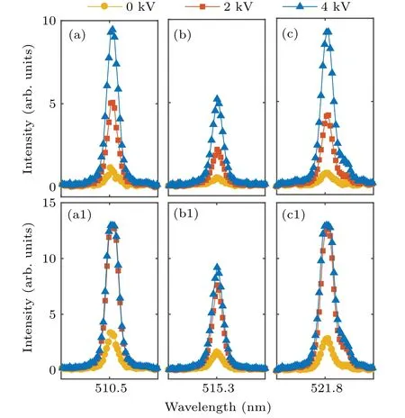
Fig.3. Comparisons between spectral intensities without((a)–(c))and with((a1)–(c1))NPs(6 ppm)and different discharge voltages.
Figure 4 shows the spectral intensity of Cu (I) with a wavelength of 515.3 nm. The voltages used are 0,2,and 4 kV.When nanosecond laser irradiates the surface of the brass sample, many electrons and atoms escape from the sample surface to form gaseous cluster plasma. The plasma resistance is smaller than that of air, and two electrodes are connected by plasma to form a closed circuit. A strong electric field is formed between the two electrodes, and the air is broken down, resulting in SD. More particles absorb energy and are excited to higher energy levels. Therefore, it is observed that the spectral intensity obtained with SD is significantly higher than that without SD. With discharge voltage increasing, the energy deposition of plasma increases,the enhancement effect will become more evident.

Fig. 4. Evolution of Cu (I) 515.3-nm spectral intensity with nanoparticle concentration for different discharge voltages(0 kV,2 kV,and 4 kV).
In addition, the line emission is significantly improved after depositing NPs. The enhancement of NPs is attributed mainly to the three aspects below. (i) Because the size of Au NPs is small, the breakdown threshold of metallic NPs is much lower than that of bulk metal,[15]which can effectively reduce the lower limit of laser excitation of plasma.[38]The ablation efficiency is improved by reducing the ablation threshold, which is the advantage of low thermal conductivity of small-size objects under laser irradiation.[39](ii) Laser irradiates the sample surface,and each NP is equivalent to an ignition point,[14]heating the adjacent sample. (iii) The NPs attached to the sample surface make the sample surface rough,and the rough structure on the metal substrate surface has a better enrichment effect on light.[40]When irradiated by laser,it will absorb more energy,improve the coupling efficiency between laser and metal substrate,and excite more sample particles to achieve the effect of spectral enhancement. Also,with the increase of the nanoparticle concentration, the line emission under SD increases more evidently than that under SD.
When the concentration of NPs is low, the enhancement effect is relatively weak. With the increase of the concentration of NPs, the enhancement effect is positively correlated with the concentration, and the enhancement effect is pronounced. When the concentration of NPs reaches a specific value,the enhancement effect is not apparent.[41]As a result,the inter-particle distance effectively induces the collective oscillation of conduction electrons in NPs under laser irradiation,like the scenario in surface-enhanced Raman spectroscopy.[42]On the other hand,when the concentration of NPs is too high,the NPs will prevent the laser from reaching the sample surface. Many researchers have optimized the case,[41,43]which will not be described in detail here.
In addition, the increase in the concentration of NPs enhances the coupling between the laser and the sample. This is equivalent to increasing laser energy. And,the plasma shielding effect will become evident with the increase of the laser energy. So,the plasma shielding effect will occur with the increase in the concentration of NPs,which can hinder the laser and the sample surface from interacting. The plasma emission cannot increase continuously nor significantly.[41]At this time,SD plays a role;the particles in the plasma absorb energy by SD, which is equivalent to reheating the plasma and fully ionizing the particles in the plasma. The higher the discharge voltage, the more energy the plasma absorbs from spark discharge,and the more pronounced the enhancement effect.
3.2. Plasma temperature
The error of using two spectral lines to calculate the electron temperature is relatively large. The measurement error of plasma temperature is largely due to the measurement of the transition probability and intensity of the spectral line. Therefore,the two spectral line method should be rewritten and calculated with multiple spectral lines to improve the calculation accuracy. Since the sample we selected in the experiment is Cu, there are three spectral lines near 515.3 nm, and these spectral lines are relatively close to each other so that the reliable transition data can be found. For example,the difference in the upper-level excitation energy of the spectral line in Table 1 is significant,so the three spectral lines are used to calculate the plasma temperature.[44]The formula is as follows:[45]

whereλis the wavelength,Iis the intensity,gis the degeneracy,Ais the transition probability,Eis the energy,kis the Boltzmann constant,Tis the plasma temperature,andCis the intercept. Figure 5 shows a typical Boltzmann plot without and with NPs(6 ppm)at discharge voltages of 0 kV and 2 kV,respectively.

Table 1. Spectral parameters of Cu(I)lines.
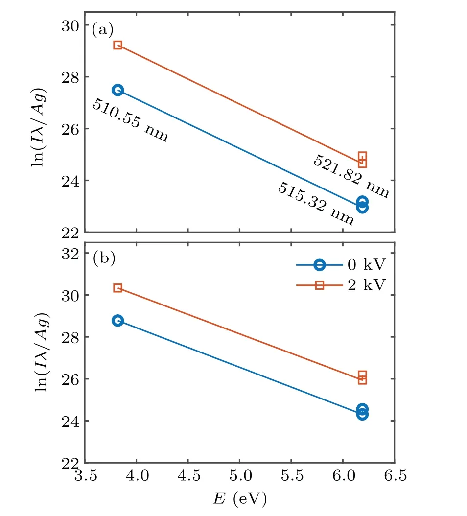
Fig. 5. Typical Boltzmann plot without (a) and with (b) NPs (6 ppm) at discharge voltages of 0 kV and 2 kV.
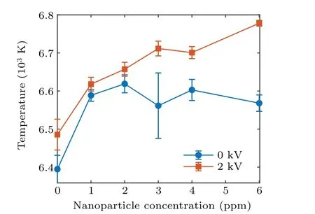
Fig.6. Evolution of plasma temperature with nanoparticle concentration at 0 kV and 2 kV.
Figure 6 displays the calculated plasma temperatures for different nanoparticle concentrations at 0 kV and 2 kV. After depositing NPs, the plasma temperature increases. The NPs enhance the interaction between the laser and the target, and increase the plasma temperature,but the enhancement effect is not linearly related to the concentration of NPs. The change in the plasma temperature with the concentration of NPs is similar to that with laser energy increasing. The plasma shielding effect is gradually strengthened with the nanoparticle concentration increasing in a range from 2 ppm to 6 ppm. So the plasma temperature slowly reaches saturation. When a voltage of 2 kV is applied to the two electrodes, the SD happens, and thus reheating the plasma. The more particles in plasma absorb the energy from SD, the plasma temperature increases.[46]And,the SD has not the plasma shielding effect;the plasma temperature increases continuously in the concentration range from 2 ppm to 6 ppm. Therefore,when the SD is combined into NE-LIBS,it is equivalent to further amplifying the enhancement effect of NPs.
3.3. Electron density
Generally, Stark broadening plays a leading role in a plasma with an electron density more than 1015cm−3.[47]The spectral line is no longer strictly dependent on the velocity nor on the temperature distribution of electrons or ions. The electron density can be estimated by using the Stark broadening of the spectral line. The Stark broadening line caused by the collision of atoms in the plasma with a large number of fastmoving free electrons is of the Lorentzian type. The broadening caused by the ion perturbation field makes the spectral line profile Gaussian,but its contribution to spectral line broadening is much smaller than that of electron collision. Therefore,the line shape is Lorentzian, and its line profile is shown in Fig.7.
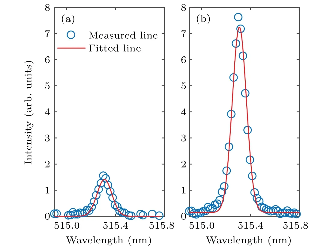
Fig. 7. Lorentz fitting of Cu (I) 515.3 nm without (a) and with (b) SD at 2 kV for 6-ppm concentration of NPs.
Figure 7 shows slight asymmetry on both sides of the spectral line. The spectral line reflects the internal change process of the plasma. It will be affected by the surrounding particles, such as the intense collisions among electrons and the effect of electric field perturbation produced by ions. The collision among electrons leads the spectral lines to broaden and the central frequency to increase. In addition, the nonuniformity of particle density in plasma is also one of the important reasons for the asymmetry of spectral lines.
At present, the Stark broadening method is an important method of obtaining the electron density of the plasma. Once the spectral line shape is determined,the electron density can be obtained from[20]

where ∆λ1/2is the width,ωis the collision parameter,neis the density. In plasma,the broadening of atomic spectral lines results from the adjacent charged particles acting on luminescent atoms,so the broadening of spectral lines is a function of electron density. As can be seen from Fig.8,comparing with simple samples,the plasma density increases significantly after adding NPs. Still, the changing trend of electron density rises up slightly with the increase of nanoparticle concentration. The reason is the same as that for the plasma shielding effect caused by the rapid expansion rate of plasma generated by nanoparticles mentioned above. It can be seen from the figure that without nanoparticles,the simple discharge has a weak effect on electron density. It is further proved from the mechanism that SD will not increase the ablation quantity of the sample. After the voltage is applied, the electron density increases linearly with the concentration of nanoparticles due to the fact that the SD fully ionizes more particles in the plasma,and thus improving the optical emission line intensity.
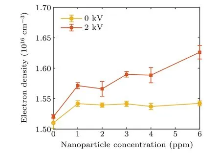
Fig. 8. Evolutions of electron density with nanoparticle concentration increasing at 0 kV and 2 kV,respectively.
4. Conclusions
The combination of electrical spark and NPs enhanced LIBS technology is introduced. The study is mainly to analyze the optical enhancement of two techniques. The evolution of the spectral intensity with nanoparticle concentration is measured. Comparing with the spectra produced by a single laser pulse,the enhancement effect of NPs is evident. But the spectral line intensity of Cu(I)for higher nanoparticle concentration does not change significantly with the concentration of NPs. The reason is that the higher concentration of NPs promotes the absorption of laser energy,and the plasma shielding effect appears earlier. The SD made up for this disadvantage.After the plasma is generated,the discharge transmits the energy to the plasma, thus the plasma is reheated, resulting in the enhancement effect. The combination of the two methods proves to be able to significantly enhance the emission intensity of LIBS technology.
Acknowledgements
Project supported by the National Key Research and Development Program of China(Grant No.2019YFA0307701),the National Natural Science Foundation of China (Grant Nos. 11674128, 11674124, and 11974138), and the Jilin Provincial Scientific and Technological Development Program,China(Grant No.20170101063JC).
猜你喜欢
杂志排行
Chinese Physics B的其它文章
- Magnetic properties of oxides and silicon single crystals
- Non-universal Fermi polaron in quasi two-dimensional quantum gases
- Purification in entanglement distribution with deep quantum neural network
- New insight into the mechanism of DNA polymerase I revealed by single-molecule FRET studies of Klenow fragment
- A 4×4 metal-semiconductor-metal rectangular deep-ultraviolet detector array of Ga2O3 photoconductor with high photo response
- Wake-up effect in Hf0.4Zr0.6O2 ferroelectric thin-film capacitors under a cycling electric field
