Clivus-involved immunoglobulin G4 related hypertrophic pachymeningitis mimicking meningioma: A case report
2022-06-27YangYuLiangLvSenLinYinChengChenShuJiangPeiZhiZhou
lNTRODUCTlON
Immunoglobulin G4 related disease (IgG4-RD) was initially noticed in patients with autoimmune pancreatitis in 2001 and formally named in 2010, classified as sarcoidosis with different manifestations in several organs and the same pathological characteristics[1,2]. The main characteristic of IgG4-RD is elevated levels of serum IgG4. Moreover, the lesions are often tumescent with abundant IgG4-positive plasma cells and fibrosis. Such inflammatory lesions can be seen in the pancreas, kidney, lungs, salivary glands, and other organs. Specifically, the conditions of IgG4-RD in the central nervous system are meningitis and hypophysitis[3]. As for the IgG4-related hypertrophic pachymeningitis (IgG4-RHP), the clinical and imaging manifestation is similar to that of meningioma, posing a challenge for preoperative diagnosis[4,5]. Additionally, the time and scope of operation should be considered carefully. Finally,this disease is related to some bacterial infections, such as tuberculosis. And we need to weight the pros and cons between these infections and corticosteroid therapy for IgG4-RD. Herein, we report a rare case with IgG4-RHP at the clivus area mimicking meningioma and discuss the relevant literature.
CASE PRESENTATlON
Chief complaints
A 40 year-old man was admitted for headache, bilateral temporal visual field defect, and limited abduction in both eyes.
History of present illness
Five years before the present admission, the patient started to experience discontinuous and aggravating headache. Owing to symptomatic deterioration, the patient was admitted to the neurology department of a local hospital. Because the patient also had a history of pulmonary tuberculosis, he was suspected of having tuberculosis meningitis and treated with anti-tuberculosis drugs at the local hospital. However, the symptom did not alleviate. Upon presentation to our hospital, the patient underwent a brain magnetic resonance imaging (MRI) scan that showed the presence of a clival lesion measuring 2.6 × 1 cmwith isointense signal on T1-weighted (T1WI) and T2-weighted (T2WI) imaging;accordingly, he was diagnosed with meningioma. The lesion was homogenously enhanced on contrast MRI with a dural tail sign (Figure 1). Because there was no cranial nerve function defect, the patients chose to undergo Gamma Knife Surgery at a dose of 11 Gy at the 45% isodose line, and regular followup was planned.
History of past illness
The patient had suffered from pulmonary tuberculosis 11 years ago and accepted standard antituberculosis treatment for 1 year.
Case 1.The two sources exhibit both uniform distribution.

Personal and family history
No other particular personal and family history was reported.
Physical examination
This patient showed right abducens paralysis, hoarse voice, bitemporal hemianopsia, and slight swallowing difficulty. No other positive signs were found.
Laboratory examinations
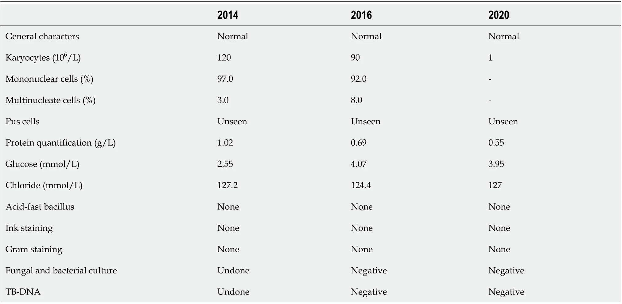
Lumbar puncture was performed and we found that the number of karyocytes (mainly mononuclear cells) and protein levels in cerebrospinal fluid had risen (Table 1).
After pathological results showed IgG4-RD, further systemic evaluation was performed to find other lesions associated with IgG4-RD. The serum IgG level was 17.20 g/L (reference range: 8.00-15.50 g/L),and the serum level of IgG4 was 1.90 g/L (reference range: 0.035-1.500 g/L). Tuberculosis associated gamma interferon release assay showed positive results with TB-IGRA (T-N) at 414.21 pg/mL.
Imaging examinations
After admission, routine laboratory testing and preoperative preparation were carried out. A repeat brain MRI scan showed that the lesion became larger, measured 3.8 cm × 2.9 cm × 2.9 cm, and compressed the adjacent brain stem (Figure 1). Further, small pneumatoceles in the upper lobe of the right lung were detected by thorax computed tomography (CT). Moreover, the examination of visual field confirmed binocular hemianopia (Figure 2). No other positive results were found.
由表4数据可以看出,铷和铌钽在各粒级中分布大致上是均匀的,没有相对富集的情况,据此可以认为铷和铌钽均匀地分布于各粒级中。
FlNAL DlAGNOSlS
The postoperative pathology confirmed the proliferation of fibrous tissue accompanied by numerous lymphocytes and plasma cells, which is displayed in Figure 3. Immunohistochemical staining showed positive results for CD138 and IgG4. Gene rearrangement test showed negative results for IgH. Thus,IgG4-RD was finally diagnosed.
长江之旅,稳居大江之上,睥睨两岸风光,乘风破浪前行。贵州西洋也正携手优秀经销商,顺应肥料行业发展浪潮,积极抓住机遇、应对挑战,向着助力中国农业发展、实现企商共赢、成就中国复合肥“航母”企业的广阔海天劈波斩浪不断前进。


TREATMENT
⑲OECD,Competitive Neutrality:Maintaining a level playing field between public and private business,Paris:OECD Publishing,2012,p.53.

For the diagnosis of IgG4-RD, solu medrol was administrated at a dose of 80 mgday, and methotrexate was administrated at 10 mg every week. Famotidine, calcium carbonate, and vitamin D3 tablets were prescribed against adverse reactions during the treatment. After discharge from the hospital, the solu medrol was tapered over 4 wk to 50 mgday.
OUTCOME AND FOLLOW-UP
The purpose of the operation was not only to perform a biopsy but also to alleviate symptoms. We know that the lesion would stretch meninges and then cause headache. Similarly, the lesion compresses cranial nerves to cause relevant symptoms. The resection can reduce meningeal tension, release compression, and finally alleviate headache and nerve deficits. Further, it is suitable to use the transnasal endoscopic approach for a clival lesion in IgG4-RHP. When the lesion is too broad to remove completely, it is sensible to leave some parts in order to maintain the integrity of the dura mater, which can prevent severe complications such as cerebrospinal fluid leakage and intracranial infection.
设备安装完成使用至今,基本实现了桥吊大车与集卡车辆的定位系统、桥吊远程监控系统、桥吊远程操控系统、码头入口闸机与集卡旋锁联动系统功能,达到了桥吊远程智能化操控的目标,基本达到了预期效果。待改进完善后,可进一步推广使用。同时建议开展桥吊小车定位系统的开发研究。
DlSCUSSlON
传统的假日购物季通常从黑色星期五开始,即感恩节第二天,然后一直持续至圣诞节。在这期间,零售商会打折,购物者则去寻找礼物、在商店排队、匆忙跑过通道以获得最佳优惠。这是以前的常态,当然在一些地区现在仍是常态,不过由于百货店和在线零售商不断更改折扣日期,在很多地方,购物季早在感恩节之前便已经开始,而且成为动态事件,没有固定的日期。
IgG4-RD of the CNS is mainly related to IgG4-related hypertrophic pachymeningitis and hypophysitis. Among them, IgG4-RHP is relatively rare, with the primary clinical manifestation of headache and other nerve function disabilities. Moreover, it was apparent that the cranial nerve function could partially recover once the disease was in remission. At the first onset of the disease,multi-organ disease is not widespread (57%)[6]. Therefore, regular follow-up and systemic evaluation is crucial.
2.2.1 师德水平 有的教师师德缺失,对待学生态度不端正,甚至辱骂、体罚学生,这必然导致师生关系紧张甚至对立。有些教师偏护优秀学生,对“差生”态度差,甚至冷嘲热讽或置之不理,使“差生”产生心理阴影。有些教师上课不严格要求自己,做不到为人师表,难以树立威信;有些教师备课不认真,消极怠工,举止粗俗,使得一些学生消极模仿或厌恶排斥。
Through this case, we summarize the differential diagnoses of IgG4-RHP, such as meningioma,tuberculosis meningitis, fungal meningitis, and metastatic tumor. Furthermore, the complete MRI images showed the lesion alteration during treatment. However, there are limited reports of this rare disease in the literature. Higher evidence-based studies are needed to promote the diagnosis and treatment of IgG4-RHP.
Other diseases, such as metastatic tumors and fungal infections, should also be considered. It was observed that metastatic tumors could spread and proliferate along the meninges, causing various severe symptoms. In this situation, the history of malignant tumor provided clues to the diagnosis.Likewise, a CNS fungal infection can show similar features, which can be identified by examining the cerebrospinal fluid.
Measuring the serum concentration of IgG4, radiological examination, and pathological screening are important for diagnosis. It is difficult to distinguish IgG4-RHP and meningioma before the operation and pathologic examination. The serum level of IgG4 can facilitate diagnosis, but it does not always show an increase. As reported by Wallace[12], the sensitivity and specificity of serum IgG4 were 90% and 60%, respectively. Moreover, the negative predictive value and positive predictive value of the serum IgG4 assay were 96% and 34%, respectively, which could be helpful and convenient to exclude the diagnosis of IgG4-RD related to the CNS[12]. It is also helpful to distinguish tuberculosis and IgG4-RD based on the fact that serum IgG4 does not significantly increase in tuberculosis[13]. Further,imaging results could be a crucial clue for preoperative diagnosis. Lumbar puncture provides the necessary information for differentiation from CNS infections and malignant tumors. IgG4 levels in cerebrospinal fluid have been reported to be elevated[14]. However, the concentration of IgG4 in cerebrospinal fluid could not distinguish this disease from other inflammatory pachymeningitis[6].
The patient underwent transnasal endoscopic approach resection which aimed to partially remove the lesion for pathology analysis and alleviate the headache caused by meningeal tension. During the operation, we found that the lesion extended to the sphenoid sinus and nasopharynx without a clear boundary. Notably, the local mucosa was edematous and tight. The clivus bone had been partially damaged, and the clivus epidural was thicker. The intraoperative frozen section examination revealed the proliferation of spindle cells accompanied by many lymphocytes and plasma cells.
Radiology examination plays an essential role in diagnosis. The lesion could be observed as linear dural thickening or a bulging mass. The linear dural thickened lesion appears both in the brain and spine. The tumoral lesion is frequently located in the clivus area. The heterogeneity was observed on MRI because of active inflammation. Typically, T1WI MRI would exhibit a hyperintense or isointense signal. Hypertrophic pachymeningitis usually shows thickening meninges and hypointensity on T2WI MRI, while it would become relative hyperintense when the inflammation aggravates[3,4,6,7,15]. The lesion would be homogenously enhanced on enhanced MRI. In this case, the lesion showed an isointensite signal on T1WI and T2WI and was homogenously enhanced on contrast MRI with a dural tail sign. CT showed that the skull was involved apparently and the lesion appeared hyperdense when contrast-enhanced CT was performed. In case of a meningioma, CT frequently displays that the lesion is isodense or has slightly higher density with a round, leafy, or flat shape[3,6]. Calcification becomes visible in some tumors[6]. Meningioma has similar characteristics as an IgG4-RHP lesion. T1WI often shows isointense or mildly hypointense signal, and T2WI usually shows isointense or mildly hyperintense signal. Besides, the meningioma could be markedly characterized by the tail of the meninges.
It is advisable to focus on some characteristics to help distinguish between meningioma and IgG4-RHP. We noticed that the symptoms of IgG4-RHP were severe and diverse, while those of meningioma were not as varied. These symptoms were due to inflammatory irritation and compression of the adjacent nerves and dura mater[16]. Another characteristic of IgG4-RHP was that the tail signal was broader than meningioma on MRI for the diffuse inflammation along with the dura mater. The meningioma lesion seems relatively confined and phymatoid compared with IgG4-RHP. Moreover, the IgG4-RHP lesion frequently involves extracranial parts.
3.裸车销售+电池租赁。这种模式是电动汽车生产商只出售裸车,由能源供应商提供电池租赁服务。这种情况下,电动汽车的电池集中于能源供应商,所以当其进行管理时具有规模效应,成本低,效率高,并且专业程度高。这种情况下大大降低了消费者的购车成本和用车成本,使车与电池产生问题的责任分离,并且电池的使用寿命和使用效率得到提高。但是由于不同的车型对电池的规格和充电要求的不同,短期内难以实现电池的标准化、统一化,进而难以快速的建设基础充电设施,阻碍了电动汽车的推广发展。但是,这个问题在电池生产商和电动汽车生产商的合作协商下,在政府法律法规和政策的要求下,电池和充电设施的标准化会得以解决。
CNS tuberculosis is another antidiastole. Patients with tuberculous meningitis often have a fever,headache, and focal neurological symptoms. And tuberculous meningitis is often secondary to pulmonary or intestinal tuberculosis. As for radiology examination, CT often exhibits nodular or punctate calcifications and hydrocephalus, and enhanced scans are often accompanied by meningeal strengthening. MRI frequently shows a hypointense T1WI signal and hyperintense T2WI signal. The enhancement scan could display irregular bar or nodular strengthening lesions of the meninges.Cerebrospinal fluid is essential for the diagnosis of tuberculous meningitis. Moreover, TB-IGRA could facilitate this diagnosis.
The patient confirmed that his headache and hoarse voice gradually improved after 1 mo. The follow-up was arranged 3 mo after the operation, which showed that the abduction movement could be achieved for binocular vision. Brain MRI showed that the residual lesion obviously shrunk (Figure 1). The change for bilateral visual fields is displayed in Figure 2.
Glucocorticoids and immunosuppressants can be used for the non-surgical treatment, such as prednisolone (0.6 mg/kg/d) for 4 wk. The dose of steroid was gradually decreased through 3-6 mo and the dose was finally maintained at 2.5 to 5.0 mg/d for 3 years[17]. Other immunosuppressants should be considered, such as methotrexate, cyclophosphamide, mycophenolate mofetil, and azathioprine[6,7].Another consensus recommended utilizing calcium carbonate and vitamin D3 tablets to prevent glucocorticoid-induced osteoporosis[18,19]. Additionally, it is essential to exclude some latent infections before using glucocorticoids and immunosuppressants. In this case, the patient had a history of tuberculosis and we performed the chest CT and TB-IGRA to ensure the absence of any current underlying infection. In future, when similar patients with the imaging characteristics described in this report are encountered, measurement of serum IgG4 levels may be helpful for diagnosis.
CONCLUSlON
IgG4-RHP is a relatively rare disease that seems complicated to diagnose preoperatively. The purpose of surgery is to obtain the specimens required for pathological examination and plan the follow-up treatment. It is essential to perform a rigorous follow-up and systematic assessment of the whole body.
FOOTNOTES
Yu Y collected the data, contacted with the patient, and wrote the manuscript; Lv L wrote and revised the manuscript; Chen C and Yin SL made the revision to the primary manuscript; Jiang S and Zhou PZ supervised the whole work and made the operation.
反应堆压力容器为核电站反应堆冷却剂系统的主设备之一,固定和包容堆芯及堆内构件,使核燃料的裂变反应限制在一个密封的空间内进行。它和一回路管道共同组成高压冷却剂的压力边界,是防止放射性物质外逸的第二道屏障之一。在筒体法兰上钻有58个螺孔,用以安装螺栓与顶盖密封,螺栓拧入过程中如果发生螺栓咬死的情况,处理起来比较困难,且影响较大。
IgG4-RD is a condition that affects multiple organs, and its clinical manifestations often vary across different organs. Reportedly, several kinds of bacterial infection can be causative factors for this disease related to stimulation with Toll-like receptor ligands[6,7]. Several previous studies have also reported the comorbidity of IgG4-RD with tuberculosis, as seen in our patient[8-11].
1·3·5 Project for Disciplines of Excellence-Clinical Research Incubation Project, West China Hospital,Sichuan University, No. 2019HXFH018.
Informed written consent was obtained from the patient for publication of this report and any accompanying images.
The authors declare that they have no conflict of interest to disclose.
The authors have read the CARE Checklist (2016), and the manuscript was prepared and revised according to the CARE Checklist (2016).
This article is an open-access article that was selected by an in-house editor and fully peer-reviewed by external reviewers. It is distributed in accordance with the Creative Commons Attribution NonCommercial (CC BYNC 4.0) license, which permits others to distribute, remix, adapt, build upon this work non-commercially, and license their derivative works on different terms, provided the original work is properly cited and the use is noncommercial. See: https://creativecommons.org/Licenses/by-nc/4.0/
China
Yang Yu 0000-0002-9901-1310; Liang Lv 0000-0002-5524-7014; Sen-Lin Yin 0000-0003-2241-3749; Cheng Chen 0000-0002-7540-1306; Shu Jiang 0000-0002-6700-7560; Pei-Zhi Zhou 0000-0002-8017-5833.
Fan JR
Wang TQ
Fan JR
猜你喜欢
杂志排行
World Journal of Clinical Cases的其它文章
- Stem cells as an option for the treatment of COVID-19
- Development of clustered regularly interspaced short palindromic repeats/CRISPR-associated technology for potential clinical applications
- Prostate sclerosing adenopathy: A clinicopathological and immunohistochemical study of twelve patients
- Effectiveness and postoperative rehabilitation of one-stage combined anterior-posterior surgery for severe thoracolumbar fractures with spinal cord injury
- Construction and validation of a novel prediction system for detection of overall survival in lung cancer patients
- Identification of potential key molecules and signaling pathways for psoriasis based on weighted gene coexpression network analysis
