Experimental investigation on the radiation background inside body counters
2022-05-13YuWangYuanYuanLiuBinWuXiangPengMengJianPingChengYingWangLiJiaoWangYunShiXiaoQinJianCaoJianFengZhangFeiTuo
Yu Wang · Yuan-Yuan Liu · Bin Wu · Xiang-Peng Meng · Jian-Ping Cheng ·Ying Wang · Li-Jiao Wang · Yun-Shi Xiao · Qin-Jian Cao · Jian-Feng Zhang ·Fei Tuo
Abstract In vivo measurement of radioactivity based on various body counters is arguably the leading measure used to determine the distribution and activity of radionuclides in human subjects,such as I-131 in the thyroid,Am-241 in the lungs, and Pb-210 in the skull. Throughout the measurements, the radiation background is the key factor that determines the sensitivity of the counter. Therefore, to facilitate in vivo measurements, a well-designed shielding room is required to create a low-background environment.However, because the compositions of the radiation background are quite complicated, the respective contributions from each source remain obscure, which places a considerable burden on seeking an optimized design of shielding rooms that strikes the optimum balance between the construction cost and background suppression effect.In this study, we conducted a systematic experimental investigation on the radiation background outside and inside four representative body counters with assorted designs using a variety of radiation detectors, including high-purity germanium detectors, CdZnTe detector, radon emanometer, and gamma-ray dosimeter. By carefully controlling the experimental conditions and synergetic analysis of the measurement results, in conjunction with previous studies, we separated and determined the relative contributions induced by environmental radiation (4%),airborne radon and its daughters (2%), the normal radioactivity of human subjects arising from K-40 (58%),cosmic rays(12%),and radioactivity in shielding materials and measuring instruments (24%). Furthermore, based on these results, we discuss practical guidelines to design a shielding room for body counters.
Keywords Body counter · Radiation background ·Shielding room · In vivo measurement
1 Introduction
Situations such as prolonged exposure to naturally occurring radionuclides(e.g.,radon),improper operation of radioactive sources, and leakage of nuclear materials may considerably increase the risk of radioactive substances entering the body through inhalation, ingestion, and absorption,thereby resulting in internal radiation exposure.To quantitatively evaluate the radiation dosages for individual organs and tissues under such circumstances, it is critical to accurately determine the distribution of radionuclides in the body and the corresponding radioactivity. There are two main techniques for this purpose, namely in vitro and in vivo measurements [1].
In the former,the excreta(such as urine and feces)of the person of interest are collected and the radioactivity is measured. This technique often exhibits high detection sensitivity, which can be further improved by certain pretreatments of the samples, for example, decontamination.However, because the collection and pretreatment of samples are usually time-consuming, the processing speed of each measurement is often limited. In addition, the measurement results only reflect the radioactivity of the sample,that is,excreta,based on which certain biodynamic models can be applied to reconstruct the distribution of radionuclides inside the organs and tissues; this indirect approach is subject to uncertainties.
In contrast,in vivo measurements can directly determine the activity of radionuclides in targeted anatomical parts.This technique employs detectors with high energy resolution to directly detect and count the characteristic gamma rays (or characteristic X-rays) released by radionuclides from the focused region (e.g., organs or bones) of the test subject in a low-radiation background environment (created by a shielding room, as discussed below), which are then converted the counting statistics into the activity of the corresponding radionuclides through calibration,which employs a physical or computational phantom[2].Devices enabling such in vivo measurements are often referred to as body counters.There are assorted body counters that focus on various anatomical parts,such as thyroid counters,lung counters, and whole-body counters. Following emergencies, such as radioactive or nuclear accidents, potentially exposed persons are often surveyed through in vivo measurements to screen for internal radiation contamination because this method is direct, fast, sensitive, and convenient [3]. For example, two days after an occupational incorporation event at the Radioactive Waste Treatment and Disposal Facility (Hungary) in 2013, using a wholebody counter, three persons involved were found to experience Am-241 exposure on the order of 1 kBq [4]. In another example,following the Fukushima Daiichi Nuclear Power Station accident, screening based on a thyroid counter indicated that two heavily exposed first responders carried a large amount of I-131 in their thyroid [5]. In addition to applications in these emergent situations,in vivo measurements can also be used for retrospective health assessment of persons subjected to low dose rate but long-term internal irradiation. For example, naturally occurring airborne Rn-222 can gain access into the human body through breathing, posing a substantial risk of lung cancer to personnel working in high-radon environments[6], such as uranium mines. In vivo measurement of Pb-210 in the living skull is a promising technique to reconstruct the cumulative exposure to Rn-222. Pb-210 is a relatively stable decay product of Rn-222 and can accumulate in bones. Therefore, it is believed that the radioactivity of Pb-210 in the skeleton can be a good indicator of the total radon intake [7].
It is conceivable that the accuracy of in vivo measurements provides an important basis for the evaluation of subsequent internal radiation doses. However, these measurements are inevitably subject to certain radiation background interference. If the radiation background level is high and accompanied by large fluctuations, it would naturally lead to substantial difficulty in correctly identifying and counting the photoelectric peaks of targeted radionuclides from the measured spectrum. The sources of radioactive background are complex, including radioactivity in the construction materials [8, 9] (e.g., cement and marbles), shielding materials (discussed next), measuring instruments and their accessories, airborne radon, cosmic rays, and K-40 of the tested person. Such complexity and diversity in the sources of radiation background pose a significant challenge to the task of background suppression.The most commonly used measure relies on passive shielding, where thick metallic walls with various compositions and thicknesses are employed to surround the measurement environment; this enclosed environment is often referred to as a shielding room. For example, the whole-body counter and lung counter of the Human Monitoring Laboratory (Radiation Protection Bureau,Canada) were set in a chamber made of steel of 20 cm thick and lead of 6.3 mm thick [10]. The whole-body counter at the National Institute of Radiological Science in Japan was built with steel of 20 cm thick and lead of 3 mm thick [11]. A low-background whole-body counter was built with pre-World War II steel shielding materials of 15 cm thick at the Waste Isolation Pilot Plant [12]. The exterior shielding of the whole-body counter at the University of Ioannina Medical Physics Laboratory was made of lead bricks and pre-World War II iron plates of 2.0 cm thick [13].
The aforementioned examples indicate that researchers have made significant efforts and employed a variety of designs in shielding walls to create a low-radiation background environment to facilitate in vivo measurements.Furthermore, the construction of shielding walls accounts for the major costs of building body counters.Therefore,an optimized design that strikes a balance between its construction cost and the return benefit of background suppression has long been sought. Consequently, it is necessary to understand the contributions from each source discussed earlier and, accordingly, seek efficient measures for suppression. However, studies that critically analyze shielding designs from the perspective of comparing their background reduction effects and understanding contributions from different sources are scarce, which motivates the current study.
In this study, we performed the first comprehensive measurements of the radiation background outside and inside four sets of body counters in China. We conducted systematic analyses of the results to determine the background components. The results presented herein provide important guidelines to design shielding rooms for body counters.The remainder of this paper proceeds as follows:In Sect. 2, the relevant key features of four studied body counters,including internal space and shielding design,and the experimental instruments and methods are elaborated.In Sect. 3, the measurement results are presented and analyzed and comparisons are made by considering the shielding effects from four body counters. Finally, conclusions are presented in Sect. 4.
2 Experimental conditions
2.1 Four sets of body counters
The major building blocks of body counters consist of a shielding room that is commonly made of metallic walls,one or multiple radiation detector(s), a bed or a chair that positions the person for measurement, and other accessories. The structure of the metallic walls of the shielding room is critical for determining the background level for in vivo measurements. In this study, we focus on four sets of body counters, referred to as A, B, C, and D hereafter.The characteristic features of each shielding room are listed in Table 1 and reiterated below.
The internal dimension of counter A is approximately 1.8 m × 1.1 m × 1.7 m, and the shielding material from outside to inside is steel of 15 cm thick, lead of 1.5 cm thick, copper of 0.5 cm thick, and polyethylene of 0.5 cm thick;the internal dimension of counter B is approximately 2 m × 1 m × 1.46 m, and the shielding material is steel of 10 cm thick, lead of 0.3 cm thick, copper of 0.6 cm thick,and polyethylene of 0.4 cm thick from the outside to the inside; the internal dimension of counter C is approximately 2.2 m × 1.7 m × 2.1 m, and the shielding material is steel of 20 cm thick and lead of 2 cm thick from outside to inside;and the internal dimension of counter D is approximately 2.18 m × 2.08 m × 1.22 m, and the shielding material is steel of 15 cm thick.
2.2 Measuring instruments
High-purity germanium(HPGe)detectors:Each counter is inherently equipped with multiple HPGe detectors that measure the gamma rays emanating from radionuclides residing in the human body, as shown in Table 2. In counter A, there are four OrtecTMGEM-S9430P4 detectors, and the energy resolution is reported to be 1.75-2.30 keV at 1.33 MeV; in counter B, there are two CanberraTMBE6530 detectors,and the energy resolution is reported to be approximately 2.0 keV at 1.33 MeV; in counter C,there are two OrtecTMGEM40P4 detectors,and in counter D,there are four CanberraTMBE3825 detectors.Owing to the differences in the models and manufacturers of the HPGe detectors associated with each body counter,the responsive energy ranges and sizes of the Ge crystals of the four counters are not the same. For fair comparisons,we focused on the energy range of 20-1470 keV. This is because the highest energy of the gamma ray that is our major concern is 1.46 MeV,which stems from K-40 and is ubiquitous in ordinary materials and even in the human body. Moreover, we normalized the counting rate by the mass of the Ge crystals by following the common practice in rare-event experiments. The unit of counting rate for HPGe detectors is thus expressed in terms of counts/(keV kg s), abbreviated as cpkks, which indicates the counts per second per kg of Ge crystal per keV of the energy interval. In addition, because the HPGe detectors cannot be readily detached from the body counters, we could not measure the background outside the shielding room using the same HPGe detectors. Consequently, the CdZnTe detector introduced below was employed in this study.

Table 1 Characteristic features of the shielding room for 4 sets of body counters

Table 2 HPGe detectors in each body counter
CdZnTe detector (CZT detector): We also performed energy-resolved measurements based on a H3DTMS400 CZT detector, as shown in Fig. 1a, because it is compact(21 cm × 9 cm × 15 cm) and thus can be conveniently transferred between places and positions. It contains four CZT crystals and works at room temperature[14,15].The responsive energy ranged from 50 to 3000 keV, and the energy resolution was approximately 5 keV at 662 keV.Because the volume of the CZT crystal is limited (approximately 16 cm3in total), the intrinsic detection efficiency is relatively low compared to that of the HPGe detectors.
Emanometer: A DurridgeTMRAD7 emanometer(Fig. 1b) was used to measure the airborne radon concentration and its decay products. It is equipped with a drying tube to reduce the humidity of the inlet air, thereby improving the accuracy of the measurement results.
Dosimeter: A Thermo FisherTMFH40G dosimeter(Fig. 1c) was used to measure the gamma-ray dose rate.Throughout the measurements, the cylindrical FHZ672E-10 unit composed of a large plastic scintillator served as the main sensitive volume interacting with the incoming gamma radiation.
2.3 Experimental method
The sources of background radiation can be largely grouped into five categories [16]: (1) environmental radiation, such as radioactivity in terrestrial and construction materials, (2) radioactivity in shielding materials, (3)radioactivity in the constituent materials of detectors and accessories, (4) naturally occurring airborne radionuclides,and (5) cosmic rays. For in vivo measurements, the (6)K-40 that is abundantly present in human subjects also adds to the background. It is desirable to understand the contribution from each source such that efficient measures can be sought to achieve optimized background suppression at a reasonable cost. Consequently, we conducted the measurements under three conditions: (1) We performed the measurements inside and outside the shielding room in the absence of a test subject to study the background suppression effect of the shielding walls on environmental radiation, (2) by turning the ventilation ON and OFF, we varied the concentration of airborne radon and its daughters and analyzed their contribution to the background, and (3)similarly, we investigated the background arising from K-40 in human subjects by comparing the results with and without a test subject. Owing to the limitations of the experimental conditions, the measurements were conducted on a feasible basis for each body counter,as shown in Table 3.

Fig. 1 (Color online) Measuring instruments: a CZT detector, b emanometer, and c dosimeter

Table 3 Experimental conditions and measuring instruments used for the three groups of experiments
3 Results and discussion
3.1 Inside and outside the shielding room
The measurement results inside and outside the shielding room without a test subject for each body counter are shown sequentially below.
3.1.1 Background measurement results for counter A
In counter A, the average gamma dose rate with ventilation ON is 5.17 nSv/h. Using the inherently equipped HPGe detector, the measurement time is 7 h, and the total counting rate is 1.38 cps/kg.The measured energy-resolved spectra are shown in Fig. 2a. The measurement time using the mobile CZT detector is 9 h, where the total counting rate is 1.34 cps inside the shielding room and 49.08 cps outside the shielding room. The measured energy-resolved spectra are shown in Fig. 2b, where the blue line denotes the measurement outside the shielding room, and the red line denotes the measurement inside the shielding room.
3.1.2 Background measurement results of counter B
In counter B, the average gamma dose rate with ventilation ON is 6.34 nSv/h. Using the inherently equipped HPGe detector,the measurement time is 12 h,and the total counting rate is 1.75 cps/kg.The energy-resolved spectrum is shown in Fig. 3a. The measurement time using the mobile CZT detector is 18 h, and the total counting rate is 1.39 cps inside and 72.52 cps outside. The measured spectra are shown in Fig. 3b, where the color code is the same as in Fig. 2b.
3.1.3 Background measurement results of counter C
In counter C, the average gamma dose rate with ventilation ON is 4.21 nSv/h. Using the inherently equipped HPGe detector,the measurement time is 22 h,and the total counting rate is 2.05 cps/kg.The energy-resolved spectrum is shown in Fig. 4a. The measurement time using the mobile CZT detector is 3 h, and the total counting rate is 1.13 cps inside the shielding room and 27.96 cps outside.The energy-resolved spectra are shown in Fig. 4b.

Fig. 2 (Color online) Energy-resolved spectra measured on counter A: a measurement using the inherently equipped HPGe detectors inside the shielding room and b measurements using the CZT detector inside (red) and outside (blue) the shielding room. The arrows mark the photoelectric peaks belonging to various radionuclides

Fig. 3 (Color online) Energy-resolved spectra measured on counter B: a measurement using the inherently equipped HPGe detectors inside the shielding room and b measurements using the CZT detector inside (red) and outside (blue) the shielding room. The arrows mark the photoelectric peaks belonging to various radionuclides

Fig. 4 (Color online) Energy-resolved spectra measured on counter C: a measurement using the inherently equipped HPGe detectors inside the shielding room and b measurements using the CZT detector inside (red) and outside (blue) the shielding room. The arrows mark the photoelectric peaks belonging to various radionuclides
3.1.4 Background measurement results of counter D
In counter D, the average gamma dose rate with ventilation ON is 4.74 nSv/h. Using the inherently equipped HPGe detector, the measurement time is 1 h, and the total counting rate is 1.76 cps/kg.The energy-resolved spectrum is shown in Fig. 5a. The measurement time using the mobile CZT detector is 1 h, and the total counting rate is 1.36 cps inside and 60.03 cps outside.The energy-resolved spectra are shown in Fig. 5b.
3.2 Analysis and comparison
3.2.1 Gamma dose rate and total counting rate
In the first step, the overall qualitative indicators of the background level, such as the total counting rate and gamma dose rate, were examined and compared. For statistical analysis, we define xias the measured data from counter i,μ as the average obtained over four counters,σ as the standard deviation, and CV as the coefficient of variation. The equations used for the statistical analysis are as follows:

Fig. 5 (Color online) Energy-resolved spectra measured on counter D: a measurement using the inherently equipped HPGe detectors inside the shielding room and b measurements using the CZT detector inside (red) and outside (blue) the shielding room. The arrows mark the photoelectric peaks belonging to various radionuclides

The comparisons are shown in Fig. 6. Panel (a) shows the dose rates measured using the dosimeter inside and outside the shielding room,while Panel(b)shows the total counting rate measured using the CZT detector under the same conditions. Because the HPGe detectors are not readily detachable from the body counters, we could not use them to measure the background outside the shielding room. Therefore, Panel (c) only shows the total counting rate measured using the inherently equipped HPGe detectors inside the shielding room.
The background levels outside the four shielding rooms vary greatly.Specifically,the gamma dose rates outside the shielding rooms are 96.9 nSv/h, 87.3 nSv/h, 50.5 nSv/h,and 91.0 nSv/h for counters A, B, C, and D, respectively,and the corresponding CV is 0.22; the lowest and highest counting rates from the CZT detector are 27.96 cps and 72.52 cps, respectively, and the corresponding CV is 0.31.In contrast,the background levels inside the shielding room are similar. The lowest and highest gamma dose rates are 4.21 nSv/h and 6.34 nSv/h, respectively, and the corresponding CV is 0.15;the highest and lowest counting rates from the CZT detector are 1.39 cps and 1.13 cps, respectively, and the corresponding CV is 0.08; and the highest and lowest counting rates from the HPGe detector are 2.05 cps/kg and 1.38 cps/kg,respectively,and the corresponding CV is 0.14. The above comparisons indicate that although the background levels outside the shielding rooms vary considerably, the counterparts inside the four body counters are similar. The underlying reasons for these observations are as follows.
On the one hand, the radiation outside the shielding room is dominated by the environmental radioactivity originating from radionuclides in the soil and construction materials. For instance, a study reported that surveyed building bricks contain up to 724.90 Bq/kg K-40,70.91 Bq/kg U-238, and 22.25 Bq/kg Th-232 and that the cement contains up to 348.17 Bq/kg K-40, 86.71 Bq/kg U-238, and 7.19 Bq/kg Th-232 [17]. Because the concentrations of these natural radionuclides in the soil and construction materials vary significantly, the background levels outside the corresponding shielding rooms also differ.
On the other hand, the composition of the radiation background inside the shielding room is much more complicated because the contribution from the external environment can be severely attenuated by the thick shielding wall[18],and the contribution from other sources becomes appreciable.A simulation study showed that lead of 12 cm thick (equivalent to steel of approximately 17 cm thick) is sufficient to reduce the environmental gamma rays by a factor of approximately 1200 [19]. Based on this result,because the equivalent steel thicknesses of the four shielding rooms are 17.8 cm, 11.2 cm, 22.9 cm, and 15 cm,the environmental gamma radiation level should be reduced by approximately 1700, 200, 14,000, and 600 times, respectively. Suppose the background radiation outside the shielding room is ascribed to the environmental gamma radiation; that is, the following analysis assumes the counting rates of 49.08 cps, 72.52 cps, 27.96 cps, and 60.03 cps measured from the CZT detector for counters A,B, C, and D exclusively resulted from the environmental radiation. In that case, the corresponding counting rates inside the shielding room arising from the attenuated environmental radiation would be approximately 0.03 cps,0.36 cps, 0.001 cps, and 0.10 cps. However, in actual measurements, the counting rates inside the shielding rooms using the CZT detectors are 1.34 cps,1.39 cps,1.13 cps,and 1.36 cps,which suggests that the background from environmental gamma radiation after attenuation by the shielding room only accounts for a small portion of the total background inside the shielding room;furthermore,it can be deduced that environmental gamma radiation should roughly account for 2.2% (0.03/1.34), 25.9% (0.36/1.39),0.1%(0.001/1.13),and 7.4%(0.10/1.36)of the background inside the shielding rooms for each of these counters.Their average is approximately 10%, and the standard deviation is approximately 10%.This crude analysis does not aim for an accurate percentage;however,it is intended to show that other background sources contribute significantly to the background inside the shielding room.

Fig. 6 (Color online) Overall indicators of the radiation background inside and outside the shielding rooms of four body counters:a gamma dose rate measured using the gamma dosimeter inside(brown) and outside (purple) the counters, b total counting rate measured using the CZT detector inside(dark blue)and outside(red)the counters, and c total counting rate measured using the HPGe detector inside the counters
Studies have demonstrated that the main sources of background inside shielding rooms are cosmic rays and radiation from radionuclides in shielding materials and measuring instruments. Specifically, the experimental study of Kaye et al. suggests that approximately 30% of detector counting is attributable to cosmic rays and approximately 60% originates from the radioactivity of shielding materials under reasonably thick shielding and good ventilation conditions [20]. These results can be understood as follows.First,approximately 60%of cosmic rays are energetic muons produced from the interaction between primary cosmic rays originating from outer space and the Earth’s atmosphere [21]. Because the average kinetic energy of these muons is as high as 3 GeV [22],they can readily penetrate the shielding materials to reach the detector [23], thus directly contributing to the background. In addition, by interacting with the shielding materials, these muons can yield secondary radiation, for example, δ-electrons and bremsstrahlung radiation, which can further induce additional background [24]. This component is challenging to remove and requires special treatment beyond ordinary passive shielding. Muon veto is a good example, as it exploits the strong penetrating capability of muons [25]. In this technique, another ancillary detector (plastic scintillators in most cases) is placed near the main detector (the HPGe detectors). It relies on coincident events to identify the traces of muons for removal. Here, the coincident event means that both the ancillary and the main detector receive energy from muons simultaneously or within a narrow time window.However,because plastic scintillators and supporting electronic modules that exploit coincident events are expensive, the muon veto technique is commonly used to create a lowbackground environment at a small scale and is thus rarely used for in vivo measurements.Second,shielding materials inevitably contain naturally occurring radionuclides, even if they are carefully selected. For instance, steel and lead are among the most commonly used shielding materials.In low-background steel, for example, in a single lot of battleship steel produced prior to 1945, the activity concentrations of Ra-226 and Th-232 were observed to be 13 mBq/kg and 8 mBq/kg, respectively [26].
Based on the above analyses, ①environmental gamma rays are the dominant source of background outside the shielding room, which can be efficiently suppressed using steel of approximately 15 cm thick or materials with equal mass thickness; ②when the thickness of steel is increased beyond 15 cm, it gradually becomes inefficient at further reducing background because the cosmic rays and radioactivity from shielding materials and measuring instruments then become the major components;and ③the radioactivity from shielding materials is difficult to avoid;however, the background contribution from muons can be suppressed through active shielding, such as muon veto techniques.
3.2.2 Energy-resolved spectrum
Although the overall background levels inside the four sets of body counters are quite close in terms of counting and gamma dose rates, the energy-resolved spectra illustrated in Figs. 2, 3, 4 and 5 are remarkably distinct.
The first noticeable difference is reflected in the photoelectric peaks in the spectrum. For better identification,arrows are used to mark the names of the nuclides corresponding to these peaks. Because the energy resolution of the CZT detector is much inferior to that of the HPGe detector, the peaks are subject to much wider broadening,and the CZT detector also exhibits a lower intrinsic detection efficiency than the HPGe detector owing to its smaller sensitive volume. Both factors result in fewer photoelectric peaks that can be unambiguously resolved from CZT measurements. Furthermore, the lowest responsive energy of our CZT detector was 50 keV,which explains the steep drop below this energy level. We only identified and analyzed the peaks from the spectra measured using the HPGe detector for the above reasons.
The number of resolvable characteristic peaks in the background spectrum measured from counters A,B,C,and D is 11, 11, 28, and 3, respectively. The spectrum from counter D has the fewest peaks, which is attributed to the poor counting statistics resulting from the short measurement time (1 h). In contrast, the spectrum from counter C has the most peaks, and the underlying reasons are threefold:First,the measurement time of counter C is relatively long(22 h),which results in good counting statistics and is thus advantageous for peak recognition; second, the innermost material from shielding is lead, which yields a handful of characteristic gamma and X-rays from radioactive lead isotopes in the energy range of 40-100 keV (as shown in Fig. 4a); and third, although counters A and B employ lead as the shielding material, they supplemented lead with a copper lining, which attenuates lead-borne gamma rays or X-rays. In addition to the number of peaks, the counting rates in the regions of interest (ROIs) for several common radionuclides for the four counters also vary,as shown in Table 4,which directly influences the corresponding minimum activities detectable from in vivo measurements [27].
This unexpected observation indicates that the specific ordering of the shielding materials is critical in determining the detailed background spectrum inside the shielding room. As shown in Table 1, steel, lead, and copper are among the most commonly used shielding materials. In particular, lead exhibits a higher density and is thus more efficient in attenuating environmental gamma rays. Furthermore,supplementing 20-cm-thick steel shielding with a thin (~3 mm) lead lining can significantly suppress the low-energy portion (~<500 keV) of the background spectrum through photoelectric absorption [28]. Nonetheless, because lead contains a relatively high abundance ofradioactive isotopes, such as Pb-210, and can emit characteristic X-rays with energies as high as 70 keV,applying additional low-z materials inside lead is recommended, as this low-z material absorbs the gamma rays or X-rays characteristic of lead and then emits photons with lower energies. In this vein, there is a graded-Z lining design[29], where materials with decreasing atomic numbers are sequentially added to the steel shielding, for example,lead-cadmium-copper.However,because cadmium is very expensive,it is often omitted,which explains the design of counter C.

Table 4 Counting rate in the ROIs pertinent to several common radionuclides for in vivo measurement
3.3 Airborne radon and daughters
There are two main sources of radon inside the shielding room. First, Ra-226, which is ubiquitously present in shielding materials,can yield radon[30].Second,the radon gas from the outside can diffuse into the shielding room.Among the decay products of Rn-222, Pb-214 and Bi-214 are the main gamma emitters [31]. They can attach to aerosols, dust particles in the air, and the surface of the detector, thus generating a radiation background.
To study the background arising from airborne radon and daughters, we used ventilation to control the concentration of radon and daughters. Among the four counters,only counter B shows an appreciable difference between the conditions when the ventilation is ON and OFF; as shown in Table 5, when the ventilation is ON, the radon concentration is 10.8 Bq/m3and the total counting rate measured using the HPGe detector is 1.75 cps/kg; and when the ventilation is OFF, the radon concentration is 114.4 Bq/m3and the total counting rate measured using the HPGe detector is 2.34 cps/kg. The radon concentration differs by 103.6 Bq/m3, and the total counting rate differs by 0.59 cps/kg under the two ventilation conditions.Therefore, the counting rate using the HPGe detector resulting from the unit concentration of radon and daughters (1 Bq/m3) is 5.69 × 10⁻³cps/kg. Based on this estimation, we can infer that for counter B, when the ventilation is OFF, the radon-led counting rate is 0.65 cps/kg, which accounts for 27.6% of the total counting rate.The radon-led counting rate obtained with activated ventilation is 0.06 cps/kg,which accounts for 3.4%of the total counting rate.
Next, we detailedly analyzed the energy-resolved spectrum resulting from airborne radon and daughters. We subtracted the background spectrum measured using the HPGe detector with ventilation ON from its counterpart with ventilation OFF and illustrated the results in Fig. 7.Similarly,we identified and marked 10 photoelectric peaks originating from three characteristic gamma rays of Pb-214, five characteristic gamma rays of Bi-214, and two characteristic X-rays from Pb and Bi, which show good agreement with previous studies. In addition, because the characteristic peaks of radon and daughters are widely dispersed in the spectrum, their contribution can cover a wide energy range and even distort the spectral shape at low energies [32].
The above analyses suggest that ventilation is an efficient measure to lower the concentration of radon and daughters inside the shielding room and is thus critical in body counters.
3.4 Background from human subjects
Radioactivity in the human body originates from naturally occurring radionuclides in tissues, organs, and bones.It was reported that an adult male weighing 70 kg contains approximately 140 g of potassium, including approximately 4000 Bq of K-40, which can emit 1.46 MeV gamma rays (10% branching ratio in a decay) and 1.31 MeV beta particles (90% branching ratio in a decay)[33]. Although the human body also contains approximately 3000 Bq of C-14, this radionuclide only emits 156 keV beta rays, which can rarely escape from human subjects.In addition to K-40 and C-14,other radionuclides have extremely low concentrations[34].Therefore,K-40 is the only noticeable radioactive source in human subjects.
To study the background arising from a human subject,we conducted a background measurement in counter D loaded with a test subject. The measurement time was 30 min. The subject laid flat, and the detector incidence window was close to the chest of the subject. The subject was an adult male weighing 65 kg. For comparison, we also ran a control measurement without the test subject under the same conditions, including the same measurement duration and ventilation conditions.
The total counting rates measured using the HPGe detector are 4.21 cps/kg and 1.76 cps/kg in the presence and absence of the subject, respectively. Based on this difference, the background counting rate induced by the subject in counter D is 2.45 cps/kg, accounting for 58.2%of the total counting rate when a test subject was present.This estimation is consistent with the results of previous studies.

Table 5 Radon concentration and background counting rate in counter B when the ventilation is ON and OFF

Fig. 7 Result of subtracting the energy-resolved spectrum measured inside counter B using the HPGe detector with ventilation ON from its counterpart with ventilation OFF
Next, we detailedly analyzed the energy-resolved spectrum resulting from K-40 in the test subject.We subtracted the background spectrum measured without the test subject from its counterpart with the test subject,and the results are shown in Fig. 8. The figure shows the 1.46 MeV characteristic gamma ray from K-40. Furthermore, the counting rate in the low-energy region is very high.Specifically,the counting rate in the energy range of 20-400 keV(Fig. 8)is 1.83 cps/kg,accounting for approximately 75%of the total counting rate. These low-energy portions arise from bremsstrahlung radiation of 1.31 MeV beta particles and multiple Compton scattering of 1.46 MeV gamma rays[35, 36].
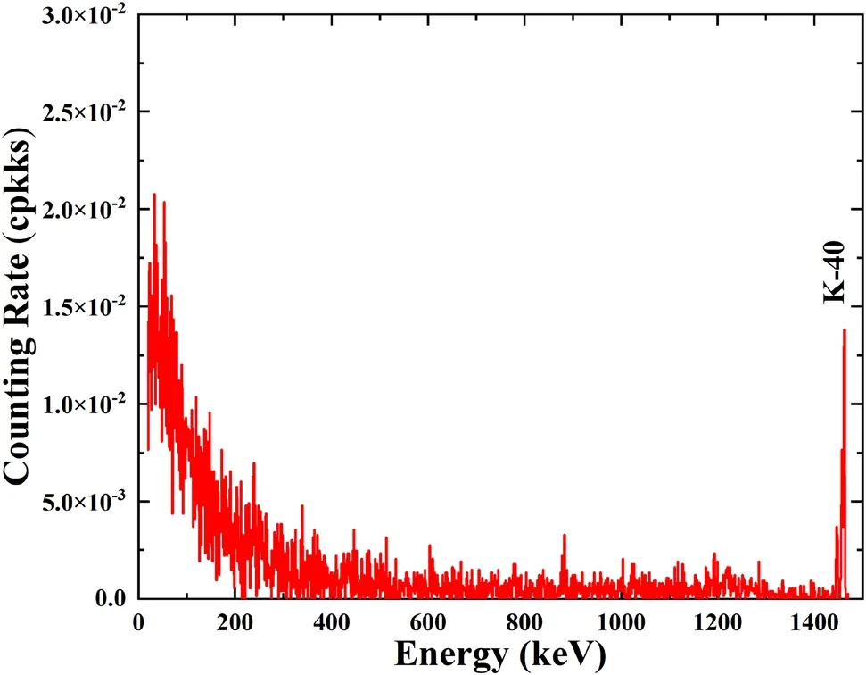
Fig. 8 The energy-resolved spectrum resulting from K-40 in the test subject measured inside counter D, which is obtained by subtracting the energy-resolved spectrum measured using the HPGe detector without a tested subject from its counterpart with a tested subject
The above analyses indicate that radioactivity in human subjects is the dominant source of background inside the shielding room for in vivo measurements. Unfortunately,there are no effective measures to suppress this background. The Phoswich detector, composed of two or more layered scintillators, was employed for this purpose; it relies on coincident events when both scintillators simultaneously receive energy to remove high-energy photons.However, our recent study indicates that this detector is useful in suppressing 1.46 MeV gamma-led background but is highly inefficient in removing 1.31 MeV beta-led background (bremsstrahlung radiation) because 1.31 MeV beta-led background is overwhelmingly distributed toward low energies (peaking at approximately 100 keV) [37]. A potential resolution is to model the shape and intensity of the background from a human subject based on state-ofthe-art computational phantoms. With such a computational phantom, in conjunction with a Monte Carlo simulation, the phantom can be readily tuned to reproduce the body type and K-40 content of the test subject. With this accurately simulated background spectrum,it is possible to achieve a background reduction in the post-measurement analysis.
4 Conclusion
In this study, we conducted a comprehensive experimental investigation on the radiation background outside and inside the shielding room of four sets of body counters to understand the contribution from each source.The major findings are as follows. First, the radiation levels outside the shielding room are drastically different among the four counters;however,the counterparts inside are qualitatively the same. Furthermore, our simple calculation indicates that attenuated environmental gamma rays should account for approximately 10% of the background inside the shielding room when no test subject is present.Second, by controlling ventilation, we observed that the background from airborne radon and daughters in shielded rooms contributed approximately 3% of the background when a human subject was absent. Third, by comparing the background measurements in the presence and absence of a test subject,we found that K-40 in a human subject contributed approximately 58% of the background in total. Unfortunately, owing to the constraints of the experimental conditions, the respective backgrounds contributed by the cosmic rays and radioactivity in the shielding materials cannot be separated in our experiment.Therefore,we refer to the results of Kaye et al., which suggest that approximately 30%of the background originates from cosmic rays and approximately 60%originates from the radioactivity of the material under similar conditions without a test subject.Finally, in a synergetic analysis of all these results, the relative contribution from each background source in the body counter can be obtained, as shown in Fig. 9.

Fig. 9 (Color online) Contribution of each background source for a body counter
In addition, this study provides a series of useful guidelines to design shielding rooms for in vivo measurements, as summarized below. First, a steel thickness of 15 cm is suitable to balance the construction cost and shielding effect. Second, radioactivity in shielding materials is inevitable, yet the material in the innermost layer should be carefully chosen. Suppose that lead is used to supplement steel for shielding. In this case, the addition of another lower-Z material to the interior of lead, such as copper, is recommended to absorb characteristic gamma rays or X-rays from the radioactive isotopes of lead.Third,the employment of active shielding may be considered,such as muon veto techniques, to achieve an ultralow background environment by removing the cosmic ray-induced background. Fourth, ventilation is a necessary and effective method of suppressing the radon-induced background in shielding rooms with poor air circulation. Fifth,K-40 from human subjects is the dominant source of background in in vivo measurements. There is no better measure to suppress this background source.In conjunction with a state-of-the-art computational phantom,we consider employing Monte Carlo simulations to model the characteristics of this background and rely on post-measurement spectrum analysis to reduce its impact.
Authors’ contributionsAll authors contributed to the study conception and design.Material preparation,data collection,and analysis were performed by Yu Wang, Yuan-Yuan Liu, Bin Wu, Xiang-Peng Meng, and Jian-Ping Cheng. The first draft of the manuscript was written by Yu Wang, and all authors commented on previous versions of the manuscript. All authors read and approved the final manuscript.
杂志排行
Nuclear Science and Techniques的其它文章
- Anisotropy flows in Pb-Pb collisions at LHC energies from parton scatterings with heavy quark trigger
- Development and application of a multi-physics and multi-scale coupling program for lead-cooled fast reactor
- A novel approach for radionuclide diffusion in the enclosed environment of a marine nuclear reactor during a severe accident
- Investigation of combined degrader for proton facility based on BDSIM/FLUKA Monte Carlo methods
- Anisotropic flow in high baryon density region
- Effect of relaxation time on the squeezed correlations of bosons for evolving sources in relativistic heavy-ion collisions