A novel aptamer-based histochemistry assay for specific diagnosis of clinical breast cancer tissues
2021-11-06MeiLiuLeiXiTingTanLianJinZhifeiWangNongyueHe
Mei Liu,Lei Xi,Ting Tan,Lian Jin,Zhifei Wang**,Nongyue He,*
a State Key Laboratory of Bioelectronics, National Demonstration Center for Experimental Biomedical Engineering Education (Southeast University), School of Biological Science and Medical Engineering, Southeast University, Nanjing 210096, China
b School of Chemistry and Chemical Engineering, Southeast University, Nanjing 211189, China
c Department of Pathology, The First Affiliated Hospital of Nanjing Medical University, Nanjing 210029, China
d Department of Pathology, Hunan Provincial Maternal and Child Health Care Hospital, Changsha 410008, China
e Economical Forest Cultivation and Utilization of 2011 Collaborative Innovation Center in Hunan Province, Hunan Key Laboratory of Biomedical Nanomaterials and Devices, Hunan University of Technology, Zhuzhou 412007, China
1 These authors contributed equally to this work.
ABSTRACT Pathological detection using immunohistochemistry(IHC) has become an indispensable process in the diagnosis confirmation of various cancers.However,the production of monoclonal antibodies is always very complex, expensive and time-consuming, and the batch differences are significant due to the corporeity and health statuses of animals may be different.In this work, an aptamer-based histochemistry (aptahistochemistry) assay was developed using a DNA aptamer for specific diagnosis of clinical breast cancer tissue sections.This aptahistochemistry assay can specifically distinguish Luminal A breast cancer molecular subtype from Luminal B(HER2+),HER2-enriched,and triple-negative breast cancer molecular subtypes, as well as para-carcinoma tissue, mastitis tissue and normal breast tissue.The accuracy of this aptahistochemistry assay for the diagnosis of Luminal A breast cancer was as high as 80%, which showed a great potential for clinical pathological diagnosis applications.
Keywords:Aptamer Immunohistochemistry Breast cancer Molecular subtype
In the clinical medicine, pathological detection using immunohistochemistry (IHC) has become an indispensable process in the diagnosis confirmation of various cancers [1-3].To conduct IHC,monoclonal antibodies are usually employed to bind with the cell-specific biomarkers expressed in the tumor cells of formalinfixed paraffin-embedded (FFPE) tissue sections.However, the production of monoclonal antibodies is always very complex,expensive and time-consuming, and the batch differences are significant due to different corporeity and health statuses of animals used[4,5].Therefore,to surmount those shortcomings of monoclonal antibodies, many efforts have been exploited to synthesize new cell-specific molecular probes to replace the monoclonal antibodies used in the IHC.
The emergence of aptamers has lighted the hope to establish novel and progressive aptamer-based histochemistry (aptahistochemistry) assays for more precise and low-cost pathological detection of tumor biomarkers in the cancer cells of FFPE tissue sections of cancer patients.Aptamers are functional short singlestranded non-coding RNAs or single-stranded DNAs developed by the systematic evolution of ligands by exponential enrichment(SELEX) method in vitro, which have the ability to specifically recognize various targeting molecules by forming various secondary structures [6-11].They are figuratively called “chemical antibodies” due to their generation by chemists and similar molecular functions to monoclonal antibodies.Moreover,aptamers also display several advantages over antibodies such as higher specificity and affinity, better stability, easier modification,and non-immunogenicity [12-18].Therefore,aptamer selection and applications have been one of the hottest research fields in the past years,which have attracted great attention in bioanalysis[19-25], molecular diagnosis [26-28], bioimaging [29-31], drug delivery[32-36]and even tissue engineering[37-39].In addition,aptamers have also been employed for pathological detection based on histochemistry or fluorescence staining [40,41], which show a great potential in the pathological diagnosis of cancer tissue sections.
In our previous work, a DNA aptamer MF3Ec specifically targeting Luminal A breast cancer molecular subtype was developed [42], which showed a great potential for the precise discrimination of Luminal A breast cancer molecular subtype by direct fluorescence staining.However, the results showed that there were strong background signals,and its non-specific binding to the nucleus is significant.Herein, to further decrease the background signals and increase the signal-to-noise rates, and exploring methods to reduce the non-specific binding of aptamers,aptamer MF3Ec was employed for the establishment of an aptahistochemistry assay to replace the monoclonal antibody with respect to achieve precise and accurate pathological diagnosis of Luminal A breast cancer molecular subtype.Meanwhile, the specific detection ability of this aptahistochemistry assay among breast cancer molecular subtypes,para-carcinoma tissue,mastitis tissue,and normal tissue was also systematically evaluated.Those results demonstrated that this aptamer MF3Ec-basd aptahistochemistry assay was able to distinguish Luminal A breast cancer subtype from the Luminal B (HER2+), the HER2-enriched, the triple-negative,para-carcinoma tissue,mastitis tissue,and normal tissue, despite its limited ability in the differential diagnosis between Luminal A and Luminal B (HER2-) breast cancer subtypes.Collectively, aptamer MF3Ec was demonstrated to be a promising molecular probe to establish an aptahistochemistry assay for clinical pathological diagnosis of Luminal A breast cancer molecular subtype.
To conduct the experiments, 140 cases of clinical FFPE breast cancer tissue sections from patients ranging from 40 to 60 years old, including 30 cases of Luminal A, 20 cases of Luminal B(HER2-), 20 cases of Luminal B (HER2+), 20 cases of HER2-enriched,20 cases of triple-negative,10 cases of breast cancer paracarcinoma tissue,10 cases of mastitis tissue sections and 10 cases of normal breast tissue sections, were used.All those tissue sections were obtained from the First Affiliated Hospital of Nanjing Medical University (Nanjing, China) with signed informed consents obtained from either the patients or from the next of kin.Biotin-labeled aptamer MF3Ec (5′-biotin-ACGACCCGATAAGTGCATTAGCACGTCCGAGAAAGGCCAGACGGGTCACACAGAGTTA-3′)and random library (5′-biotin-ACG-rN (r=52)-TTA-3′) were purchased from Sangon Biotech, Shanghai, CN to bind with the cell targets of FFPE tissue sections.HRP-conjugated streptavidin(Sangon Biotech,Shanghai,China)was used for specific binding to the biotin-labeled aptamer or random library.Bovine serum albumin (BSA, Sangon Biotech, Shanghai, China) was used for blocking.The binding buffer used in those experiments was phosphate buffered saline(PBS,pH 7.4)supplemented with 4.5 g/L glucose,5 mmol/L MgCl2,0.1 mg/mL yeast tRNA,and 1 mg/mL BSA,and the washing buffer was phosphate buffered saline(PBS,pH 7.4)supplemented with 0.05% Tween-20.
As illustrated in Scheme 1, the tissue sections were deparaffinized 3 times in xylene for 20 min and washed with a graded ethanol series and distilled water for 5 min respectively.Subsequently, the tissue sections were heated in 0.01 mol/L boiling citrate buffer (pH 6.0) for 4 min and washed with PBS buffer for antigen retrieval.Then,the tissue sections were incubated with 3%hydrogen peroxide for 20 min to block endogenous peroxidase.Afterwards,PBS buffer containing 5 mg/mL of BSA was applied and incubated with the tissue sections at 37°C for 60 min to reduce non-specific staining, followed by incubating with 250 nmol/L of biotin-labeled aptamer probe or random library in binding buffer at 4°C for 30 min.The tissue sections were then washed with washing buffer for 3 times and incubated with 200 μL of the HRPconjugated streptavidin at room temperature for 30 min before being treated with 200 μL of 3,3′-diaminobenzidine (DAB)peroxidase substrate solution at room temperature for 3 min.Finally,hematoxylin solution was added for counterstaining of the cell nucleus in the tissue sections, after which the sections were sealed, and observed by a fluorescence microscope (Olympus BX51).

Scheme 1.Schematic illustration of the aptahistochemistry assay for specific diagnosis of clinical breast cancer tissues.
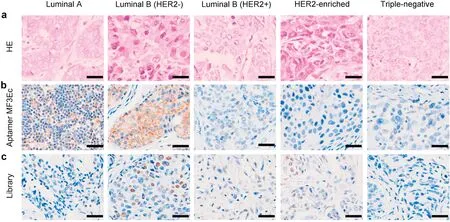
Fig.1 showed the specific pathological diagnosis ability of this aptamer MF3Ec-based aptahistochemistry assay among different breast cancer molecular subtypes.For tissue sections treated with biotin-labeled aptamer MF3Ec, there were significant strong brown signals exhibited on the cytomembranes of Luminal A breast cancer tissue sections, while there was no or negligible brown signals displayed on the cytomembranes of Luminal B(HER2+), HER2-enriched, triple-negative breast cancer tissue sections.However, there were also some strong brown signals displayed on the tumor cytomembranes of Luminal B (HER2-)breast cancer tissue sections.As negative controls,all those tissue sections treated with biotin-labeled random library displayed no obvious brown signals on the tumor cytomembranes.Those results indicated that aptamer MF3Ec was able to distinguish clinical Luminal A breast cancer subtype from Luminal B (HER2+), HER2-enriched and triple-negative breast cancer tissue sections by this established aptahistochemistry assay, despite its inability to differentiate Luminal A and Luminal B (HER2-) breast cancer subtypes.

Fig.1.Specific diagnosis of breast cancer tissue sections of different molecular subtypes.(a)Tissue sections were selected with hematoxylin and eosin stain for morphological confirmation.(b)Tissue sections were incubated with aptamer MF3Ec.(c)Tissue sections were incubated with random library.All pictures were taken under a fluorescence microscope with ×400 magnification.Scale bar = 100 μm.
We hypothesize that part of Luminal B(HER2-)breast cancers also express the same biomarker expressed on the tumor cytomembranes of Luminal A breast cancers.Due to the high heterogeneity of breast cancer,breast cancer can be classified into four major molecular subtypes: Luminal A, Luminal B, HER2-enriched and triple-negative[43].However, the current biomarkers are not enough for further depth subtyping of breast cancer and each of those four major molecular subtypes can also be further classified into more secondary subtypes.Therefore,additional cellspecific biomarkers for the classification of breast cancer molecular subtypes are needed to be discovered and identified.As a paradigm,Poudel et al.stratified Luminal A breast cancer into five subtypes by well-characterized and cancer-associated heterocellular signatures including stem, mesenchymal, stromal, immune,and epithelial cell types[44].Most recently,triple-negative breast cancer was reported to be further classified into five molecular subtypes by evaluating the expression patterns of androgen receptor (AR), CD8, FOXC1, and DCLK1 biomarkers using IHC[45].Besides, part of Luminal A breast cancers can also be reclassified as Luminal B [46].Therefore, to further identify Luminal A and Luminal B (HER2-) breast cancer molecular subtypes, the most important is to discover more cell-specific biomarkers for further depth and more precise subtyping of both breast cancer subtypes.Cell-SELEX can generate cell-specific aptamers without knowing of the specific targets in advance on the cell membranes, which can contribute to the biomarker discovery and identification by aptamer-based biomarker discovery technology [47,48].Finally, owing to the limited specificity of biomarkers,multiplex aptamers are strongly suggested to be used in combination to achieve better classification.
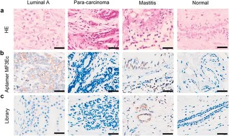
To further evaluate the pathological diagnosis ability of this aptamer MF3Ec-based aptahistochemistry assay among Luminal A breast cancer molecular subtype and para-carcinoma tissue,mastitis tissue, and normal tissue, those tissue sections were treated as described above.Fig.2 showed that only Luminal A breast cancer tissue sections treated with biotin-labeled aptamer MF3Ec exhibited prominent brown signals on the tumor cytomembranes, other tissue sections including all the negative controls treated with biotin-labeled random library only displayed negligible brown signals on the tumor cytomembranes.Those results indicate that this aptamer MF3Ec-based aptahistochemistry assay also has the ability to differentiate Luminal A breast cancer tissue from para-carcinoma tissue, mastitis tissue, and normal tissue.

Fig.2.Differentiating Luminal A breast cancer molecular subtype from para-carcinoma tissue, mastitis tissue, and normal tissue.(a) Tissue sections were selected with hematoxylin and eosin stain for morphological confirmation.(b)Tissue sections were incubated with aptamer MF3Ec.(c)Tissue sections were incubated with random library.All pictures were taken under a fluorescence microscope with ×400 magnification.Scale bar = 100 μm.
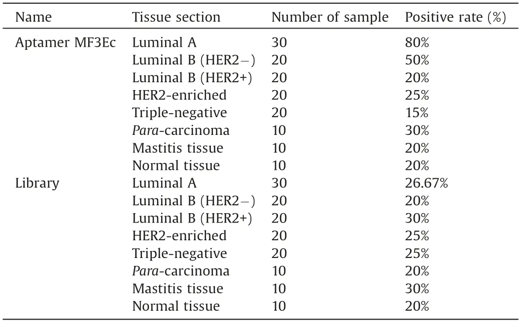
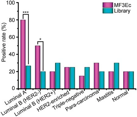
Statistical analysis was conducted to validate the accuracy of this aptahistochemistry assay for the precise pathological diagnosis of breast cancer tissues.As shown in Fig.3 and Table 1, the Luminal A breast cancer tissue sections treated with aptamer MF3Ec have a positive rate of 80%,which was significantly higher than those treated with random library whose positive rate was only 26.67%.While Luminal B (HER2+), HER2-enriched, triplenegative breast cancer tissue sections, para-carcinoma tissue sections,mastitis tissue sections and normal breast tissue sections treated with aptamer MF3Ec exhibited positive rates of lower than 30%,which were comparable to those treated with random library.For Luminal B (HER2-) breast cancer tissue sections, the positive rate (50%) of tissue sections treated with aptamer MF3Ec was considerably higher than those treated with random library(20%).Those results indicate that this aptamer MF3Ec-based aptahistochemistry assay can achieve specific diagnosis of Luminal A breast cancer tissue section from Luminal B (HER2+), HER2-enriched, triple-negative breast cancer tissue sections, paracarcinoma tissue sections, mastitis tissue sections and normal breast tissue sections.

Table 1 Statistical analysis of the aptahistochemistry assay for specific diagnosis of breast cancer tissues.

Fig.3.Statistical analysis of the aptahistochemistry assay for specific diagnosis of clinical breast cancer tissues.*P < 0.05, ***P < 0.001.
Compared with previously reported aptahistochemistry assays for the pathological diagnosis of tumor tissue sections, who displayed limited specificity and could not achieve differential diagnosis among cancer molecular subtypes [40,41,49,50], this aptahistochemistry assay exhibited preferable selectivity and can achieve specific diagnosis of Luminal A breast cancer subtype among different breast cancer molecular subtypes.However, the performances of this aptahistochemistry assay need to be further improved compared with clinical used IHC, despite its favorable potential application for Luminal A breast cancer diagnosis.Firstly,its accuracy for Luminal A breast cancer tissue sections is 80%,which was slightly lower than the clinical used IHC,meaning there is a false negative rate of 20%.Besides, the false positive rate of random library is as high as 26.67%.We hypothesize that this is attributed to the non-specific binding of single-stranded DNA oligonucleotides, despite the prominently decreased background signals and non-specific binding of this established aptahistochemistry assay,when compared with the previous reported direct fluorescence staining tests.However, due to the fast penetration and electrostatic forces between aptamers and nucleoproteins,there is still some weak non-specific binding to the cell nucleus.Therefore,how to circumvent the nonspecific-binding of aptamers in clinical tissue testing is one of the great challenges for the clinical applications and industrialization of aptamers.To further reduce the non-specific binding of aptamers, one strategy is to achieve the modified and functionalized aptamers to improve their structure stability in complex clinical samples and maintain their specific recognition ability.Another strategy is to develop new blocking methods to strongly inhibit the non-specific interaction between aptamers and nucleoproteins [15,51,52].Finally, to decrease the non-specific binding and improve the diagnosis accuracy of aptahistochemistry assays, the most important is to improve the specificity and binding affinity of aptamers,which in turn requires the development of advanced and high-efficient SELEX methods.
In conclusion, aptamer MF3Ec was employed to establish an aptahistochemistry assay for specific diagnosis of clinical breast cancer tissues of different molecular subtypes in this work.The aptamer MF3Ec-based aptahistochemistry assay can specifically distinguish Luminal A breast cancer molecular subtype from Luminal B (HER2+), HER2-enriched, and triple-negative breast cancer molecular subtypes, as well as para-carcinoma tissue,mastitis tissue and normal breast tissue with increased signalnoise rates, low background, decreased non-specific binding and comparable high accuracy of 80% for the diagnosis of Luminal A breast cancer compared with the previously reported direct fluorescence staining methodology.In addition, this aptahistochemistry assay is more specific, faster and cheaper than the traditional IHC,which can be accomplished within 4 h.Collectively,all those results demonstrate that this aptamer MF3Ec-based aptahistochemistry assay is a high-efficient and low-cost method for the clinical pathological diagnosis of breast cancer molecular subtypes.
Declaration of competing interest
The authors declare that they have no known competing financial interests or personal relationships that could have appeared to influence the work reported in this paper.
Acknowledgments
This work was supported by the National Key Research and Development Program of China (No.2017YFA0205301), the National Natural Science Foundation of China (Nos.61527806,81902153 and 81771976), and the Scientific Research Foundation of the Graduate School of Southeast University (No.YBPY1881).
杂志排行
Chinese Chemical Letters的其它文章
- Molecular recognition triggered aptazyme cascade for ultrasensitive detection of exosomes in clinical serum samples
- Synthesis of sponge-like TiO2 with surface-phase junctions for enhanced visible-light photocatalytic performance
- Zn-based metal organic framework derivative with uniform metal sites and hierarchical pores for efficient adsorption of formaldehyde
- High active amorphous Co(OH)2 nanocages as peroxymonosulfate activator for boosting acetaminophen degradation and DFT calculation
- Effects of the molluscicide candidate PPU06 on alkaline phosphatase in the golden apple snails determined using a near-infrared fluorescent probe
- A lysosomal polarity-specific two-photon fluorescent probe for visualization of autophagy
