Rare autonomic nervous system dysfunction: Alternating bilateral mydriasis
2021-09-09PengXuJingWuLidaSuFangQianHuqiangWanYueYuMinLiYinghongHuManHuang
Peng Xu, Jing Wu, Li-da Su, Fang Qian, Hu-qiang Wan, Yue Yu, Min Li, Ying-hong Hu, Man Huang
1 Department of Neuroscience Care Unit, the Second Affiliated Hospital of Zhejiang University School of Medicine, Hangzhou 310009, China
2 Department of General Intensive Care Unit, the Second Affiliated Hospital of Zhejiang University School of Medicine, Hangzhou 310009, China
3 Department of Intensive Care Unit Severe Burn Wards, the Second Affiliated Hospital of Zhejiang University School of Medicine, Hangzhou 310009, China
Dear editor,
Pupillary abnormalities, i.e., mydriasis, pupillary arefl exia, and/or anisocoria, may indicate the occurrence of acute clinical events in intensive care units (ICUs), which are particularly seen in severe patients with brain and/or cervical spinal cord injury. The pathophysiology of benign episodic mydriasis is still not completely clear. We present a patient with severe burns presented with temporary alternating bilateral pupil enlargement accompanied by paroxysmal increases in respiration, heart rate, and blood pressure.
CASE
A 67-year-old male patient was admitted to the hospital for multiple burns throughout the body for over one day, who suff ered 98% of the entire body area with deep II and III degree skin burns from tanker explosion, including 100% deep II-III degree burns on the face, neck, and chest. He had been a hypertension patient for more than a decade, but within normotension by taking antihypertensive drugs intermittently. After the 72 days’ treatment, the patient developed bilateral unequal pupils, with the left pupil diameter of 2.0 mm and the right pupil 4.0 mm (Figure 1A), whose eyes normally responded to direct and indirect light. The degree of bilateral pupil disparity was less severe in the dark environment. All the above symptoms indicated that the right pupil was abnormal. However, there was no obvious ptosis or eye movement disorder on both sides. The patient had a clear consciousness and responded to calls. At the same time, there was no involuntary movement or abnormal changes in muscle tension on both sides. Two hours later, the size of both pupils recovered to 2.0 mm. No abnormalities were found on the emergency cranial computed tomography (CT) scan.
On the 79thday after admission, the disorder developed again in the bilateral pupils, with the same symptom but left pupil of 4.0 mm and right pupil of 2.0 mm (Figure 1B), and also recovered within two hours. However, seven hours later, the pupils appeared again with unequal size: the left pupil of 2.0 mm and the right pupil of 4.0 mm. The patient was clear and had no localization signs. The head CT re-examination showed the same result as before, and CT angiography revealed carotid and cerebral arteriosclerosis. Again, his pupils returned to the normal diameter of 2.5 mm after two hours, with sensitive light reflection. The head and cervical spine magnetic resonance imaging (MRI) on the second day also showed no new lesions.
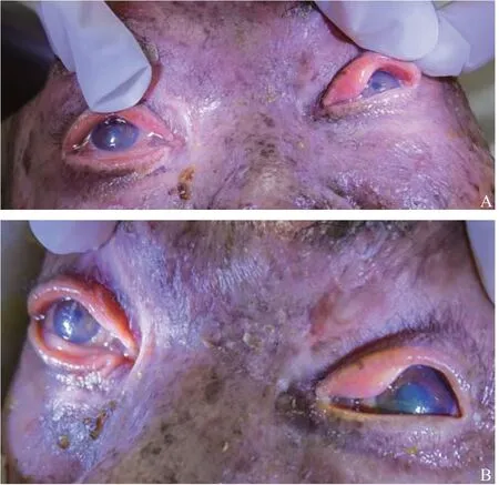
Figure 1. Bilateral unequal pupils of the patient. A: at 72 days after admission; B: at 79 days after admission.
Ophthalmic examination: no abnormalities of the iris or conduction were found, the direct light refl ection was the same as the indirect light reflection, and no object ghost was ignored. Pharmacological tests with instillation of two drops of 10% cacaine can cause bilateral pupil dilation, while 1% aclonidine eye drops cause a strong contraction of the smaller pupil, and 0.125% pilocarpine eye drops do not shrink the dilated pupil. When the size of the patient’s pupils changed, the patient’s breathing, heart rate, and blood pressure raised by 20%. Unilateral sweating was difficult to observe due to severe burns on the face, neck, and chest skin.
DISCUSSION
A recent case report showed that bilateral pupillary dilation might be associated with autonomic nervous system dysfunction.[1]The authors believe that this is mainly caused by sympathetic and/or parasympathetic nerve stimulation, accompanied by compensatory contralateral inhibition. It should be highlighted that some patients may have a different response with a sympathetic outburst, and physiologic anisocoria can occur in every three individuals.[2]Also, acute anisocoria had been reported with some drugs.[3]In this patient, the pupil contraction disorder and sympathetic excited performance were observed, which were transient, paroxysmal, and recurrent.
It has been reported that alternating bilateral pupillary disparity is associated with cervical spinal cord injury[4]or severe traumatic brain injury,[5]but has never been noticed in case of severe burns. Although we haven’t fully understood the mechanism of this phenomenon, the current hypothesis is that this is another form of Horner’s syndrome.[6]Besides, the differential diagnosis of this patient should be considered as follows: (1) changes of the recovery period after oculomotor nerve palsy, which often manifest as alternating unilateral pupil dilation and contraction; (2) Horner’s syndrome often seen in varieties of diseases, which could compress and/or destroy the pathways from the sympathetic nerve center to the orbit, leading to mydriasis, enophthalmos, ptosis, facial anhidrosis on the affected side, and unilateral miosis ; (3) Pourfour du Petit (PdP) syndrome, which is characterized by dilated pupils, enlarged eye clefts, hyperhidrosis, eyelid contraction, neck pain and swelling, and some palpable neck masses.
CONCLUSIONS
In general, this case shows a patient with reversible episodic bilateral mydriasis, which not only happens in one pupil, but in both pupils alternately, and is capable of self-recovery. It may be related to metabolism, pain, and stress. The researchers proposed the term “seesaw dysautonomia”[7]to describe this phenomenon.
Funding:This study did not receive any funding.
Ethical approval:The research was approved by the hospital ethics committee.
Conflicts of interests:The authors declare that they have no competing interests.
Contributors:PX, JW, and LDS analyzed and interpreted the patient data regarding autonomic nervous system dysfunction. FQ, HQW, YY, and ML performed the specialized examination and imaging analysis of the patient, and were major contributors in writing the manuscript. YHH and MH checked and revised the paper. All authors have read and approved the fi nal manuscript.
杂志排行
World journal of emergency medicine的其它文章
- Filter implantation for double inferior vena cava: A case report and literature review
- Thoracic aortic rupture due to airbag deployment
- Immediate oral amiodarone re-challenge following the development of parenteral-induced acute liver toxicity
- Drug-induced erythroderma in patients with acquired immunodefi ciency syndrome
- Clinical characteristics and risk factors of Talaromyces marneff ei infection in human immunodefi ciency virusnegative patients: A retrospective observational study
- The cell phone in the twenty-fi rst century: Risk for addiction or ingestion? Case report and review of the literature