Fibula pro-tibia vs standard locking plate fixation in an ankle fracture saw bone model
2021-08-23TosanOkoroKarHaoTeohHiroTanaka
Tosan Okoro,Kar Hao Teoh,Hiro Tanaka
Tosan Okoro,Department of Arthroplasty,Robert Jones and Agnes Hunt Orthopaedic Hospital NHS Foundation Trust,Oswestry SY10 7AG,United Kingdom
Kar Hao Teoh,Department of Orthopaedic Surgery,Princess Alexandra Hospital NHS Trust,Harlow CM20 1QX,United Kingdom
Hiro Tanaka,Department of Orthopaedics,Royal Gwent Hospital,Newport NP20 2UB,United Kingdom
Abstract BACKGROUND Locking plate fixation in osteoporotic ankle fractures may fail due to cut-out or metalwork failure.Fibula pro-tibia fixation was a technique prior to the advent of locking plates that was used to enhance stability in ankle fractures by achieving tri or tetra-cortical fixation.With locking plates,the strength of this fixation construct can be further enhanced.There is lack of evidence currently on the merits of tibiapro-fibula augmented locking plate fixation of unstable ankle fractures.AIM To assess if there is increased strength to failure,in an ankle fracture saw bone model,with a fibula pro-tibia construct when compared with standard locking plate fixation.METHODS Ten osteoporotic saw bones with simulated supination external rotation injuries were used.Five saw bones were fixed with standard locking plates whilst the other 5 saw bones were fixed with locking plates in a fibula pro-tibia construct.The fibula pro-tibia construct involved fixation with 3 consecutive locking screws applied across 3 cortices proximally from the level of the syndesmosis.All fixations were tested in axial external rotation to failure on an electromagnetic test frame(MTS 858 Mini-Bionix test machine,MTS Corp,Eden Praire,MN,United States).Torque at 30 degrees external rotation,failure torque,and external rotation angle at failure were compared between both groups and statistically analyzed.RESULTS The fibula pro-tibia construct demonstrated a statistically higher torque at 30 degrees external rotation(4.421 ± 0.796 N/m vs 1.451 ± 0.467 N/m;t-test P =0.000),as well as maximum torque at failure(5.079 ± 0.694N/m vs 2.299 ± 0.931 N/m;t-test P = 0.001)compared to the standard locking plate construct.The fibula pro-tibia construct also had a lower external rotation angle at failure(54.7 ±14.5 vs 67.7 ± 22.9).CONCLUSION The fibula pro-tibia locking plate construct demonstrates biomechanical superiority to standard locking plates in fixation of unstable ankle fractures in this saw bone model.There is merit in the use of this construct in patients with unstable osteoporotic ankle fractures as it may aid improved clinical outcomes.
Key Words:Unstable ankle fractures;Pro-tibia fixation;Improved stability;Simulated biomechanical analysis;Osteoporotic fractures;Ankle injuries
INTRODUCTION
In osteoporotic bone,there is unsatisfactory fixation strength with uni-cortical cancellous fixation for distal fibula fractures[1,2],which can lead to loss of fixation as well as delayed or non-union[3].The ways to try to obviate these risks include the use of locking,posterior plating,or non-locking constructs with adjunct fixation.One such example of the latter is the use of tri- or tetra-cortical fixation with fibula pro-tibia(syndesmotic)screws[1].In comparison to the same construct without additional screws,fibula pro-tibia fixation has demonstrated a 9% increase in torque to failure,24% increase ability to withstand external rotation,and a 34% increase in energy before failure of the construct[4].This technique adds little operative time,is inexpensive,and is a technically straightforward method to increase the stability of the construct[4].
In unstable bi-malleolar ankle fractures,the talus remains attached to the lateral malleous[5].Reducing the medial malleolus alone may prevent anatomical repositioning of the talus,as in some cases the lateral malleolus cannot be accurately reduced when it impinges on the proximal fibular fragment.Repositioning of the talus can be achieved by forcibly internally rotating the ankle in such cases,but this stretches the fibular collateral ligament.When external immobilization is discontinued,the lateral ligaments remain in a stretched position and slight to moderate talar instability,which predisposes to development of late degenerative arthritis,may be the result[5].
The lateral malleolus appears therefore to be the key to the anatomical reduction of displaced bi-malleolar fractures,and restoring the integrity of the lateral malleolus restores the integrity of the ankle[5].
Being able to maintain the integrity of the lateral malleolar fixation in osteoporotic bone in therefore important.This study aims to biomechanically assess whether there is an increased strength to failure with a fibula pro-tibia construct when compared with standard locking plate fixation for ankle fractures in an ankle fracture saw bone model.
MATERIALS AND METHODS
Materials
Ten osteoporotic saw bones(Pacific Research Laboratories Inc.,Vashon,WA,United States)with simulated supination external rotation injuries were used in this study.
Fracture simulation
A lateral malleolar ankle fracture was simulated with an osteotomy at the lateral malleolus(oblique orientation,starting medially at the level of the tibial plafond),and extending distally and laterally at a 45° angle[4].Figure 1 illustrates the simulated lateral malleolar fracture.
Fracture fixation
Five of the lateral malleolar osteotomies were fixed in a standard fashion using a fibular locking plate(Stryker Variax locking plate;Stryker,Mahwah,NJ,United States;Figure 2A)whilst fibula pro-tibia fixation was utilized in the other 5 models [fibular locking plate(Stryker Variax locking plate;Mahwah,NJ,United States)] with tricortical fixation.Tri-cortical fibula pro-tibia fixation entailed the use of 3 consecutive fully threaded cortical 3.5mm locking screws placed proximal to the lateral malleolar osteotomy at the level of the tibio-fibular syndesmosis(Figure 2B).
Biomechanical testing
Each model was then subjected to biomechanical analysis after being mounted on a resin and tested on an electromagnetic test frame(MTS 858 Mini-Bionix test machine,MTS Corp,Eden Praire,MN,United States),Figure 3,with measurement of torque(N/m)at 30 degrees external rotation,maximum failure torque(N/m)and external rotation angle(°)at failure.

Figure 1 Simulated lateral malleolar sawbone ankle fracture.
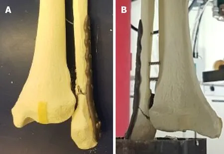
Figure 2 Sawbone lateral malleolar fixation construct.A:Sawbone lateral malleolar fracture treated with standard locking plate;B:Sawbone lateral malleolar fracture treated with locking plate in fibula pro-tibia configuration.

Figure 3 Fibula pro-tibia construct of ankle fracture sawbone model mounted on the electromagnetic test frame(MTS 858 Mini-Bionix test machine,MTS Corp,Eden Praire,MN,United States).
Statistical analysis
The student’st-test was used to analyze differences between both groups with aPvalue <0.05 taken as statistically significant.
RESULTS
Fibula pro-tibia vs standard locking plates(torque assessment)
The mean torque for the fibula pro-tibia constructs at 30° external rotation was 4.421 ±0.796 N/m,which was significantly higher than that obtained for the standard locking plate fixation constructs 1.451 ± 0.467 N/m(ttestP= 0.000).This difference was also noted in the maximum torque to failure(tibia-pro-fibula 5.079 ± 0.694 N/mvsstandard locking plate fixation 2.298 ± 0.931 N/m;ttestP= 0.001).
The torque values of each construct for the above parameters are detailed in Tables 1 and 2.

Table 1 Torque(N/m)recorded for fibula pro-tibia and standard locking plate constructs(torque at 30 degrees external rotation)
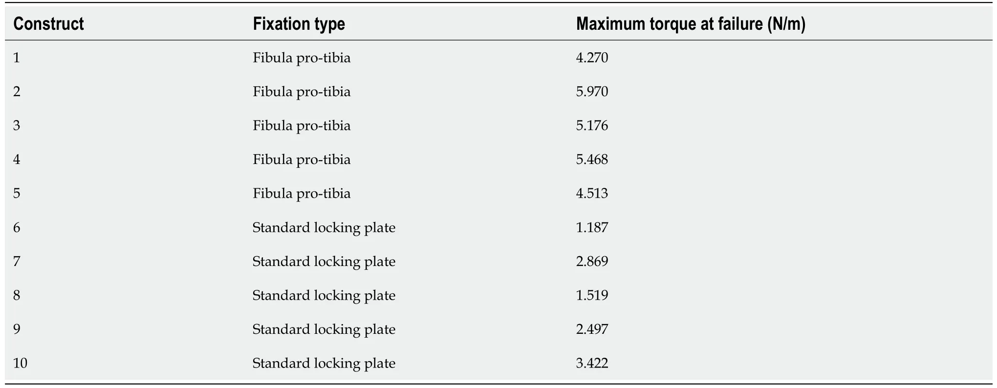
Table 2 Torque(N/m)recorded for fibula pro-tibia and standard locking plate constructs(maximum torque at failure)
Fibula pro-tibia vs standard locking plates(angle to failure)
There was a lower mean external rotation angle to failure for the fibula pro-tibia construct(54.7° ± 14.5°)compared to standard locking plate fixation 67.7° ± 22.9°,but this was not statistically significant;ttestP= 0.313.
DISCUSSION
Open reduction and internal fixation for an unstable ankle fracture in young patients is relatively predictable with excellent outcomes[6].However the management of ankle fractures in the elderly remains less predictable,secondary to the various comorbidities associated with elderly patients such as osteoporosis,diabetes,cardiovascular,and peripheral vascular disease[3].
A recent trial demonstrated superiority of tibio-talo-calcaneal(TTC)nailing over standard locking plate fixation in the elderly in terms of having a low risk of complications,an earlier return to previous level of mobility,and the allowance of an immediate return to full weight bearing[6].A limitation to the use of the TTC nail in routine practice is the risk of proximal peri-prosthetic fractures,as well as the need for the availability of a senior trauma surgeon or foot and ankle specialist to obtainoptimal outcomes[7].
This study demonstrates that the fibula pro-tibia locking plate construct has biomechanical superiority to standard locking plates in a saw-bone model.There is increased torque at 30 degrees external rotation as well as a higher torque at maximum failure of the construct.The reduced maximal external rotation angle at failure of the construct is most likely due to the increased rigidity of the fixation,which is potentially beneficial for osteoporotic bone.
A limitation of this study is that it was performed on sawbones not cadaveric bone.Such data may therefore not be readily transferable to a clinical scenario,as we have performed an isolated analysis of a lateral malleolar fracture.The data we have shown however gives an objective assessment of the difference in biomechanical properties between the two constructs.Another limitation of the study is that we have not used a model that accounts for a bi-malleolar fracture pattern.The lateral malleolus is key to the anatomical reduction of displaced bi-malleolar fractures,and restoring the integrity of the lateral malleolus restores the integrity of the ankle[5].The use of fibula pro-tibia fixation in this study demonstrates that there is up to approximately 3 times the level of torque achieved at 30 degrees external rotation and twice the failure torque in comparison to standard locking plate fixation.Initiating use of the adjunctive technique whilst performing such fixation is inexpensive,adds little operative time,and is not technically demanding.We propose the 3,3,3 rule for use of this adjunctive technique;Fixation with 3 screws across 3 cortices starting 3 cm above the tibial plafond.
The fibula pro-tibia construct utilizes the combined pull-out strength of locking screws to ensure a more biomechanical stronger construct.By using a tricortical fixation,it also ensures that the fixation is not as rigid as non-locking tetracortical fixation and provides some syndesmosis micro movement.There is therefore not a need for removal before weight bearing of the patient.
The increased biomechanical strength of the fibula pro-tibia construct demonstrates that there is merit to its use in patients with unstable osteoporotic ankle fractures.Future research is required to evaluate if its use would aid improved clinical outcomes in this important group of patients.
CONCLUSION
This study compared a fibula pro-tibia construct to standard locking plate fixation in an ankle fracture saw bone model.The fibula pro-tibia construct demonstrated biomechanical superiority and there is merit to consideration of its use in patients with unstable osteoporotic ankle fractures.
ARTICLE HIGHLIGHTS
Research background
The lateral malleolus is key to the anatomical reduction of displaced bi-malleolar fractures,and restoring its structural integrity restores the integrity of the ankle.Various fixation techniques have been utilized in osteoporotic bone to ensure lateral malleolar integrity.
Research motivation
Biomechanical assessment of whether there is an increased strength to failure with a fibula pro-tibia construct when compared with standard locking plate fixation for ankle fractures in an ankle fracture saw bone model.
Research objectives
To compare a fibula pro-tibia construct to standard locking plate fixation in a saw bone model using biomechanical parameters.
Research methods
After simulation of supination/external rotation injuries in a series ofn= 10 sawbones,n= 5 were fixed with the fibula pro-tibia construct andn= 5 were fixed with the standard locking plate.Biomechanical analysis was performed to assess torque(N/m)at 30 degrees external rotation,maximum failure torque(N/m)and external rotation angle(°)at failure.Students t test was used for comparison of both groups.
Research results
The fibula pro-tibia construct was biomechanically superior to the standard locking plate in torque at 30 degrees external rotation,and maximum failure torque.There was no statistically significant difference in the external rotation angle at failure.
Research conclusions
There is merit to considering the use of the fibula pro-tibia construct in fixation of bimalleolar ankle fractures in view of its biomechanical superiority over standard locking plates.
Research perspectives
Future research should evaluate the clinical significance of these findings.
ACKNOWLEDGEMENTS
We would like to thank King I and Lane H of Cardiff University School of Engineering for their assistance in providing access to the biomechanical testing equipment.
杂志排行

World Journal of Orthopedics的其它文章
- Safety and efficacy of surgical hip dislocation in managing femoral head fractures:A systematic review and meta-analysis
- Complications in growth-friendly spinal surgeries for early-onset scoliosis:Literature review
- Limb lengthening with PRECICE magnetic nail in pediatric patients:A systematic review
- Performance of alpha-defensin lateral flow test after synovial fluid centrifugation for diagnosis of periprosthetic knee infection
- Thromboelastography in elective total hip arthroplasty
- Calcar-guided short-stem total hip arthroplasty:Will it be the future standard? Review and perspectives