Clickable rhodamine spirolactam based spontaneously blinking probe for super-resolution imaging
2021-03-14ZengjinLiuYingZhengTingXieZihnChenZhenlongHungZhiweiYeYiXio
Zengjin Liu,Ying Zheng,Ting Xie,Zihn Chen,Zhenlong Hung,b,Zhiwei Ye,Yi Xio
a Drug Research Center of Integrated Traditional Chinese and Western Medicine,Affiliated Traditional Chinese Medicine Hospital,Southwest Medical University,Luzhou 646000,China
b State Key Laboratory of Fine Chemicals,Dalian University of Technology,Dalian 116024,China
Keywords:Super resolution imaging Spontaneously blinking pKa Rhodamine spirolactam
ABSTRACT Spontaneously blinking probe,which switches between dark and bright state without UV or external additives,is extremely attractive in super resolution imaging of live cells.Herein,a clickable rhodamine spirolactam probe,Atto565-Tet,is rationally constructed for spontaneously blinking after biorthogonal labelling and successfully applied to super resolution imaging of mitochondria and lysosomes.
The advent of super resolution imaging (SRI) techniques has enabled nanoscale monitoring of biomacromolecules or organelles dynamics with nanometer precision,providing insightful details for biological study [1,2].In contrast to other SRI techniques,such as stimulated emission depletion (STED) [3],structure illumination microscopy (SIM) [4],single molecule localization microscopy(SMLM) [5]based SRI technique does not require sophisticated optical configurations,but achieves state-of-art resolution at molecular scale.
However,SMLM demands the photo-switching between dark and bright states,which yet relies largely on bioincompatible imaging enhancing buffer or UV activation light.In 2014,a promising probe,HMSiR,with spontaneously blinking characteristics was first developed by Uno and co-workers [6].This fluorophore utilized the intramolecular spirocyclization reaction to achieve dark-bright transformation which is extremely compatible for living cells imaging.Following this seminal work,a variety of spontaneously blinking fluorophore based on intramolecular cyclization reaction or intermolecular nucleophilic addition was developed [7].Remarkably,Liu and Xu,predicted a pKacycle value ≥5.3 as the lower threshold for spontaneously blinking rhodamine dye [8].Since most rhodamine spiroamides exhibits low pKa,they are promising candidates for developing the spontaneous probe.Yet,most of them has a low pKavalue less than 5,providing a unstatisfactory slow blinking rate.

Fig.1.The structure of Atto565-Tet and biorthogonal labelling with BCN tethered tag.
The pKavalue of spiroamides is influenced by both electron and steric effect of theN-substituents on the spirolactams [9–11].It is urgent to exploit these effects for tuning pKavalue rhodamine spiroamides to preferable region for living-cell super-resolution imaging.
By connecting (6-methyl-1,2,4,5-tetrazin-3-yl)methanamine[12]to the spirolactam site of a bright rhodamine fluorophore Atto565 (Fig.S1 in Supporting information),Atto565-Tet was constructed as a novel probe for specific labeling and supperresolution imaging of live cells (Fig.1).The tetrazine unit of Atto565-Tet quenches the fluorescence to minimize background,and offers a universal biorthogonal handle to selectively label BCN tethered tags [13].Therefore,Atto565-Tet was constructed as a novel probe for specific labelling and super-resolution imaging of live cells.

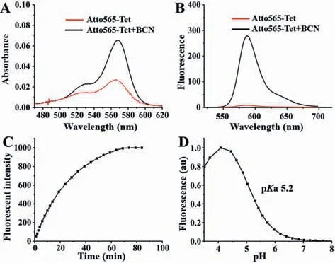
Fig.2.The change of the UV–vis absorption (A) and emission (B) spectra of Atto565-Tet (5.0 μmol/L) in phosphate buffer (pH 4.2) before and after biorthogonal labelling.(C) Fluorescence enhancement of Atto565-Tet (5.0 μmol/L) upon reaction with 10-fold BCN at pH 4 in aqueous buffer.(D) The pH dependent fluorescence of Atto565-Tet (5.0 μmol/L) after biorthogonal labelling.
As shown in Figs.2A and B,Atto565-Tet does not absorb light apparently in visible region and maintains in non-fluorescent state with a low fluorescence quantum yield of 0.05 in PBS buffer (pH 4.2).In contrast,after biorthogonal labeling reaction with BCN(bicyclo[6.1.0]non-4–yne),the ring-opened form of Atto565-Tet exhibits strong visible absorption peaked at 565 nm and a high fluorescence quantum yield of 0.46 (pH 4.2).
The fluorogenicity of Atto565-Tet during biorthogonal reaction was investigated by recording its fluorescent intensity at 588 nm(Fig.2C).Up to 19-fold fluorescence enhancement was observed after reacted with BCN.Then the pH-dependent equilibrium of the closed and opened isomers was measured by fluorescence spectroscopy.The pKavalue was determined to be 5.2 (Fig.2D),which indicated that the closed isomer was predominant over the opened isomer.

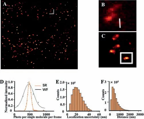
Fig.4.The utility of Atto565-Tet for super-resolution imaging of lysosome in live HeLa cells.(A) Super-resolution image.(B) Wide-field image of boxed area.(C)Super-resolution image of boxed area.(D) Histograms of the number of photons per single-molecule event.(E) Histograms of the localization precision.(F) Transverse profile of a lysosome along the white line in B and C.Scale bars in A:3 μm.
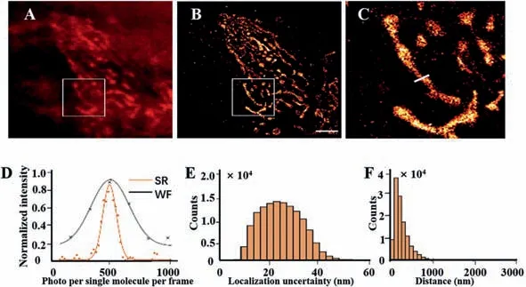
The favorable pKavalue implied that Atto565-Tet could spontaneously blinking without UV activation at physiological conditions [8].The encouraging photophysical characteristics of Atto565-Tet promoted us to evaluate it in super resolution imaging in living cells.Following a recently reported TPP-BCN [14],which is targeted to mitochondria specifically,we synthesized TPP-BCN for labelling of mitochondria in living cells and subsequently attachment of Atto565-Tet by biorthogonal reaction.SMLM imaging in standard PBS buffer (pH 7.4) demonstrated that Atto565-Tet exhibited spontaneous blinking behavior and did not require UV or external thiol additives to switch it between closed and opened state.The SMLM image was constructed from 10,000 frames,collected during 100 s with a rate of 100 Hz.Compared with the wide-field image (Figs.3A and B),the SMLM imaging gave much higher resolution and revealed detailed structural information of mitochondria(Fig.3C).We were able to determine the widths of mitochondria to beca.132 nm (Fig.3D).The average localization uncertainty is 25.3 nm with an average single-molecule brightness of 277 photon counts/10 ms (Figs.3E and F),which was comparable with previous reportedf-HM-SiR [14].The excellent performance of Atto565-Tet in super-solution imaging should be ascribed to the suitable pKato achieve spontaneously blinking and the precise targetability endowed by high efficiency of biorthogonal labeling reactions of tetrazine with BCN tags.

Fig.3.The utility of Atto565-Tet for super-resolution imaging of mitochondria in live HeLa cells.(A) Wide-field image and (B) super-resolution imaging.(C) Enlarged map of the boxed area of images A and B.(D) Histograms of the number of photons per single-molecule event.(E) Histograms of the localization precision.(F) Transverse profile of a mitochondria along the white line in C.Scale bars in B:3 μm.
It was generally accepted that spontaneously blinking dye based on intramolecular cyclization is restriction to non-acidic organelles such as mitochondria and cytosol [6].However,we are eager to evaluate its applicability in acidic organelles such as lysosome,which have a pH value between 4.5 and 5.5.In order to selective lable lysosome,we constructed a bridge ligand Lyso-BCN (Fig.S2 in Supporting information),with piperazine moiety for specific accumulation in lysosome [15,16].HeLa cells were first treated with Lyso-BCN for 30 min,followed by the addition of Atto565-Tet.To our surprise,sparsely distributed of single molecule signal was observed.The SMLM image was constructed from 10,000 frames during 100 s at a rate of 100 Hz.Again,the SMLM imaging gave much higher resolution than wide-field image (Figs.4A-C).The full width of a lysosome estimated from super-resolution was about 123 nm (Fig.4D),and the average localization uncertainty is 22 nm (Fig.4E) with an average single-molecule brightness of 589 photon counts/10 ms (Fig.4F).Considering the pKacycle value of Atto565-Tet was 5.2,photons emitting from the resulted biorthogonal labelling product should be overlapped with each other rather than sparse distributed.This blinking phenomenon is ascribed to the affinity equilibrium of the targeting ligand lyso-BCN between the neutral microenvironment (such as the lysosomal membrane and the cavity of membrane proteins) and the acidic lumen.This microenvironment change shifts the spirocyclization of our probe,which further providing the sparse single-molecule signals.
In summary,we have developed a novel spontaneously blinking probe with a desirable pKavalue that well suited for SMLM.The dye exhibited self-blinking behavior without UV activation or external additives to induce it switch between dark and bright state.Our dye was successfully implied to super resolution image of mitochondria and lysosomes at satisfactory precision.
Declaration of competing interest
The authors declare no conflict of interest.
Acknowledgments
This work was supported by the National Natural Science Foundation of China (Nos.21421005,21576040,21776037,22004011),China Postdoctoral Science Foundation (Nos.BX20200073 and 2020M670754),and Dalian Science and Technology Innovation Fund (No.2020JJ25CY014).
Supplementary materials
Supplementary material associated with this article can be found,in the online version,at doi:10.1016/j.cclet.2021.04.038.
杂志排行
Chinese Chemical Letters的其它文章
- Progress in mechanochromic luminescence of gold(I) complexes
- Spinel-type bimetal sulfides derived from Prussian blue analogues as efficient polysulfides mediators for lithium-sulfur batteries
- Alopecuroidines A-C,three matrine-derived alkaloids from the seeds of Sophora alopecuroides
- Boronic acid-containing diarylpyrimidine derivatives as novel HIV-1 NNRTIs:Design,synthesis and biological evaluation
- Diaminodiacid bridge improves enzymatic and in vivo inhibitory activity of peptide CPI-1 against botulinum toxin serotype A
- Peptide stapling with the retention of double native side-chains
