Study on Quantitative Analysis Method of Methanol Raman Spectra in OilBy Extraction Technology
2020-09-18WANGCongZHANGJiayiSONGShijieGAOJunyingSUBing
WANG Cong, ZHANG Jiayi, SONG Shijie, GAO Junying, SU Bing
(1 State Grid Shandong Electric Power Company Dezhou Power Supply Company, Shandong, 253074, China; 2 State Key Laboratory of Power Transmission Equipment & System Security and New Technology Chongqing University, Chongqing, China)
Abstract: Accurately detecting the concentration of methanol dissolved in transformer oil is of great importance for diagnosing the aging degradation of insulating paper. In this paper, acetonitrile were selected as extractants, and mixed with transformer oil samples containing different concentrations of methanol to complete the sample pretreatment work. The concentration of methanol in transformer oil samples was determined by Raman spectroscopy. The quantitative analysis method based on the principal component analysis of peak intensity of Raman spectra and the quantitative analysis method based on principal component analysis of peak area of Raman spectra were compared. The results show that the accuracy of fitting and error calculation using Raman spectral peak area as quantitative analysis parameter is higher, especially in the low concentration case concerned, it is helpful for us to detect trace methanol dissolved in transformer oil.
Key words: State monitoring; Methanol; quantitative analysis; Extraction, Raman spectroscopy
1 Introduction
Transformer is the core equipment of power transmission and distribution system, and its reliability is of great significance to ensure the stability and security of power system. The average operating life of power transformers is about 30 years, and its service life is affected by various factors including load, manufacturing process and operating environment. Aging will cause the mechanical properties of the main insulation of the transformer to decline, so that when the transformer suffers a sudden external short circuit fault, the coil is easily deformed, which will lead to mechanical damage to the insulation paper, loss of insulation capacity and ultimately cause an accident. The content of methanol in transformer oil is one of the important characteristics to evaluate the aging state of transformer oil-paper insulation.
The production of methanol has an influential relationship with the decomposition of the 1,4-β-glycosidic linkage to the cellulose glucose unit. In addition, methanol has the same Arrhenius factor value (Aa)[1,2]due to the production of 1, 4-glycosidic bond as that of the ruptured 1, 4-glycosidic bond, so methanol can be detected more rapidly than furfural contained in transformer oil[3,4].
According to research, the main methods for detecting methanol concentration in transformer oil are headspace gas chromatography (HS-GC) and gas chromatography-mass spectrometry (GC-MS). Both have high sensitivity and reliability, but all require very complicated pre-processing steps, which take a long time to implement. These two methods have certain limitations for our on-site samples.
The Raman effect means that when a substance is irradiated with a single-color excitation light, a series of wavelengths larger or smaller than the incident wavelength of the Raman scattered light are generated. The displacement (wavenumber) of these Raman scattering lines with respect to the excitation light corresponds to the type of the substance group, and the intensity reflects the number of molecules of the substance to be tested. Therefore, Raman spectroscopy can be used for qualitative and quantitative analysis of molecules. In this paper, the LRS method is used to detect the dissolved methanol concentration in transformer oil. However, the actual transformer oil contains a variety of substances and the methanol concentration is relatively low. How to improve the detection sensitivity is the key to using LRS technology to detect the methanol concentration in transformer oil.
Confocal Raman technology has high intensity of excitation and efficiency of collection, and its own microscope objective of Raman probe can be better for detecting trace liquid. In this paper, the LRS liquid test platform has been set up in the laboratory based on the light path of confocal Raman. In order to furtherly improve the detection sensitivity, extraction technology was applied to LRS detection experiments. And the Raman peak at 1030 cm-1in the extract was selected for quantitative analyses. Besides, the analytical process based on spectral data could be summarized into three steps, including spectral preprocessing, establishing regression models, and analyzing samples using regression models. The Raman spectrum obtained from the spectrometer contains interference information such as fluorescence background, detector noise, laser power fluctuations, etc. in addition to the Raman signal of the analyte. In general, equipment improvements do not completely eliminate the above interference. Therefore, before establishing the regression model, some mathematical methods are needed to eliminate various interference factors in the spectrum and highlight the characteristic signals of the samples. Two quantitative methods were established by least squares method, one is the quantitative method between Raman characteristic peak area and concentration, and the other is the quantitative method between characteristic peak intensity and concentration. The results show that it is better to establish a quantitative method based on the characteristic peak area and concentration, and the goodness of fit reaches 0.98374.
2 Experimental
2.1 Experimental Facilities
The detection platform for liquid dissolved in the transformer oil based on microscopic Raman technology is illustrated in Fig.1. A confocal laser Raman spectroscopy liquid testing platform was established in the laboratory[5].The platform is equipped with a 532 nm solid-state laser with a maximum power of 100 mW. The low nose diode pumped laser can operate in TEM00 mode with a spectral width of 0.001 pm. The convergence and Raman scattering of incident light were collected by a 50× long-focal length objective. These signals have a high spatial resolution to avoid interference from the entrance window. Three flare gratings (1800/500, 600/500 and 1200/750 nm) were installed in the ANDOR spectrometer with a focal length of 500 mm. The refrigeration temperature could reach 85 °C back-thinned CCD output port to connect to the spectrometer.
2.2 Sample preparation
The pure methanol and 25# transformer mineral oil(41.6% naphthenic, 50.0 % paraffinic, and 8.4 % aromatic)used in this work was provided by ChuanRun Lubricant Company, China .In order to eliminate the influence of different initial conditions, the mineral oil was pretreated by degassing and drying . Pour 100 mL methanol into a 500 mL conical flask. Then, 200 mL transformer oil was added to the volumetric flask, stirred for 10 min, and stored for 24 hours without light. After 24 hours, the solution stratification was observed. According to the data, the density of transformer oil is 0.895 g/ cm3, and the density of methanol is 0.7918 g/ cm3. The excess methanol solution not melted into the upper layer is sucked away by the glue head dropper, and the saturated methanol transformer oil sample is obtained.
2.3 Extraction
The transformer oil in operation contains a variety of substances. The dissolved concentration of methanol in transformer oil is relatively low. Separating methanol can eliminate the influence of other substances contained in transformer oil and the like. Thereby, the detection lower limit of the LRS method is further improved. Therefore, we apply the extraction technology to the pretreatment of oil samples.
Acetonitrile is selected as the extraction based on the principle that the same substance is more soluble. A 100 mL sample of oil was taken from a measuring cylinder and poured into a conical flask with a lid. Then, 10 mL of acetonitrile was measured by a pipette, and the oil sample was mixed and stirred for 5 minutes with a magnetic stirrer. The measured oil sample is allowed to stand, and the liquid surface is layered in about 10 minutes, and the supernatant liquid can be used for test detection.
2.4 Quantitative Analyses
Raman spectroscopy directly reflects the vibrational structure information of the material molecules, and was used to qualitatively analyze the material properties and molecular structure information in the early days. With the rapid development of optical measurement technology, the amount of information contained in Raman spectroscopy is becoming more and more abundant, which makes Raman spectroscopy technology suitable for more accurate quantitative analysis applications. The quantitative analysis technique based on Raman spectroscopy has been developed for about 60 years[6,7]. From the beginning, the single characteristic peak corresponds to the linear analysis of a single substance, and the simultaneous analysis of multiple components in complex system, the analysis limit is also reduced from 10% to 100 ppm[8].
According to the classical electromagnetic field theory, the Raman scattering intensityI(v) of a molecule at a certain frequencyvcan be expressed as:
(1)

3 Results and Discussions
3.1 Raman Characteristic Peaks of Methanol extracted in Acetonitrile
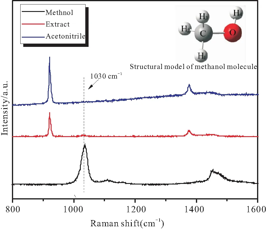
Fig.1. shows the Raman spectrum of acetonitrile, the Raman spectrum of the extract and the Raman spectrum of pure methanol. The use of acetonitrile as an extraction may affect the mode of the vibrating methanol molecule. The displacement and methanol Raman peak intensity may also change accordingly. Using comparative analysis, there were two distinct Raman peaks when the methanol concentration was 1030 and 2829 cm-1in the extract. This experiment uses these two Raman peaks as the basis for qualitative analysis of methanol. Compared to the Raman peak of pure methanol, the Raman peak of the extract is shifted to 2827 cm-1, which is mainly due to the overlap of Raman spectra of acetonitrile and methanol between Raman signals, which does not help to our research. For our qualitative analysis of methanol and we only need to select a Raman spectrum peak for quantitative analysis, so this experiment chooses 1030 cm-1as a Raman characteristic peak.
3.2 Quantittative Analyses
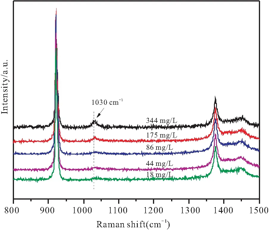
As shown in Fig. 2. The spectra of different concentrations of methanol were determined. Raman spectroscopy noise reduction, baseline cancellation, and normalized pretreatment can reduce the effects of fluorescent background, instrument noise, and cosmic rays. The intensity values and peak areas of Raman peaks under different concentration conditions were obtained respectively, and a mathematical quantitative analysis model of methanol concentration was established. As shown in the figure, the intensity of the Raman peak changes significantly at 1030 cm-1, and the intensity and area of the Raman peak decrease as the methanol concentration decreases.

Fig.1 Experimental Raman spectra of acetonitrile, extract, and methanol

Fig.2 Raman spectra of methanol at different concentrations dissolved transformer oil
The least squares method of methanol concentration of transformer oil was established by linear fitting curve of methanol concentration of transformer oil and Raman characteristic peak intensity at 1030 cm-1. It can be seen from Fig. 3(a) that there is a significant linear relationship between the Raman peak intensity at 1030 cm-1and the methanol concentration, and the fitness is 0.89975. The linear regression equation of this method is:
y=2776.81+7.93x
(2)

Fig.3 Relationship between methanol concentrations and the areas and intensity of Raman peak at 1030 cm-1
Based on the linear fitting curve of methanol concentration of transformer oil and Raman characteristic peak area at 1030 cm-1, the least square method of methanol concentration of transformer oil was established. According to Fig. 3(b), there is a more obvious linear relationship between the Raman peak area at 1030 cm-1and the methanol concentration. The fitting degree is 0.98374, which is higher than fitting degree between the Raman peak strength and methanol concentration. The linear regression equation of this method is :
y=149247.02+1429.39x
(3)
The results show that the accuracy of the fitting method and the error calculation by the characteristic peak area and concentration is higher, and the goodness of fit reaches 0.98374.
4 Conclusions
In this paper,the in situ measurement of methanol dissolved in the transformer oil was realized using the Raman detection platform for liquid. The vibration mode of the main Raman peak of methanol in the acetonitrile extract was identified. And the Raman peak at 1030 cm-1in the extract was used for quantitative analysis. The feasibility of using the LRS method to detect the concentration of ethanol in transformer oil was verified. Using the extraction technique, the detection limit of the LRS method reached 11.2 mg/L. Two quantitative methods for the Raman characteristic peak intensity value and concentration at 1030 cm-1and the Raman characteristic peak area and concentration were established by least squares method. The experimental results show that it is more accurate to establish quantitative method fitting and error calculation by using characteristic peak area and concentration. The experimental results show that the LRS method can effectively detect the concentration of methanol in transformer oil. According to the detection characteristics, the limitations of LRS detection can be further improved by optimizing the extraction parameters and using optical devices with better performance. The LRS method provides a new method for the rapid detection of methanol in transformer oil.
Acknowledgment
This work was supported by the National Natural Science Foundation special fund joint fund project (U1766217), the science and technology project of the national Power Grid Corp (SGS00200FCJS1700228), and the construction plan of the innovation team at the Colleges of Chongqing (CXTDX201601001).
