Spontaneous pneumothorax in a single lung transplant recipient-a blessing in disguise:A case report
2020-09-14
Himanshu Deshwal,Division of Pulmonary,Critical Care and Sleep Medicine,New York University Grossman School of Medicine,New York,NY 10016,United States
Subha Ghosh,Imaging Institute,Section of Thoracic Imaging,Cleveland Clinic,Cleveland,OH 44195,United States
Kathleen Hogan,Olufemi Akindipe,Charles Randall Lane,Atul C Mehta,Respiratory Institute,Cleveland Clinic,Cleveland,OH 44195,United States
Abstract
BACKGROUND
End-stage chronic obstructive pulmonary disease (COPD) is one of the common lung diseases referred for lung transplantation.According to the international society of heart and lung transplantation,30% of all lung transplantations are carried out for COPD alone.When compared to bilateral lung transplant,singlelung transplant (SLT) has similar short-term and medium-term results for COPD.For patients with severe upper lobe predominant emphysema,lung volume reduction surgery is an excellent alternative which results in improvement in functional status and long-term mortality.In 2018,endobronchial valves were approved by the Food and Drug Administration for severe upper lobe predominant emphysema as they demonstrated improvement in lung function,exercise capacity,and quality of life.However,the role of endobronchial valves in native lung emphysema in SLT patients has not been studied.
CASE SUMMARY
We describe an unusual case of severe emphysema who underwent a successful SLT 15 years ago and had gradual worsening of lung function suggestive of chronic lung allograft dysfunction.However,her lung function improved significantly after a spontaneous pneumothorax of the native lung resulting in auto-deflation of large bullae.
CONCLUSION
This case highlights the clinical significance of native lung hyperinflation in single lung transplant recipient and how spontaneous decompression due to pneumothorax led to clinical improvement in our patient.
Key words:Native lung hyperinflation;Single lung transplant;Pneumothorax;Bronchoscopic lung volume reduction;Endobronchial valve;Case report
I NTRODUCTI ON
End-stage chronic obstructive pulmonary disease (COPD) is one of the common lung diseases referred for lung transplantation[1].Indications for a lung transplant in COPD include poor functional status (BODE index of 7-10),severely reduced lung function(forced expiratory volume 1 <20% of predicted),hypercapnia (PaCo2 ≥ 55 mm Hg)and hypoxemia with decreased diffusion capacity (DLCO <20%)[2].According to the International Society of Heart and Lung Transplantation,30.6% (18233/6400) of all adult lung transplants have been carried out for COPD alone[3].When compared to bilateral lung transplant (BLT),single-lung transplant (SLT) has similar short-term and medium-term results for COPD patients[1].
For patients with severe upper lobe predominant emphysema,lung volume reduction surgery (LVRS) is an excellent alternative,which results in an improvement in functional status and long term mortality[4].In 2018,endobronchial valves were approved by the Food and Drug Administration for severe upper lobe predominant emphysema as they demonstrated improvement in lung function,exercise capacity,and quality of life[5].However,the role of endobronchial valves in native lung emphysema in SLT patients has not been studied.
We describe an unusual case of severe emphysema who underwent a successful SLT 15 years ago and had a gradual worsening of lung function suggestive of chronic lung allograft dysfunction.However,her lung function improved significantly after a spontaneous pneumothorax of the native lung resulting in the auto-deflation of large bullae.
CASE PRESENTATION
Chief complaints
A 74-year-old female presented to the hospital with acute left-sided chest pain,neck swelling and shortness of breath for one hour.In the emergency department,she appeared in respiratory distress and was placed on supplemental oxygen.
History of present illness
She was in her usual state of health,when she developed acute onset left-sided severe chest pain that was sharp in nature and non-radiating.It was worse with inspiration and had no relieving factors.It was associated with significant shortness of breath and neck swelling that prompted her to seek medical attention.
History of past illness
She had a past medical history of End-Stage COPD (FEV1-15%) with severe diffuse emphysema and underwent a successful single right lung transplant in July 2004(Figure 1).Her medications included tacrolimus,mycophenolate mofetil,and prednisone for immunosuppression and sulfamethoxazole-trimethoprim,valacyclovir and itraconazole forPneumocystis,cytomegalovirus and fungal infections prophylaxis respectively.Her peak FEV1 on post-operative follow up was 65%.She had no evidence of acute rejection on early surveillance bronchoscopies.
Unfortunately,in 2015,eleven years following the transplantation,she developed acute complicated diverticulitis requiring urgent right hemicolectomy and ileostomy.Her postoperative course was complicated by frequent episodes of dehydration and acute kidney injury.She also had two bouts of lower lobe pneumonia involving the allograft and acute tubular necrosis that eventually progressed to chronic kidney disease.On subsequent follow-ups,she complained of worsening dyspnea and cough.Her spirometry revealed a gradual decline in FEV1 from a peak of 65% of predicted to a nadir of 51% (Figure 2).In November 2018,she presented with worsening renal function,volume overload,and recurrent left-sided pleural effusions (Figure 3).Hemodialysis was initiated,and therapeutic thoracentesis was performed with 1200 cc transudative fluid removal.Despite the optimization of her volume status,she continued to have worsening dyspnea and lung function along with recurrent leftsided pleural effusions.We excluded ongoing acute cellular rejection by transbronchial biopsies,and a diagnosis of chronic lung allograft dysfunction prompted replacing mycophenolate with sirolimus.She also had a tunneled pleural catheter placed in left pleural space for recurrent effusions.She was placed on inhaled corticosteroids and long-acting bronchodilators with minimal improvement.By December 2018,she was on supplemental oxygen at 4 L/min by nasal cannula for chronic hypoxemic respiratory failure.
Personal and family history
She had no significant family history.Prior to lung transplantation she was a lifelong smoker with 40 pack year smoking history and subsequently quit in 2003.She denied any illicit drug use or alcohol use.
Physical examination upon admission
Her blood pressure was 90/60 mmHg and pulse was 100/min and oxygen saturation was 86% on 4 liters of oxygenvianasal cannula.On inspection she was using accessory respiratory muscles and appeared in respiratory distress.She had significant subcutaneous emphysema with crepitus extending up to the neck.On auscultation of the chest,there were absent breath sounds on the left hemithorax.
Laboratory examinations
Initial laboratory examination was largely insignificant with normal hemoglobin,white blood cell count,platelets,liver function and electrolytes.She had history of chronic kidney disease and her blood urea nitrogen and creatinine were 48 mg/dL and 2.3 mg/dL respectively.
Imaging examinations
A chest radiograph revealed a large left hydropneumothorax with an indwelling pigtail drainage catheter in the left pleural space.The native left lung was partially collapsed.There was subcutaneous emphysema in the left chest wall (Figure 4).
FINAL DIAGNOSIS
She was promptly diagnosed with a spontaneous pneumothorax due to rupture of a large bullae in her native emphysematous lung.
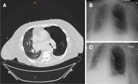
Figure1 Computed tomography (prone position) of the chest (A) and chest radiographs AP upright (B) and portable (C),show native left lung and transplanted right lung.
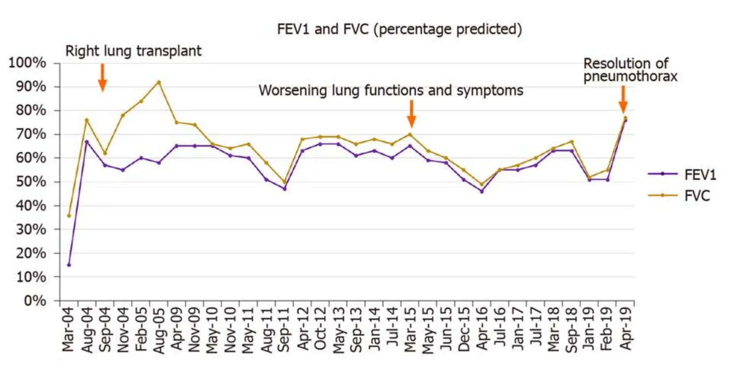
Figure2 Linear graph demonstrating the FEV1 and FVC (percentage predicted) for the patient on follow-ups.
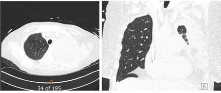
Figure3 Computed tomography chest (Axial and coronal reformatted images) show a large left pleural effusion with near-complete opacification of the left lung parenchyma.
TREATMENT
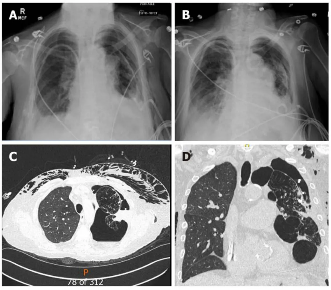
Figure4 The native left lung is partially collapsed,there is subcutaneous emphysema in the left chest wall.
We placed her pre-existing tunneled pleural catheter to suction with subsequent improvement in her symptoms.She further needed an additional pigtail pleural catheter placement in the left pleural space for a persistent hydropneumothorax.However,she became extremely dyspneic when taken off suction,prompting the placement of a Heimlich valve to the pleural catheter.Repeat chest x-ray demonstrated improvement in hydropneumothorax,and both pigtail and pleural catheters were clamped (Figure 5).Just before discharge,she again experienced respiratory distress,and both catheters were placed back on suction.This time,her symptoms improved significantly,and the indwelling tunneled pleural catheter was removed.She was discharged home with the pigtail catheter in-situ.Remarkably,on her way to a followup appointment,her pigtail catheter fell out of the pleural space,with no clinical consequences.
OUTCOME AND FOLLOW-UP
On a follow-up visit in April 2019,she reported a marked improvement in her dyspnea and no longer required supplemental oxygen.At the same time,she demonstrated a 25% improvement in her FEV1 (51% to 76%) (Figure 2),and the chest x-ray revealed the expansion of the right allograft as a result of marked deflation of large bullae in the left lung (Figure 6).
DISCUSSION
COPD is one of the common indications for lung transplantation.Over 18233 transplants have been carried out for COPD alone[3].A single lung transplant may be considered for COPD as it offers shorter waitlist time,shorter surgical time,and technical ease.Single lung transplant for COPD has similar short-term and mediumterm outcomes compared to BLT.However,long term outcomes are better with BLT[3].Several factors may play a role in this difference,with a critical factor being the diseased native lung.
Native lung complications can develop in up to 13.8% of patients with SLT and are associated with worse prognosis[6].Infections and malignancy may lead to alterations in the immunosuppressive regimen,and as a result,put the transplanted lung at risk of rejection.The emphysematous native lung can also compress the allograft and lead to its decreased perfusion,subsequently leading to graft dysfunction[7].
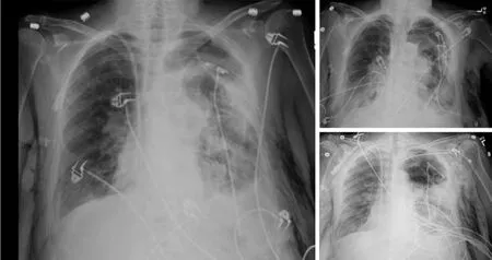
Figure5 Serial portable chest radiographs show replacement of a left pleural drain within a large left hydropneumothorax with compressive atelectasis of portions of the native left lung.
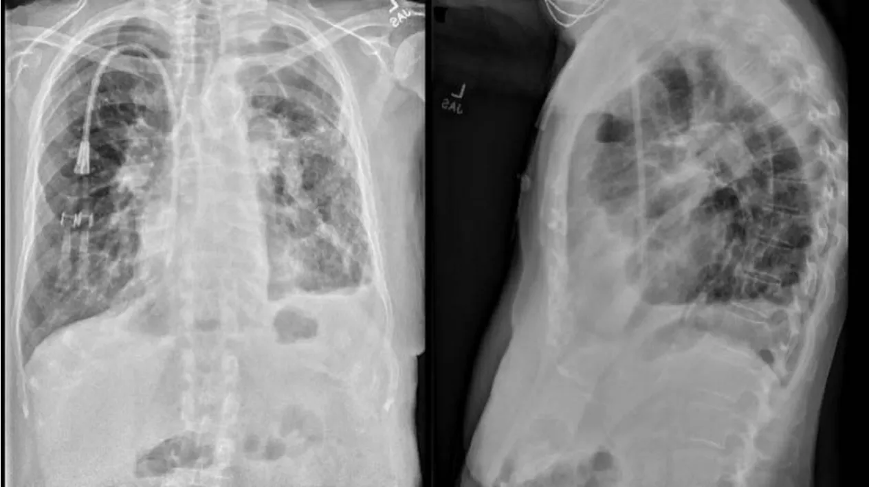
Figure6 Chest radiographs (PA and lateral views) demonstrate auto-deflation of large left upper lobe bulla resulting in subsequent reexpansion of the previously atelectatic left lung as well as improved aeration of the transplanted right lung.
Kinget al[6]studied the role of native lung pneumonectomy in SLT patients with native lung complications[6].They found a trend towards improved survival in patients undergoing pneumonectomy compared to those managed non-surgically (4.3 yearsvs2.4 years,respectively).However,the result did not reach statistical significance (P=0.128)[6].Native lung hyperinflation may lead to impairment of gas exchange and worsen respiratory function among recipients of SLTx for emphysema.Several cases have been described with improvement in lung function,exercise capacity,and symptoms after lung volume reduction surgery,bullectomy,or pneumonectomy of the native lung[8,9].
While the role of LVRS is well established in patients with severe upper lobe predominant emphysema,some case reports and retrospective studies highlight the benefit of LVR in severe emphysema of native lung in SLT patients[4,10,11].However,LVRS is associated with significant morbidity (20% to 29.8% pulmonary and cardiac morbidity respectively),and operative mortality (5.5%) and careful patient selection is warranted[12].
Less invasive approaches have since been described for lung volume reduction in severe emphysema.The use of bronchoscopic LVR has also demonstrated efficacy in improving lung function,exercise capacity,and quality of life.The use of endobronchial one-way valve placement has led to significant improvement in exercise capacity and lung function[13,14].A meta-analysis conducted by Iftikharet al[15]demonstrated the non-inferiority of Bronchoscopic LVR compared to LVRS[15].In 2018,the Food and Drug Administration approved the endobronchial valve Zephyr(Pulmonx.Inc.,Palo Alto,CA,United States) for Bronchoscopic LVR in severe emphysema[16].Other novel techniques such as lung volume reduction coils,bronchial thermal vapor,and installation of bio-degradable gel or polymers have also demonstrated efficacy in severe diffuse emphysema,yet,they remain experimental in the United States[17-19].Perchet al[20]conducted a case series of 14 patients with native lung hyperinflation in SLT patients and demonstrated significant improvement in lung function and symptoms with endobronchial valves[20].The study demonstrated the feasibility of endoscopic treatment for native lung hyperinflation in SLT patients,but prospective trials are required to assess its real efficacy.Our case highlights a phenomenon of auto-deflation of a large bulla leading to spontaneous lung volume reduction of the native lung and subsequent improvement in allograft lung function,thus highlighting the importance of considering LVR in SLT patients.While native lung hyperinflation was considered a possibility for her clinical decline,this phenomenon occurred even before considering her for LVR.Surgical lung volume reduction may be challenging in these patients due to co-morbid conditions and prior thoracic surgery (SLT),but bronchoscopic LVR is an attractive alternative.The real challenge lies in patient selection and identifying which patient will benefit most from it.Pulmonary function tests alone are unable to distinguish CLAD from native lung hyperinflation as a cause of a drop in lung function.The presence of CLAD in allograft would also reduce the likelihood that the graft will expand and improve function after native lung LVR.From our experience,certain clinical features can assist in patient selection for LVR in SLT patients.The patient should have apical predominant severe emphysema in the native lung.A SPECT scan can help identify areas with perfusion and poor ventilation that can be targeted for LVR.It is imperative to rule out CLAD in the transplanted lung before considering LVR.
Like any other intervention,bronchoscopic LVR is associated with certain complications that include valve migration,post-obstructive pneumonia,pneumothorax,and bleeding[20].It is thus imperative to discuss risks and benefit with the patient in detail.
CONCLUSION
Native lung pathology can impede the function of the allograft and lead to an erroneous diagnosis of allograft rejection or chronic lung allograft dysfunction.Spontaneous pneumothorax involving the native lung is yet another sequela of SLT.In our patient,hyperinflation and air trapping in the native emphysematous lung led to chronic respiratory failure following SLT.Incidentally,the patient developed a spontaneous pneumothorax with a natural volume reduction of the native lung leading to improvement in overall lung function.Among symptomatic recipients of SLT,the contribution of native lung hyperinflation and air trapping should prompt consideration of endobronchial valve therapy albeit with appropriate patient selection criteria.
杂志排行
World Journal of Clinical Cases的其它文章
- Recommendations for perinatal and neonatal surgical management during the COVID-19 pandemic
- Clinical applicability of gastroscopy with narrow-band imaging for the diagnosis of Helicobacter pylori gastritis,precancerous gastric lesion,and neoplasia
- Identification of APEX2 as an oncogene in liver cancer
- Restenosis after recanalization for Budd-Chiari syndrome:Management and long-term results of 60 patients
- Comparison of microendoscopic discectomy and open discectomy for single-segment lumbar disc herniation
- Clinical characteristics of patients with COVID-19 presenting with gastrointestinal symptoms as initial symptoms:Retrospective case series
