Traditional investigation and management for recurrent hemarthrosis after total knee arthroplasty:A case report
2020-09-14XiaoGengYangLiXuanHeHuaTian
Xiao Geng,Yang Li,Xuan He,Hua Tian
Xiao Geng,Yang Li,Xuan He,Hua Tian, Orthopedic Department,Peking University Third Hospital,Beijing 100191,China
Abstract
Key words:Total knee arthroplasty;Recurrent hemarthrosis;Arthroscopy;Open exploration;Case report
INTRODUCTION
Recurrent hemarthrosis is a complication of total knee arthroplasty(TKA)[1],with an incidence of 0.3% to 1.6%,and appropriate management should be implemented as early as possible to minimize the risk of severe consequences[2-5].No consensus is available on the criteria for diagnosis or treatment of this disease[6].Kindsfateret al[7]described a case series of 30 patients and their findings were broadly reflective of the subsequent literature.In the present case,various investigations and managements were performed,and the clinical effects of these interventions were analyzed separately.
CASE PRESENTATION
Chief complaints
A 50-year-old man,with a history of hypertension,presented to the Orthopedic Department of our hospital for non-traumatic acute left knee pain and swelling associated with a warmth sensation 14 mo after TKA.
History of past illness
He had a history of hypertensio n,but was not regularly taking any anticoagulant or antiplatelet medication.
History of present illness
He had a bilateral knee pain due to end-stage primary knee osteoarthritis for over 10 years and underwent TKA of the left knee in 2013 after failure of conservative treatment.There were no complications intraoperatively.A pneumatic tourniquet was used during surgery at a pressure of 300 mmHg after exsanguination.The implant used was a Press Fit Condylar prosthesis P.F.C.®SIGMA®(DePuy Synthes,West Chester,United States).A single intra-articular drainage without blood recovery system was used,and was removed 24 h postoperatively.The drainage volume was 300 mL.Postoperative rehabilitation was uneventful,and the outcome was satisfactory.Thromboprophylaxis was administered with sodium enoxaparin(6100 IU per day),starting on the day of surgery and lasting 2 wk.The patient was discharged on postoperative day 7.Clinical tests and radiographs were normal 3 mo after TKA.The range of motion of the left knee was 0° extension to 125° flexion,and the femorotibial angle was 178°.The Hospital for Special Surgery Scoring System(HSS)score improved from 50 points to 96 points(with 30 points of pain,20 points of function,16 points of mobility,10 points of force,10 points of deformity and 10 points of stability).The Western Ontario and McMaster Universities Osteoarthritis Index(WOMAC)score reduced from 62 points to 7 points.However,14 mo postoperatively,the patient experienced acute pain in the left knee while he was sleeping.This onset of pain was associated with immediate swelling of the knee and a sensation of warmth in the area of pain.
Physical examination
Local findings in the left knee showed that the range of motion was limited to 0°extension and 80° flexion,with positive patellar ballottement and no knee instability.The HSS score had decreased to 71 points and the WOMAC score was 55 points.
Laboratory examinations
Aspiration of the knee joint yielded 40 mL of dark,bloody fluid(Figure 1).No organisms were isolated,and culture was negative for bacteria over a 72-h period.Culture was sent only once to microbiology because the patient's symptoms were disappeared 10 h after non-surgical treatment was implemented.
Imaging examinations
Radiography showed excellent alignment and no prosthetic loosening(Figures 2A and B).Ultrasonography revealed hydrarthrosis of the left knee.
FINAL DIAGNOSIS
The diagnosis of the presented case is hemarthrosis after TKA.
TREATMENT
At the beginning,the patient received non-surgical treatment including compression,cryotherapy and immobilization was.The symptoms resolved 10 h later and he could walk home.But repeated similar episodes occurred in the following 2 mo.The patient was finally diagnosed with recurrent hemarthrosis after TKA.The patient presented to our department for definitive diagnosis and treatment,and arthroscopic exploration was performed.A tourniquet was initially used.After thorough lavage of the joint,there was no visible bleeding(Figure 3A).However,progressive hemorrhage blurred the view soon after releasing the tourniquet,and it was difficult to determine the source of the bleeding(Figure 3B).Therefore,open exploration was performed.Pulsatile bleeding was discovered in the medial sulcus close to the upper pole of the patella(Figure 4).The bleeding artery was about 1-1.5 mm in diameter.According to the anatomy of the knee and the structure of the blood vessel,the bleeding source was identified as the medial superior genicular artery.The artery was ligated with three sutures.There was no sign of bleeding during intraoperative flexion and extension of the knee.
OUTCOME AND FOLLOW-UP
The patient was symptom-free in the 15 mo after surgery.The left knee had a range of motion of 0° extension to 120° flexion,and the standing femorotibial angle was 177°.The HSS score returned to 92 points and the WOMAC score was 5 points in 24 mo after surgery.We also followed up the patient at 48 mo after surgery through video communication since he could not come back to our hospital.At that time,the WOMAC sore remained 5 points and knee function were perfect(Figure 5).
DISCUSSION
More recently,Raviet al[6]reviewed previous literatures and suggested that the potential causes of recurrent hemarthrosis after TKA varied and involved systemic,local and iatrogenic etiologies.They also described an algorithm for the diagnosis and treatment of recurrent hemarthrosis after TKA.It was reported previously that the blood vessel could be directly injured during procedures such as resection of the proximal tibia or posterior femoral condyles,posterior capsule release,posterior cruciate ligament removal,and screw insertion[8].In addition,a low power injury can cause the formation of an arteriovenous fistula,arterial aneurysm,or pseudoaneurysm,creating the potential for future blood vessel rupture[8-11].Drexleret al[12]reported an atypical tibial defect in open surgery and biopsy results revealed highgrade epithelioid angiosarcoma,which was treated by amputation above the level of the knee.In addition,hemarthrosis may be caused by anticoagulation medication such as warfarin,aspirin,rivaroxaban,or clopidogrel[13].Some rare causes like hemophilia,an inherited disease with a deficiency of factor VIII(hemophilia A)or factor IX(hemophilia B)[14,15],and PVNS,a proliferative tumor of synovial tissue,may also induce hemarthrosis[16-18].Routine blood tests including inflammatory markers and a coagulation profile are essential in cases of swollen and painful knees after TKA[5].In 1996,Worlandet al[3]emphasized the necessity of aspiration as an important way to differentiate the cause of knee swelling after TKA.Ultrasound can present a luminal turbulence with a“Yin-Yang” sign which can easily detect arterial aneurysms,pseudoaneurysms,and arteriovenous fistulae[5].Radiography can evaluate the position of components and determine whether the components are loose.Magnetic resonance imaging is valuable for detecting pathological changes of the synovium[19].Angiography is a very useful method for diagnosing and treating recurrent hemarthrosis in both early and late cases.Vascularized synovium could present as a“tumor blush” sign in angiography[20].All anticoagulant medication should be stopped after consultation with related experts.Cryotherapy,aspiration,and immobilization can resolve the symptoms and stop the bleeding temporarily or definitively[4,7].Although some cases will fail in conservative therapy and require surgical therapy,non-surgical therapy should be given first[5].Open surgical repair such as synovectomy is a direct approach to explore the bleeding area and ligate the blood vessel or remove the hypertrophic synovium[2,3].However,open surgery is associated with operation-related complications such as iatrogenic injury,more blood loss,and longer rehabilitation.Arthroscopic synovectomy is a minimally invasive approach that allows dissection of hypertrophic synovium with less bleeding and trauma than open surgery.However,arthroscopic synovectomy is not always successful.Suzukiet al[21]reported a case of recurrent hemarthrosis in which the source of bleeding could not be determined in an unclear arthroscopic view,and was treated with open exploration,which was same as in our case.Therefore,angiographic embolization has been regarded as a more optimal choice in cases of vascular injury and of synovial impingement.Compared with open exploration and arthroscopic surgery,angiographic treatment is a less invasive therapy with lower risk of complications and easier rehabilitation[19,20].In the present case,the patient did not undergo angiography,and the cause of the bleeding could not be determined preoperatively.In fact,the source of bleeding was suspected to be synovium impingement according to recently reported cases[5,7],and the ultrasound examination.However,open exploration revealed a bleeding artery,which strongly suggested that the cause of hemarthrosis was rupture of a pseudoaneurysm of the medial superior genicular artery.Arthroscopy was initially performed and was found to be inadequate compared with angiography.A clear scope is essential to determine the source of bleeding,unfortunately,it was difficult to visualize the effusion or jet point before the view became bloody.

Figure 1 Aspiration of the knee joint resulted in 40 mL of dark,bloody fluid.
CONCLUSION
The present case indicated the following perspectives.Firstly,angiography is essential in cases of hemarthrosis after TKA.Secondly,arthroscopy may not always be adequate as it may not provide a clear view.Lastly,traditional open exploration is still effective and sometimes necessary.

Figure 2 Radiography showed excellent alignment and no prosthetic loosening.
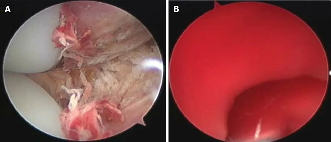
Figure 3 Arthroscopic explorations.A:After thorough lavage of the joint,there was no visible bleeding;B:It was difficult to determine the source of the bleeding after releasing the tourniquet.
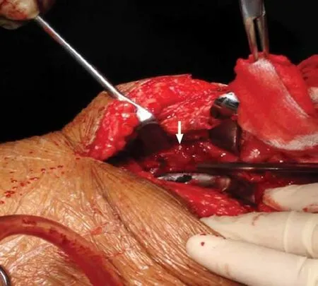
Figure 4 Pulsatile bleeding was discovered in the medial sulcus close to the upper pole of the patella.
Figure 5
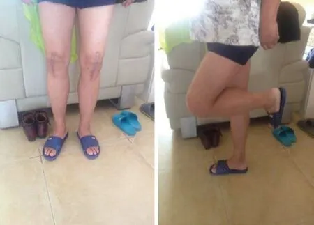
Figure 5 The Western Ontario and McMaster Universities Osteoarthritis Index score was 5 points and knee function was perfect.
杂志排行

World Journal of Clinical Cases的其它文章
- French Spine Surgery Society guidelines for management of spinal surgeries during COVID-19 pandemic
- Prophylactic and therapeutic roles of oleanolic acid and its derivatives in several diseases
- Macrophage regulation of graft-vs-host disease
- Antiphospholipid syndrome and its role in pediatric cerebrovascular diseases:A literature review
- Remotely monitored telerehabilitation for cardiac patients:A review of the current situation
- Keystone design perforator island flap in facial defect reconstruction