Effects of vascular endothelial growth factor supplementation and alginate embedding on human oocyte maturation in vitro
2020-06-23FarhangAbedMortezaFallahKarkanMasoumehMajidiZolbinPegahNaghizadehFereshteAliakbariHosseinYazdekhasti
Farhang Abed, Morteza Fallah-Karkan, Masoumeh Majidi Zolbin, Pegah Naghizadeh, Fereshte Aliakbari✉,Hossein Yazdekhasti
1Men's Health & Reproductive Health Research Center, Shahid Beheshti University of Medical Sciences, Tehran, Iran
2Laser Application in Medical Science Research Center, Shahid Beheshti University of Medical Sciences, Tehran, Iran
3Shohada-e-Tajrish Hospital, Shahid Beheshti University of Medical Sciences, Tehran, Iran
4Pediatric Urology and Regenerative Medicine Research Center, Children's Medical Center, Tehran University of Medical, Tehran, Iran
5Department of Anatomy, Faculty of Medicine, Tehran University of Medical Sciences, Tehran, Iran
6Department of Molecular Physiology and Biological Physics, Center for Membrane and Cell Physiology, University of Virginia, Virginia, United States
ABSTRACT
KEYWORDS: Alginate; Infertility; Human oocyte; Maturation;Vascular endothelial growth factor✉To whom correspondance may be addressed. E-mail: fereshtehaliakbary@yahoo.com;hosein.yazdekhasti@yahoo.com
1. Introduction
The in vitro maturation technique is a fertility preservation method that has recently found a special place amongst the new methods of assisted reproductive technologies[1]. Favorable conditions are very important for the in vitro maturation technique which is used in basic biotechnology and infertility research. Oocyte maturity under laboratory conditions is a simple, hormone-free process that facilitates the study of oocyte maturation and in vitro fertilization[2,3]. Advantages of this technique are as follows: no need of follicle-stimulating hormone (FSH) in treatment, reduction of drug administration, and prevention of over-stimulation of the ovary in women with polycystic ovary syndrome. Also, this technique leads to transferring more embryos to compensate for lower fertility. However, the rate of pregnancy and live birth by the in vitro maturation method using germinal vesicle (GV) oocyte obtained from the antral follicle is low[4].
The three-dimensional system, as one of the culture methods, was used for in vitro maturation[5]. The most important factor in a threedimensional culture system is the selection of a suitable matrix with suitable biomechanical characteristics, high water, and gelation properties[6]. Alginate is a natural biopolymer, mainly extracted from brown algae and less frequently from bacteria. It forms 40%of the dry weight of algae and is present in the extracellular matrix of these algae, combined with calcium, magnesium and sodium cations[7]. Alginate, due to its hydrophilicity and its abundance of water properties, can provide an extracellular field material as suitable bedding for cell growth[8]. Alginate gel could support the three-dimensional architecture of the follicle. On the other hand, it can facilitate cell growth and release of food in the medium. These features make this material suitable for texture design[9].
Vascular endothelial growth factor (VEGF) is a member of the transforming growth factor β superfamily and it is composed of a subunit molecular in mass of 23 kDa that plays an important role in endothelial cells, ovulation and embryo implantation[10]. The most important source of VEGF in the ovarian follicle is granulosa cells[11]. Previous studies showed that adding VEGF into the in vitro maturation culture of bovine oocytes could promote the growth of oocyte development[12]. Some studies argued that VEGF can increase the amount of oxygen supply in oocyte and granulosa cells and thus increase the fertilization[13,14]. Also, others believed that VEGF was directly associated with the number of injected gonadotropins and also associated with embryonic puberty[14].Therefore, the present study was designed to investigate the effects of VEGF supplementation and alginate on human oocyte maturation in vitro and embryo cleavage.

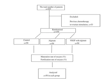
Figure 1. The study flowchart. VEGF: vascular endothelial growth factor.
2. Materials and methods
2.1. Study population
The study group consisted of women with infertility problems who referred to the fertility center of the Mahdieh hospital from 2016 to 2018 in Tehran, Iran. The total number of patients consisted of 203 women with infertility problems who were under assisted reproductive technology treatment (Figure 1). Some patients underwent several in vitro maturation cycles during different phases of the menstrual cycle: 14 patients underwent two oocyte retrievals,6 patients underwent three oocyte retrievals, and 1 patient underwent four oocyte retrievals. All types of infertility problems were included and maternal age was between 25 and 40 years old. The exclusion criteria were previous chemotherapy or any ovarian stimulation.
2.2. Oocyte retrieval
Oocyte retrieval was done according to the center’s standard for in vitro maturation practice 38 h after a subcutaneous administration of 10 000 IU of human chorionic gonadotropin. For retrieval of oocytes, a 19-gauge single lumen needle was used. Follicles (18-22 mm) were observed during ovulation induction. The follicular fluid including oocytes was aspirated and collected from the ovary in a 15 mL conical tube. The collected oocytes were assessed with a stereo microscope (Olympus, Japan) (×80 magnification) and were categorized to degenerated GV, intact GV, or GV breakdown based on the presence or absence of a GV and polar body. Briefly,oocyte immaturity was observed by the presence of GV and the first polar body, and mature oocytes were assessed when the first polar body was extruded. GV oocyte with at least three layers of compact cumulus cells and with homogenous cytoplasm was selected for in vitro maturation. For each patient, GV oocytes were randomly placed in the control and treatment groups (alginate, and VEGF+alginate groups).Mechanical denudation of cumulus cells occurred after retrieval.
2.3. Preparation of alginate
Alginate solution was produced by dissolving 0.8 g of sodium alginate powder (Sigma-USA) in 100 mL of 0.9% sodium chloride solution. Then it was filtered with a 0.2 μm filter. It was better to prepare a fresh alginate solution at any time. Alginate solution was added to 105 mM calcium chloride solution by using an insulin syringe. Alginate droplets in the calcium chloride solution were formed during 15 min, and after discharging calcium chloride,alginate seeds were added into sodium chloride (0.9%) for 10 min.After the evaporation of sodium chloride, each GV was put into alginate gel bead via syringe and in vitro maturation media were added into it.
2.4. In vitro maturation
Obtained GV oocytes during this study were randomly divided into three groups. The control group (n=50) included tissue culture medium 199 (TCM 199) (Gibco), 10% fetal bovine serum (Gibco),penicillin/streptomycin 100 IU/mL (Sigma), luteinizing hormone and FSH 0.075 IU/mL (Gonal-F, Sereno); the alginate group (n=50)included above cases with alginate 8%; the alginate+VEGF group(n=50) included TCM 199 with alginate 8% and VEGF (5 ng/mL)(Sigma). GV oocytes in 50 μL droplets of each group were placed in the basic culture medium and covered with mineral oil (Ovoil,Vitrolife, Sweden). Then, they were placed in a 37 ℃ incubator with 5% CO2for 48 h. Oocyte maturity and restoration of the meiosis were studied by a stereo microscope with ×80 magnification(Olympus, Japan) in this stage. Immature oocytes were considered as oocytes unchanged in the nucleus whereas oocytes with GV breakdown and the absence of polar body were considered as MⅠoocyte. Also, oocytes with a polar body were considered as mature oocytes [metaphase (M)Ⅱ stage]. In the next stage, the culture media were replaced with sodium citrate and the MⅡ oocytes were extracted from each group for fertilization using intracytoplasmic sperm injection (her spouse’s sperm).
2.5. Intracytoplasmic sperm injection
After the culture of oocytes for in vitro maturation, MⅡ oocytes from every group were placed in 50 μL drops that had been covered with warm mineral oil. Then, 2 μL of the sperm were placed in a drop of polyvinyl pyrrolidone (Vitrolife, Sweden) in a plate containing a mature oocyte. And 16 to 18 h after injection of MⅡoocytes by processed sperm from the patient’s spouse, the presence of 2 pronuclei in the oocytes fertilized was checked. Also, the number of cleavage embryos (8-cell stage) was checked on day 3 after sperm injection. The progression of embryos during in vitro maturation process was evaluated and none of the embryos would be transmitted inside uterus.
2.6. Statistical analysis
Patients’ data were analyzed by analysis of variance and were expressed as mean±standard deviation (mean±SD). Statistical analyses of maturation and fertilization rates of oocytes were performed by using the Chi-square test in each group. Results were expressed as % and differences were considered to be significant when P<0.05. The analyses were carried out by using SPSS Statistics for Windows, version 16 (SPSS Inc, Chicago, Ill, USA).
2.7. Ethical approval
The study was approved by the Ethics Committee of Shahid Beheshti University of Medical Sciences. Ethics approval number was IR.6391.All patients gave written consent for participating in the studies.
3. Results
3.1. Patients’ data
There were no significant differences in age, body mass index,antral follicle counts and total oocytes between groups (Table 1).
3.2. Oocyte maturation
The maturity rate of GV were higher in the treat groups compared with the control group (P<0.05). Meanwhile, the maturity of GV was higher in the alginate+VEGF group compared with the alginate group (Table 2; Figure 2). The rate of MⅡ oocyte formation was higher in the alginate+VEGF group compared with the control and alginate groups.
3.3. Oocyte fertilization rate
On day 2, cleavage rates were significantly different between the matured oocytes in treat groups compared with the control group(P<0.05). The percentage of the two-cell stage and four-cell stage embryos were significantly higher in the treat groups compared with the control group (P<0.05) (Figure 3; Table 2). The percentages of the two-cell stage, four-cell stage and egith-cell stage embryos were significantly higher in the alginate+VEGF group compared with the alginate group (P<0.05) (Table 2).

Table 1. Patients’ data in the control, alginate, and VEGF with alginate groups.

Table 2. Comparison of maturation and fertilization rates of oocytes in the control, alginate, and VEGF with alginate groups (%).

Figure 2. Maturity of human immature oocytes in metaphaseⅠ(MⅠ) and metaphaseⅡ (MⅡ) stages with release polar body (arrow tip) (magnification ×40).GV: germinal vesicle.

Figure 3. 2, 4 and 8-cell embryo from the fertilization of adult oocytes in culture medium by intracytoplasmic sperm injection (magnification ×40).
4. Discussion
There are different methods for fertility preservation which can be applied based on patient’s age and status as well as the risk of ovarian involvement[15,16]. The in vitro maturation technique is one of the fertility preservation methods[17,18].
Our study compared oocyte maturity between in vitro twodimensional and three-dimensional culture systems (alginate) with VEGF involvement in. It showed that the maturity of GV oocytes is higher in the alginate system with VEGF compared with the control group.
Gonadotropin hormones were commonly used to induce oocyte maturation and ovulation in animals and humans[19]. In our study,the r-FSH hormone was used as one of the important factors in cytoplasmic and nuclear maturity of immature oocytes. Our results showed that hormones can improve the maturity of the oocytes.Similarly, Accardo et al[20] reported that in a culture medium containing a gonadotrophin hormone, the sheep oocyte maturation was higher.
Two-dimensional monolayer is a traditional cell culture methods.A three-dimensional cell culture is established environment for growing biological cells and are permitted to interact with their surroundings in all three dimensions. A three-dimensional cell culture permits cells in vitro to grow in all directions unlike twodimensional. Three-dimensional cell culture are grown in small capsules in which the cells can grow into three-dimensional cell colonies[5]. We used two different culture systems for GV in vitro maturation in order to compare the efficiency of two-dimensional and three-dimensional culture methods. Although there are different two-dimensional culture systems for oocyte culturing,two-dimensional culture methods are not able to guarantee oocyte maturity and growth. Moreover, only a few studies have reported the success of oocyte maturity in two-dimensional culture methods[21]. It shows that the in vitro oocyte culture method is not still completely established. Different scaffolds will be evaluated for oocytes maturation applicability through three-dimensional culture.Maintaining a three-dimensional system is a crucial factor for the human clinical trial.
Alginate gel components such as fibronectin collagenⅠand Ⅳ have been shown to support oocyte development in vitro. Limited studies on the mechanical properties of alginate hydrogel and its effect on maturity oocytes have been carried out and most studies have focused on the culture of follicles in the alginate environment[22].Xu et al[22] showed that by the addition of alginate to culture media,mice oocyte maturation could be successful in an alginate culture.Stouffer et al[23] indicated that alginate in culture media can support oocyte maturation to the MⅡstage. Laronda et al[24] suggested that human oocytes in three-dimensional coculture can provide better conditions for oocyte maturation. In the present study, the maturity of premature oocytes in alginate culture media is higher than that in the control group and our results are consistent with previous studies.Previous studies indicated that growth factors affect oocytes maturation in vitro culture. In our research, we were investigated VEGF and alginate effect on GV oocyte in vitro. Three-dimensional culture has been widely used recently but further studies are needed prior to clinical application. We used a 0.80% alginate system for oocyte maturation, although in previous studies 0.25% concentration of alginate was utilized for mouse follicle culture and reported that this concentration does not completely allow the entry of essential materials[25]. Matrix pore size should allow the entry of essential materials such as proteins for the maturity of the oocyte[26]. For this purpose, effective pore size should be selected; 0.80% of concentration was considered in our study.
VEGF is a growth factor involved in the formation of the embryonic circulatory system and angiogenesis. There are a few studies of the effects of VEGF on the in vitro maturation of human oocytes. VEGF has a variety of physiological functions in the reproductive system and folliculogenesis. It is stabelished VEGF increased follicular vascularization and permeability of blood vessels. AlsoVEGF concentrations at follicular fluid increase as the antrum cavity became larger. VEGF is expressed in oocytes of follicles from all developmental stages and in the cumulus cells of antral follicles in caprine[27].
In the current study, we showed that VEGF and alginate significantly increased oocyte maturation during in vitro maturation of human oocytes compared with the control group. Luo et al[28]showed that the addition of VEGF during the in vitro maturation of porcine oocytes can accelerate nuclear maturation. Biswas et al[10] demonstrated that the addition of VEGF to porcine cumulusoocyte complexes significantly increased embryos’ developmental rate. Studies demonstrated that VEGF during in vitro maturation can improve the maturation oocyte, thus it may be that the beneficial effects of VEGF on oocyte in vitro maturation[29]. Although the maturity and growth rate of GV was significantly lower in the twodimensional culture system. In this study, nuclear maturation was assessed by the first polar body extrusion (MⅡ formation). Growth and maturation rates were higher in both alginate and alginate with VEGF groups[30]. We propose that VEGF might also influence oocyte maturity through the nucleus and the cytoplasm development.Further studies are required to evaluate the development of the primary embryo after intracytoplasmic sperm injection following oocyte in vitro maturation.
In conclusion, our results showed that in vitro culture conditions might influence the oocyte maturity in the presence of VEGF with alginate. VEGF with alginate can be an effective method for oocyte maturation through the nucleus and the cytoplasm development.
Conflict of interest statement
The authors declare that there is no conflict of interest.
Authors’ contributions
Farhang Abed carried out the project; Morteza Fallah-Karkan and Masoumeh Majidi Zolbin implemented the writing of the article; Pegah Naghizadeh performed the statistical analysis in this project; Fereshte Aliakbari and Hossein Yazdekhasti designed the project idea.
杂志排行
Asian Pacific Journal of Reproduction的其它文章
- Efficacy of standard therapy with synbiotic or without synbiotic to reduce Gardnerella vaginalis, Atopobium vaginae and Megaesphaera phylotypeⅠin pregnant women with bacterial vaginosis
- Blastocyst elective single embryo transfer improves perinatal outcomes among women undergoing assisted reproductive technology in Indonesia
- Effect of double cleavage stage versus sequential cleavage and blastocyst stage embryo transfer on clinical pregnancy rates
- Antifertility effects of Azadirachta indica methanol seed extract on canine spermatozoa in vitro
- Ascorbic acid and curcumin alleviate abnormal estrous cycle and morphological changes in cells induced by repeated ultraviolet B radiations in female Wistar rats
- Effects of extender and packaging method on morphological and functional characteristics of cryopreserved Ossimi ram semen
