Image Processing of Biological Liquids Films for Medical Diagnostics
2020-05-14MaksimAleksandrovichBaranov
Maksim Aleksandrovich Baranov
Abstract—In this paper,the development of smart medical autonomous technology is considered.An example of a smart medical autonomous distributed system for diagnostics is also discussed.To develop this system for medical image analysis we review several processing methods.The use of the cuneiform dehydration method for medical diagnosis is considered.The experimental results obtained for blood serum dehydrated films are presented.The author proposes an algorithm for the primary identification of structures formed in the films and their use for automated detection of various structures for diagnostic purposes.The paper describes the first stage of image processing,i.e.the selection of filtering types for the correct identification of structural features and characteristics of the images.The results of filtering and some computational results of various types of structures in the films are presented.
1.Introduction
Modern technologies penetrate deeply into many areas of our lives.Medicine is no exception.Modern medicine uses various technological developments both for diagnosis and for the treatment of a wide variety of diseases[1],[2].
The tasks of medical diagnostics include the processing and analysis of various images[3]-[6].Existing methods of diagnostics including fluorography,ultrasound diagnostic,magnetic resonance imaging(MRI),and X-ray computed tomography(CT)are currently fairly well researched.There is another type of diagnostic method based on working with images,which is the crystallization of biological fluids[7]-[9].
Crystallographic methods are to study the structure and shape of crystals formed during the crystallization of biological fluids.There are two main crystallization methods:
1)Crystallization of biofluid without the addition of reagents;
2)Crystallization of a crystal-forming substance that is added to the bioliquid.
At the Russian Research Institute of Gerontology,a method of cuneiform dehydration was proposed as a method of dehydration of biological fluids.This method allows us to fix highly dynamic chemical bonds between the components dissolved in them and build a stable morphological picture[9].
This method of medical diagnosis today is a poorly developed method[7],[10]-[13].A significant hindrance to its development is the difficulty in interpreting diagnostic data.There is few established method for describing experimental samples[13]-[20].
As for other diagnostic methods involving image processing,there is always a risk of incorrect interpretation of the data,and as a result,making the wrong diagnosis.This can lead to the improper treatment,endangerment to the patient’s well-being,and in some cases to death.In order to avoid such situations,it is necessary to increase the accuracy of diagnosis by creating a database of diagnostic images for each type of medical diagnosis.Based on these databases,it is possible to create entire systems for automatic diagnosis.The operation of such systems is based on image processing and machine learning[15].Similar technological solutions have already been created.To build such a system,it is necessary to develop an image processing algorithm.An important task is to highlight special features in medical images.These features are unique diagnostic parameters for various types of images[4].
This article is devoted to the analysis of the features of the joint application of the method of cuneiform dehydration of biological fluids and methods of mathematical image processing for automatic diagnosis.Section 2 is about the proposed version of an automated system for medical diagnostic purposes.In Section 3,the reader will find a description of the method of crystallization of biological fluids and the results of experimental studies.Section 4 describes the principles of image processing obtained by the crystallization of biological fluids and provides examples of image processing.
2.Autonomous System for Medical Diagnostics
Today,the evaluation of various diagnostic parameters of images obtained during medical investigations takes place only on a qualitative level.It is usually done by highly qualified medical professionals.However,even this fact does not guarantee that the diagnosis is correct.In addition,the increase in the number of patients affects the work of doctors,impairing their attention and ability to work.These factors may also adversely affect the correct diagnosis.
One solution to this problem is the creation of a special system that will allow you to diagnose even for unskilled workers.This is possible thanks to the latest advances in computer technology,in particular in the field of computer vision and machine learning,which are widely used in diagnosis and therapy.The development of such a system for medical diagnosis is an incredibly difficult task.It is necessary to build an autonomous system of expert assessment of survey results and analyses.For urgent medical problems,it is necessary to eliminate the need for the ongoing involvement of a physician[21]-[23].Periodic inspection of diagnostic results by an experienced physician will still be required,but the whole problem can be solved by telemedicine methods.The implementation of offline diagnostics can be achieved with machine learning techniques.Such systems use pre-training for a certain dataset.Although they may make some diagnostic errors,the accuracy of machine learning for medical diagnosis can be made higher or comparable to the experience of medical practitioners.
It is also possible to retrain a working system periodically to update and add information necessary for effective work.It should be noted that learning is a quite complex and time-consuming process,while the performance of the already trained machine learning module is almost comparable to the information transmission time.The requirement of the distributed system implies the absence of a single processing center.This eliminates the need to build a single powerful datacenter.This will reduce the overhead expenses on the infrastructure of the building and the costs of information transmission and processing.This can be a critical factor in some situations.In addition,the distributed structure involves multiple ways of communications on different transmission channels (radio communications,fiber optics,satellite communications).The presence of multiple channels can significantly improve the system’s resistance to possible force-majeure situations(optical fibers rupture,their inability to reach the amplifiers on the satellite)[24],[25].
To summarize,we can say that,for medical diagnosis,it is possible to create some automatic system of diagnostic parameters.It will process various diagnostic data without the participation of doctors.It is assumed that this system will report the presence of certain pathologies with a probabilistic assessment at the output.
To build such a system,let us consider a promising method for medical diagnosis based on analysis of the images of films of biological liquids.
3.Biological Liquids Crystallization
So,a new method of medical diagnostics currently being developed is the crystallization of biological fluids[26],[27].Significant steps have been made in the use of methods for the analysis of drying droplets of biological fluids for diagnostic purposes.The method of cuneiform dehydration is widely used in medical diagnostics.It is based on the analysis of the structures of a dried drop of the biological fluid and allows one to identify various pathologies.In this chapter,we will consider the method of cuneiform dehydration for the purpose of medical diagnosis as one of the promising methods for building an automated system for medical diagnosis.
Currently,methods have been created that allow us to analyze the biological fluids of various body structures to identify certain pathologies.However,not all methods allow determining the morphology of a protein and its current functionality.There are fundamental possibilities for studying these aspects,provided that biological fluids are converted into the solid phase.One of these ways is to use crystallographic methods.
There are many satisfying methods of crystallography:
·The open drop method[13];
·The closed drop method[8];
·The method of cuneiform dehydration[13];
·The method of microchemical analysis;
·Sensitive crystallization processes;
·Copper chloride crystallization;
·Tesiography[8].
We chose the method of cuneiform dehydration,because this particular method is the most accessible one to study the structures in the films of biological fluids.
In this paper,we consider the use of the cuneiform dehydration method.The theoretical basis of this method was the doctrine of self-organization and behavior of complex systems proposed by the Nobel Prize winner I.Prigogine[28],[29],as well as G.Haken[30]and P.Etkins[31].This idea only starts to penetrate into the field of scientific medicine.Researchers have identified the basic laws of the formation of solid-phase structures of various biological fluids and have given the classification and detailed description of systemic and local features.It was shown that the morphological picture of biological fluid adequately reflects both physiological and pathological changes occurring in highly dynamic spatio-temporal structures of living organisms[13].Further on,the basics of the cuneiform dehydration method will be considered.
3.1.Dehydrated Self-Organization
The basis of this method of medical diagnosis is the theory of dehydration self-organization.This physical phenomenon was discovered by Professor E.Rapis[16]-[19]with Tel Aviv University.Self-organization of proteins during their dehydration in an open thermodynamic system has been intensively studied in recent years in order to determine the possibility of using this phenomenon in the problems of diagnosing various diseases[8],[16]-[19].
In general terms,the essence of the phenomenon is the formation of a complex and at the same time unique multidimensional spatial structure during the drying of aqueous protein solutions.It resembles “cells”separated by cracks in the center with spiral nuclei.Fig.1 shows typical protein self-organization structures.

Fig.1.Albumin protein films photos[15].
However,behind this simple idea,there is a whole complex of physical,physicochemical,and biological processes,the mechanisms of interaction of which are still unexplored.A large number of experimental studies have been carried out both with real biological fluids and with aqueous solutions of various proteins and salts[16]-[19],which simulate the behavior of these biological fluids under conditions of natural drying.A number of qualitative models are proposed to describe the nature of an open phenomenon in terms of chemical reactions and physical and biological processes.However,according to many researchers dealing with this problem,there is still no satisfactory theory that could eliminate the established contradictions and draw a holistic physical picture of the processes occurring when a drop of biological fluid dries out in vivo[15].
This phenomenon has subsequently been studied by many researchers,physicists,and chemists.A great contribution to the study of the processes of self-organization of protein was made by E.Rapis.She showed that for the self-organization of protein and the formation of ordered structures,the energy generated during condensation of nonequilibrium of the system protein–water in vitro is necessary and sufficient.This leads to the hypothesis that the same mechanism of protein self-organization works in a living organism,since it is known that adenosine triphosphate(ATP),associated with protein,quickly hydrolyzes(takes water),creating conditions for a high rate of dehydration of the protein-water system and its pronounced disequilibrium in vivo[14]-[17].This aspect plays a huge role in the application of the dehydration self-organization phenomenon in medical practice.
It is known that the nature of the structuring of drying biological fluid is influenced by a large number of physical,mechanical parameters,the slightest change of which causes changes in the characteristic sizes of the structural elements of the observed patterns,their location,extent,and type of symmetry.A change in physical and mechanical conditions of the test liquid is invoked to turn various pathological processes in the body.Over the years of research,significant experimental materials have been accumulated,which allow to establish a sufficiently high degree of reliability correspondence between the structure of the crystallized biological fluid and the pathological process[7]-[13].
There are a number of papers devoted to the isolation of structures in self-organized films of blood serum droplets[14]-[21].The shape of these structures may indicate certain pathologies and diseases.The most characteristic structures which were distinguished by the authors in[8]for the diagnosis of various pathologies are as follows:
·Wrinkles and plaques—intoxication;
·Leaves—sclerotic damage to blood vessels;
·Double facies—chronic intoxication.
In addition,the shape and size of the cracks of films may indicate the presence of various changes in the state of the body[7],[10],[11].There are some pieces of evidence,that the shape of film cracks can be associated with different pathologies,as follows:
·The absence of radial cracks—anomaly;
·Scallop structures—cardiovascular disease;
·Arc cracks—the violation of the elasticity of blood vessels(cracks “silver”);
·Cranial cracks—hypoxic and ischemic processes in tissues;
·Three-beam cracks—stagnation,endogenous intoxication;
·Fish scale cracks—lipid metabolism disorders.
Beyond that,there is an inverse classification,i.e.the dependence of the shape and size of structures on the content of various substances in the blood.This classification was described in detail in[8].
Depending on the concentration of the various components of blood serum,different structures can form in the biological films.The most significant components of blood serum and their structures are presented.
·Sodium chloride(salt)—dendrites and plates;
·Cholesterol—spherulites;
·Cholesterol esters—drops and the Maltese cross in crossed polaroids;
·Triglycerides—branched dendrites;
·Phospholipids(lecithin)—amorphous accumulations,drops with a Maltese cross;
·Uric acid,sodium monourate—needles.
Currently,the method of cuneiform dehydration has been already implemented in clinical practice by[13]to[16].With this method,doctors can predict a possible pathology in the patient's body.
3.2.Experiments
Currently,researchers are conducting a large number of experimental studies on the dehydration of biological fluids.For experiments,serum and blood plasma are most often used,less often salivary fluid,lacrimal fluid,and urine.In our laboratory,experiments were carried out to study the structures in the films of blood serum obtained from donors with various diseases.In addition,to determine the degree of influence of various viruses on the formation of structures in films,various vaccines were added to the initial solutions.
To prepare blood serum solutions,we used 100 μl of blood serum and 900 μl of veronal buffer(VBS).For blood serum solutions with the addition of vaccines,we used 100 μl of blood serum,800 μl of VBS,and 100 μl of the vaccine.This buffer,as well as its concentration in solutions,are designed to dilute serums and reagents for the complement binding reaction.Two milliliters of each solution were placed in Petri dishes with 28 mm in diameter(the size of a Petri dish influences thin film formation)and then loaded into a thermostat with a temperature of 36.6 °C.The samples were ready for dehydration in 48 hours.
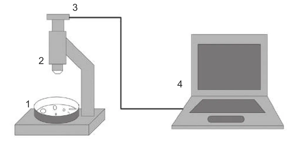
Dry clear films formed in the Petri dishes.Photos of these films were taken by microscopic USBcamera “Altami”with 3488x2616 pixels dimension.Fig.2 shows an experimental setup.

Fig.2.Experimental setup:1—experimental sample in Petri dish,2—optical microscope,3—charge coupled device(CCD)camera,and 4—computer.
3.3.Examples of Structures in Bioliquid Films
As experimental results,we obtained images of films of albumin protein solutions and serum films with the addition tick-borne encephalitis vaccine.Albumin protein films were created with various initial conditions.This is the initial volume,the concentration of protein in solution,and pH.Fig.3 shows various structures in the films of albumin protein,and Fig.4 shows films of blood serum with various compositions.

Fig.3.Albumin protein films:(a)spiral structures,(b)eyelash and spiral structures,(c)long dendrites,(d)short dendrites,(e)crooked cracks,and(f)non-define spiral structures.

Fig.4.Blood serum films:(a)pure blood serum,(b)blood serum with EGTA,(c)blood serum with tick-borne encephalitis vaccine,(d)blood serum with EGTA and tick-borne encephalitis vaccine,(e)blood serum film with tick-borne encephalitis vaccine (alternative compliment way),and (f) blood serum film with tick-borne encephalitis vaccine (alternative compliment way)with immunoglobulin(cracks “plait”).
As shown from the obtained images of albumin protein films,the structures can vary greatly depending on the conditions of the experiment.To determine the parameters of protein solutions,it is necessary to study the structures that arise as a result of self-organization.
From the images obtained,it can be seen that,depending on the presence of various substances in the blood serum,various structures form in the films as a result of self-organization.This example shows how blood serum behaves with the addition of the tick-borne encephalitis vaccine with a common and alternative complement pathway.Currently,this method is already used in medical practice.Today,the structures have been studied only at a qualitative level.In order to develop the diagnostic method,structure-recognition computing should be established.Therefore,we need to design the software for recognizing the structures(as described above)and highlighting their main features to move from qualitative analysis of films to the quantitative mathematical one.The next stage in the development of this method of diagnostics will be the creation of a unique database to assess the state of the human body by the films of biological fluids.
4.Image Processing
This section focuses on the image processing to enable the implementation of automatic medical diagnosis.
4.1.Theory
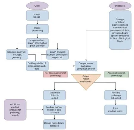
Automated image processing is the organization of a computer vision system aimed at registering structures of the interest for medical diagnostics.There are several main stages to develop the computer vision of an automatic diagnostic system.The algorithm of the image processing automatic diagnostic system is presented in Fig.5.
This algorithm includes image acquisition,its processing,and identification of metrics,on the basis of which it is possible to predict pathologies.Image acquisition is carried out using a digital camera connected to an optical microscope.The camera is controlled by a computer.Thus,the image immediately goes to the PC hard drive,after which it is processed in a program written in Python using image processing libraries,such as Open-CV,MatPlotLib,and Pillow.
4.1.1.Image Registration

Fig.5.Algorithm of the image processing automatic diagnostic system work.
The first stage includes obtaining the primary information from the photodetector,digitalizing the image,sending the information to the memory of the computing device and scaling it.It is possible to obtain two-and threedimensional arrays depending on the type of camera,corresponding to the images and containing a digital signal in each cell of the array,and corresponding to the brightness of the exposure at a point.It is possible to get a twodimensional array for the monochrome case and three sets of two-dimensional arrays for the color image.There are several options for representing color images,for example,the RGB or I palette,which are used depending on the desired result.However,in order to save computing resources,all color images are converted to monochrome images.
4.1.2.Image Filtering
In this work,filtering will be considered only in the spatial domain,which is divided into linear and nonlinear parts.Linear filtering is based on the operation of two-dimensional discrete image convolutionA(x,y)and the convolution kernelh(i,j)[32].

The convolution kernelsh(i,j)are the windows that are either pre-calculated,or the elements of the window array calculated in the process of work.For the purpose of noise reduction,the Gaussian filter is applied,defined as

whereσis the standard deviation of the pixel values of the filter window.
Non-linear filters are based on the rank and order statistics,i.e.,a median filter.Median filtering is defined as follows[32]:

whereMis a neighborhood;xandyare coordinates of pixels.
The essence of median filter work is that each element of the output image is equal to the median of the original image data located in a two-dimensional window(aperture).
4.1.3.Description and Analysis of Structures
The description of structures means the construction of broken curves by the vectorization of the image.This approach is carried out by constructing quadtrees,highlighting points describing the contour and further approximation by broken curves[33],[34].
As a result of the description of the structure,it is possible to carry out an analysis,namely,to determine their number,to calculate the area,length,center of mass of the figure,and other geometrical parameters for each structure,which allows us to form a feature vector necessary for the classification procedure.In addition to the geometric characteristics,the selection of the contours of the areas allows one to combine the colorimetric characteristics as a feature with the original image:Color,brightness gradient in points,etc.[35],[36].
It also seems possible to calculate certain mathematical parameters of structures in films of biological fluids.This is the general perimeter of contours,area of structures,aspect ratio (it is the ratio of width to height of bounding rectangle of the object),extent(extent is the ratio of the contour area to the bounding rectangle area),solidity(this is the ratio of contour area to its convex hull area),and equivalent diameter(equivalent diameter is the diameter of the circle whose area is the same as the contour area).Such analysis of films of biological fluids will allow us to move from the qualitative description to a more accurate,mathematical one.
4.1.4.Classification of Structures
Having a vector of signs,it is possible to determine the class of structure,which is the ultimate goal of medical diagnosis.Due to a well-constructed classifier,it is possible to establish a set of characteristic parameters,to determine possible pathologies by the images of the dehydration of biological liquids films.
We consider the procedures of image filtering as a part of this work,since the information that comes to the segmentation stage should have a minimum level of the noise component.Noise filtering involves two basic steps.When filtering images from noise components,it is also important to understand that all artifacts,when considering the frequency domain,have high frequency,often close to the frequency of object boundaries.Therefore,the first step is to eliminate the influence of artifacts on the general background situation.As a rule,this is done by blurring the image in the spatial domain or by processing the low-pass filter.After this,the contours and borders of the objects in the image are enhanced.Gradient methods and high-pass filters are used to implement this step.Consider the indicated steps for filtering the image separately.
4.2.Results and Discussions
Image processing took place according to the algorithm presented in Fig.6.

Fig.6.Algorithm of image processing.
The program code uses functions,such as erosion,thresholding,Gaussian blur.The determination of the contours and the calculation of their parameters directly depends on the specification of the window sizes and thresholds.Here,three test images of blood serum are shown in Fig.7.
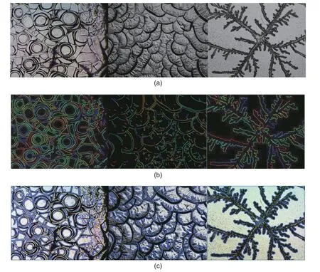
As a result of processing,the contours of the main structures were highlighted on the images,as well as individual contour strokes were depicted.Fig.8 shows the experimental results.
It was found that for all types of structures,the calculation of specified parameters can only be made with thresholds from 90 to 20.For each type of structure,depending on the size of the Gaussian blur window,the structure parameters were calculated.Data are presented in Tables 1,2,and 3,respectively.
It can be seen from the calculated parameters that basically the parameters do not change much depending on the Gaussian window,and are sometimes repeated.It has also been experimentally shown that a 5×5 erosion window size is optimal for edge detection.

Fig.7.Test images:(a)spiral structures,(b)“plait”structures,and(c)“dendrite”structures.
5.Conclusions
In this paper,a cuneiform dehydration method for medical diagnosis was described.This method is based on studying the parameters of structures in films of biological fluids.We experimentally showed that due to the presence of various pathological processes in the patient’s body,various structures are formed in blood serum films.These structures can play the role of the identification signs for the diagnosis of various diseases.
However,to describe these structures,the qualitative analysis is not enough.That is why we examined some principles of image processing in order to switch from a qualitative analysis of structures in biological fluid films to a quantitative one.As a result of the experiments,it was found that the calculated parameters of similar structures,for example,spirals and scales,were a little similar when the Gaussian blur window is 13×13,and the parameters of the dendrite structures were different from the other two types by an order of magnitude.

Fig.8.Computational results of blood serum films processing:(a)original images,(b)various contours of structures in the films,and(c)closed contours of main structures.

Table 1:Computational data of spiral structures

Table 2:Computational data of plaits structures
Thus,we can say that the mathematical parameters of structures in biological fluid films can become a fullfledged diagnostic material.For this,it is necessary to improve the image processing technique and calculate a larger number of parameters.

Table 3:Computational data of dendrites structures
Based on this diagnostic method,it is possible to create some autonomous system for medical diagnosis.This system will contain a database of diagnostic parameters of structures in films of biological fluids and will be constantly supplemented by them.Thus,when diagnosing by biological fluids films,the doctor will use this system to compare the results with many others.This will increase the likelihood of correct diagnosis.
杂志排行
Journal of Electronic Science and Technology的其它文章
- Machine Learning Application for Prediction of Sapphire Crystals Defects
- Method of Relaxation Rates Measurement in Proton-Containing Materials
- Recognition of Film Type Using HSV Features on Deep-Learning Neural Networks
- BER Performance of Finite in Time Optimal FTN Signals for the Viterbi Algorithm
- Role of Electromagnetic Fluctuations in Organic Electronics
- Utilization of Extrinsic Fabry-Perot Interferometers with Spectral Interferometric Interrogation for Microdisplacement Measurement
