Morphologies of Fibronectin Fibrils Formed under Shear Conditions and Their Cellular Adhesiveness Properties
2020-04-25PhuongThaoNguyenThiQuocPhongLeVolkerStoldtNgocQuyenTranAnhThoLeandKhonHuynh
Phuong-Thao Nguyen-Thi,Quoc Phong Le, Volker R.Stoldt,Ngoc Quyen Tran,Anh Tho Le and Khon Huynh,*
1School of Biomedical Engineering, International University,Vietnam National University,Ho Chi Minh, 70000,Vietnam
2Department of General Visceral and Pediatric Surgery,University Hospital and Medical Faculty of the Heinrich-Heine University Düsseldorf, Düsseldorf, 40225,Germany
3Graduate University of Science and Technology,Vietnam Academy of Science and Technology,Ho Chi Minh, 70000,Vietnam
4Institute of Applied Materials Science, Vietnam Academy of Science and Technology,Ho Chi Minh, 70000, Vietnam
5Faculty of Medicine, Hong Bang International University,Ho Chi Minh,Vietnam
Abstract:Fibrillar fibronectin(FFN)is a biological active form of FN which form linear and branched meshwork around cells and support cellular activities.Previous studies have demonstrated that shear stress can induce cell-free FN fibrillogenesis.In this study,we further examined the effect of shear stress conditions on morphology of formed FFN and preliminarily looked for relationship between FFN’s morphology and cell adhesion.Plasma FN at 50 μg/ml was perfused through channel slides at shear rates of 500 s-1 or 4000 s-1.Our results showed that there were four FFN structures formed:(1)FN nodules,(2)fibril in different sizes (3) with or without nodule attachment, and (4) fibrillar matrix.At 4000 s-1,FFN fibrils was formed within the first 10 min and reached the highest surface coverage only after 20 min.In contrast, FFN formation was significant more slowly at 500 s-1 at which only FN nodules and small fibrils were formed.Platelets bound on thin layer of FN and rarely found on large FN fibrils.In contrast,fibroblast stretched their shape on platform of FFN matrix and bound actively to all types of FFNs.Taken together,our data suggests a morphological dependent biological activity of FFN.
Keywords:Shear condition;fibrillar fibronectin;cell-free fibrillogenesis
1 Introduction
Fibronectin(FN)is a glycoprotein dimer with high molecular weight(440-500 kDa)and is one of the main components of extracellular matrix in different types of tissue[1-3].As its multi-domain chain gives rise to the capacity of interacting with many different types of cell and other molecule,the biological roles of this molecule to cells and body are very diverse.They can be the involvement in biological processes such as body development; tissue formation; biological activities of cells including adhesion, proliferation,migration, differentiation, or in pathophysiological processes of hemostatic and thrombotic disorders,angiogenesis,cancer metastasis, etc.[4,5].
FN are categorized into two types:cellular FN(cFN)and plasma FN.cFN is secreted,assembled into insoluble multimer fibrils (FFN) and integrated into the ECM by cells at local tissue such as fibroblast,endothelial cell, etc.[3].On the other hand, plasma FN is secreted by hepatocytes into blood stream at 200-600 μg/ml (0.4-1.2 μM) in a soluble compact dimer form.Circulating plasma FN consists of two subunits, and each of its subunits is about 220-250 kDa.Due to the compact structure, all domains that can interact with cellular receptors, ECM molecules, and other FN molecules are buried leading to inactivation of this protein.The inactivation of plasma FN may be to avoid unnecessary interactions between FN with blood cells that may lead to blood clots and vessel blockage under normal conditions[6].However, when needed, cells and platelets have the ability to use plasma FN to unfold, and assemble the molecules into FFN matrix with exposed domains and integrate into the ECM.For example, FN molecules are assembled to form FN fibrils that better support platelet adhesion on to wound site than normal plasma FN [7,8].Nevertheless, the mechanism of formation, molecular structure, the exposure of cryptic domains leading to biological activity of FN fibrils under different conditions have not been studied in detail.
Plasma FN does not spontaneously form multimeric fibrilsin vivo[9].Previous studies have clearly demonstrated that FN fibrillogensis can only occur when there are interactions between cellular integrins and FN leading to the cytoskeleton contraction of tethered cells which generates contractile forces that unfold the molecule and expose cryptic FN-binding domains and initiating the formation of FN fibrils[4,5].However, until now, there have been various chemical and mechanical approaches to synthesize FN fibrils in in vitro conditions (cell-free system) [10-14].To date, structure and function of these FFN have not yet been clearly understood.In this study, we aimed to investigate on shear-induced FN fibrillogenesis, analyzing FFN structures and characterizing their cellular interactions.
2 Results
2.1 Morphological Analysis of FFN Formed under Shear Conditions
FFNs formed into diverse shapes by shear stress under different conditions:concentration of FN,shear rate of flow and time of exposure.Microscopic analyses showed that there were four typical types of FFN matrix created under our examined conditions:(1) FFN nodules, (2) FFN fibrils arrange into dendritic structure with diameter ranging from few nm to a few 10 nm, (3) FFN fibrils into lattice, (4) large FFN matrix structure with bunches of fibrils connecting each other (Figs.1A and 1B).The structures of FFN matrix were illustrated better by fluorescence microscopy with AF488-conjugated FN(Figs.1C and 1D).
Surface morphologies of formed FFNs were characterized by SEM.Results showed that FFN variants could be classified into two groups:smooth surface and rough surface with nodule attachment(Figs.1E and 1F).In parallel experiments, FFN did not form when N-terminal 70 kDa FN fragment, an inhibitor of FN fibrillogenesis, was presented(Fig.1G).
2.2 Kinetics of FFN Formation under Low(500 s-1)and High Shear Rates(4000 s-1)
In addition to distinct fibrillar structures,we also observed differences in FFN formation kinetic between 500 s-1and 4000 s-1(Fig.2).Within 40 min of shear exposure,FN molecules deposited into FFN structures that covered around 0.9 mm2area.At shear rate of 4000 s-1,bundle of FFN fibrils was observed early during the first 10 min.These fibrils had then quickly expanded into matrix structure that reached the highest surface coverage after 20 min of shear exposure.In contrast, the process of FFN formation was significant more slowly at shear rate of 500 s-1at which only FN nodules and small fibril matrix were formed after 40 min.
2.3 Effect of FN Fibrils on Cellular Behaviors

Figure 1:(A and B) Light microscopic images of typical types of FFN matrix created under shear conditions:FFN nodules (↑), FFN fibrils arrange into dendritic structure (Δ), FFN fibrils into lattice ( ★),large FFN matrix structure with bunches of fbirils connecting each other (*).(C and D) Representative fluorescence microscopic images, and (E and F) scanning electron microscopic images of FFN fibrils.(G)In control experiments, shear induced FFN formation was inhibited in the presence of N-terminal 70 kDa FN fragment

Figure 2:Dynamic of shear-induced FN fibrillogenesis by shear rate 4000 s-1 (top) and 500 s-1 (bottom)within 40 min
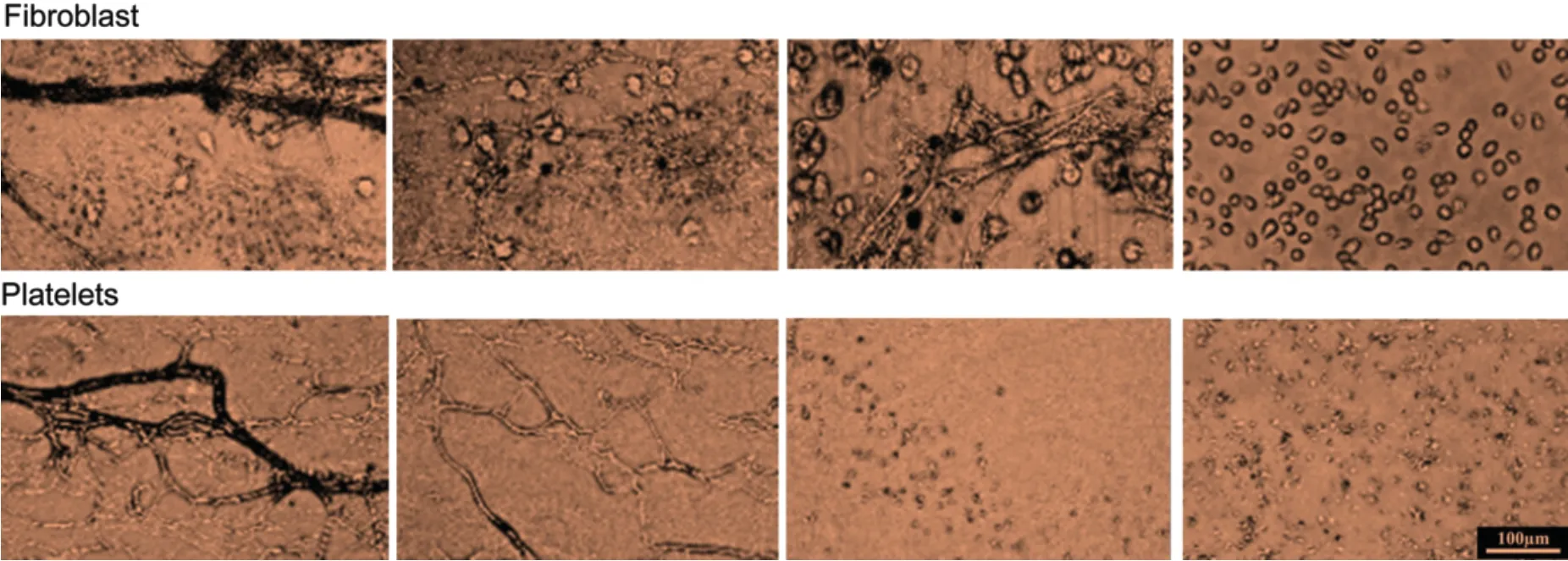
In order to test whether the FFN morphology characterizes their biological function, fibroblasts and platelet adhesion assays were performed on FFN with diverse morphologies.Our microscopic analysis showed different cell adhesion profiles on distinct FFN surfaces.In experiments with fibroblasts, cells adhesion was observed on all types of examined FFN with the strongest adhesiveness and tendency to form clusters on matrix of FFN medium-sized with nodules.In contrast, platelets did not adhere onto surfaces with medium-sized to large FFN fibril matrixes while they clustered along nodular structures and small FFN fibrils (Fig.3).In control experiments with FN surfaces, fibroblasts and platelets showed an even distribution on over the surface.

Figure 3:Cell adhesion assay revealed different fibroblast(top)and platelet(bottom)interactions of distinct FFN structures.Fibroblast adhered onto different types of FFN fibrils whereas platelets adhered only onto small FFN structures
3 Discussion
Previous studies using cone-plate rheometer have shown cell free FFN formation by shear stress [12,13].However,these studies did not analyze structures of FFN.In our current study,we observed that there were four structures of FFN that were formed under different shear conditions.Based on our results, a model of FFN formation under flow was proposed (Fig.4).FN modules tend to form under low shear rate conditions while they will develop further into FFN fibril,FFN lettuce,or FFN fibril matrix under higher shear rate conditions.

Figure 4:Proposed model of two distinct FN fibrillogenesis processes at low(500 s-1)and high shear rates(4000 s-1) leading to the formation of FN nodules and FFN matrix, respectively
FN is one of main components of the ECM in different types of tissue[15].As its multi-domain chain gives rise to the capacity of interacting with many different types of cell and other molecule,the biological roles of this molecule to cells and body are very diverse[16-18].They can be the involvement in biological processes such as body development; tissue formation; biological activities of cells including adhesion, proliferation, migration,differentiation, or in pathophysiological processes of hemostatic and thrombotic disorders, angiogenesis,cancer metastasis.However, the hallmark in understanding biological functions of FN is that these functions are extremely complex and appear to be capable of switching back and forth(dual-role)in specific cases such as hemostasis and thrombosis, or cancer progression [7,19-21].Of note, most of biological functions of FN are related to their FFN structure on which the cryptic functional binding sites of FN are exposed [7,22].We hypothesized that the functions of FN are not only dependent on its FFN form but also is affected by FFN structure.Distinct FFN structures may differ in binding site exposure.While further studies on cell-FFN interaction are needed, our cell adhesion assay data could partially confirm this hypothesis by showing that cells interact differently to FFN surfaces with different morphologies.
4 Materials and Methods
4.1 Materials
Frozen plasma bags were collected from Blood Transfusion and Hematology Hospital in Ho Chi Minh city.Gelatin-sepharose was purchased from Sigma Aldrich (Hamburg, Germany).Alexa Fluor®488 succinimidyl ester (AF488) was from Molecular Probes (Hamburg, Germany).Micro quartz cuvettes(1400 μL) were purchased form Thorlabs (Newton, New Jersey).The mouse L929 fibroblasts was purchased from the Cell Bank at ATCC.The cells were cultured in Dulbecco’s modified Eagle’s medium(DMEM; Gibco; Thermo Fisher Scientific, Inc., Waltham, MA, USA) and supplemented with 10% Fetal Bovine Serum (FBS)(Sigma Aldrich), 1%Penicillin Streptomycin (PS) (Sigma Aldrich).
4.2 Formation of FFN by Shear Stress Facility
Plasma FN was purified by affinity chromatography method with modification [7].Ibidi μ-Slide VI 0.4 (Ibidi GmbH, Martinsried, Germany) were coated with 100 μL FN (50 μg/mL) or Collagen type I(10 μg/mL) by incubation at room temperature for 4 h.FN/collagen-coated microslides were washed with PBS pH 7.4 buffer before prior to flow experiments.A volume of 15 mL of FN at 50 μg/mL was repeatedly perfused through channels of Ibidi μ-Slide by Harvard Apparatus 22 Dual Syringe Pump at flow rates of 3.8 ml/min (500 s-1) and 30.4 ml/min (4000 s-1) creating a unidirectional flow for 40 min at room temperature.In parallel experiments, perfusion was done with Alexa Fluor 488-conjugated FN or in the presence of N-70 kDa FN fragment.For further surface analysis, FFN formed under flow conditions was surface coated with gold and subjected to SEM at an accelerating voltage of 10 kV.Regions of interest(ROI) were chosen where fibrils appeared.For each sample, at least five ROIs with total of 10-20 fibrils were captured and analyzed.Results are representative of at least three independent experiments.
4.3 Cell Adhesion Assay
Before performing in vitro experiments,FFN were further sterilized by UV for 20 min and kept in clean bench to avoid contamination.In fibroblast adhesion assays,fibroblasts were seeded directly at 104 cells each slide chamber and cells were allowed to adhere for 1 hour at 37°C.Fibroblasts adhesion were tested on FFN fibrils formed under shear rate ranging from 500 s-1to 4000 s-1that showed different morphologies.After incubation, cells were washed with PBS pH 7.4 and fixed with 3.7% paraformaldehyde for further analysis.Parallel adhesion experiments were repeated with human washed platelets.For comparison of cell adhesion between different samples, at least five different ROIs were analyzed.Results are representative of three independent experiments.
Funding Statement:This research is funded by Vietnam National University Ho Chi Minh City(VNUHCM)under grant number C2018-28-01.
Conflicts of Interest:The authors declare that they have no conflicts of interest to report regarding the present study.
