Rigid ureteroscopy in prone split-leg position for fragmentation of female ureteral stones:A case report
2020-04-23KaiHuang
Kai Huang
Kai Huang,Department of Urology,Clinical Medicine College,Yangzhou University,Northern Jiangsu People's Hospital,Yangzhou 225001,Jiangsu Province,China
Abstract
Key words: Ureteroscopy;Ureteral calculi;Lithotripsy;Prone position;Female;Case report
INTRODUCTION
In recent decades,ureteroscopic lithotripsy has become the main therapy to treat urolithiasis[1],with reduced expenditure and higher efficiency.Most ureteroscopy is performed effectively and safely in the supine position.Here,we present a case of successful lithotripsy of a woman's distal ureteral stone by using rigid retrograde ureteroscopy in a modified prone split-leg position.
CASE PRESENTATION
Chief complaints
A 62-year-old woman was diagnosed with ureteral calculi through X-ray and ultrasound examination.While she underwent shock wave lithotripsy treatment twice and received lithagogue drugs,these therapies appeared to be ineffective.
History of present illness
She had a fracture of the left femoral neck at 50 years of age and did not receive proper treatment in a timely manner due to poverty.
Imaging examinations
A non-enhanced computed tomography scan of the abdomen revealed a 12 mm × 10 mm stone in the lower right ureter (Figure 1).Due to movement restriction in her left hip,surgery in the lithotomy position could not be performed.She also refused the choice of flexible ureteroscopy because of the greater economic burden.We finally decided to perform a rigid retrograde ureteroscopic lithotripsy in the prone split-leg position.
FINAL DIAGNOSIS
Ureteral calculi.
2.4 2组术后24周骨折愈合情况评估 术后24周,2组患者X线检查均有骨痂通过骨折线,患肢无纵向叩击痛,不扶拐能行走3min,对比之前X线片骨折无变形,内固定无松动开裂,无骨不连发生。
TREATMENT
The patient was oriented in the modified prone split-leg position during the operation after general anesthesia (Figure 2).A Fr 8/9.8 rigid ureteroscope was used to enter the upper ureteral tract alongside the guide wire (Figure 3).When the ureter stone was visualized at the lower ureteral position,a 20W Holmium YAG laser was used to fragment and dust the calculi.Subsequently,residual calculi exceeding 2 mm were removed using a nitinol stone basket.A F4.8 double J stent was then placed in the right ureter through a guide wire after evaluating the urinary tract for complications.An abdominal radiograph review showed perfect positioning of the double J ureteral stent with no visible calculi in the ureteral area (Figure 4).
OUTCOME AND FOLLOW-UP
At 2 d after ureteroscopic lithotripsy,the patient was discharged from our hospital and returned to our department 1 mo later for successful ureteral stent removal.

Figure1 Computed tomography scan showing a calculi in the right ureter.
DISCUSSION
The incidence and prevalence of urolithiasis,including ureteric calculi,is increasing based on international epidemiological data[2].Compared with global incidences,China has a very high incidence of urinary stones[3],which has added a considerable burden to people's health and the country's economy.Although the vital characteristics of dietary patterns and high water intake should be considered prior to any treatments,ureteroscopy performed for ureteric calculi disease has increased due to the direct effects of the rising number of urolithiasis disease cases on health resources.Since ureteroscopy was first introduced in the 1970s[4],ureteroscopes have undergone rapid development in the past century,accompanied by various improvements in their use.The working pipeline of ureteroscopy allows the insertion of laser fibers and a basket and increases the efficiency of ureteroscopy lithotripsy.Holmium laser lithotripsy has been the preferred choice to treat ureteric calculi,especially the lower ureteric stone.Although rigid ureteroscopy is the main method applied worldwide,the emergence of a flexible ureteroscope is an important development that has changed the traditional concept of endourology[5].The flexible scope can reach the upper ureter,pelvis,and renal calices and dust urinary stonesviadirect vision through the ureteral canal.However,this method is not widespread in China because of its high cost,which is also why the patient in this report ultimately opted for rigid ureteroscopy and not the initially suggested flexible ureteroscopy lithotripsy,even despite her restricted left hip movement.
All of the technological advancements have extended the application of ureteroscopy.A simultaneous approach with flexible ureteroscopy and percutaneous nephrolithotomy performed in the Galdakao-modified supine Valdivia position has been reported[6].This position has the advantage of providing a better option for complex urolithiasis in the entire upper urinary tract.It does not require the patient to change position during the operation.Hamamotoet al[7]reported that a synchronized approach with flexible ureteroscopy and minimally invasive percutaneous nephrolithotomy (mPNL) (in the prone split-leg position) achieved a 81.7% stone-free rate that was higher than mPNL (38.9%) and standard PNL (45.1%).Another report described how to manage an encrusted stent by combined endoscopic methods in the prone split-leg position[8].However,there have been no reports about rigid ureteroscopy for ureteric calculi in the prone position.In the present case,the patient's left hip movements were limited,which precluded normal ureteroscopy in the supine position.In addition,because of the economic burden,she was not willing to accept treatment by flexible ureteroscopy.Finally,after observing the angle of her hip joint activities,we performed the surgery in a modified prone split-leg position and successfully removed her lower ureteral calculi (Figure 3).
CONCLUSION
To the best of our knowledge,this is the first reported case of treating ureteral stones by rigid ureteroscopy in the prone split-leg position.Due to the different anatomical characteristics of the female urethra,we believe that rigid ureteroscopy can be used to treat ureteral calculi in certain situations.This includes situations like the present case where a patient cannot be operated on in the lithotomy position,which is especially applicable to the removal of lower ureteral stones in women.
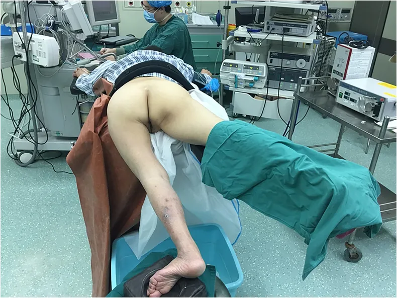
Figure2 Patient positioning in the modified prone split-leg position due to limited movement of her left hip.

Figure3 Ureteroscope enters into the right ureter alongside the guide wire.

Figure4 X-ray of kidney,ureter,and bladder shows stent position and no visible stone in ureteral area after operation.
猜你喜欢
杂志排行

World Journal of Clinical Cases的其它文章
- CD56+ lymphoepithelioma-like carcinoma of the lung:A case report and literature review
- Systemic treatment for severe concentrated sulfuric acid burns in an adult male at high altitude:A case report
- Clinical effects of apatinib mesylate for treatment of multiple brain micrometastases:Two case reports
- Disseminated histoplasmosis in primary Sjögren syndrome:A case report
- Severe venous thromboembolism in the puerperal period caused by thrombosis:A case report
- Multiple neurofibromas plus fibrosarcoma with familial NF1 pathogenicity:A case report