Mucinous adenocarcinoma of the buttock associated with hidradenitis:A case report
2020-04-08SungJinKimTaeGonKimMiJinGuSohyunKim
Sung Jin Kim,Tae Gon Kim,Mi Jin Gu,Sohyun Kim
Sung Jin Kim,Sohyun Kim,Department of Surgery,College of Medicine,Yeungnam University,Daegu 42415,South Korea
Tae Gon Kim,Department of Plastic and Reconstructive Surgery,College of Medicine,Yeungnam University,Daegu 42415,South Korea
Mi Jin Gu,Department of Pathology,College of Medicine,Yeungnam University,Daegu 42415,South Korea
Abstract BACKGROUND Mucinous adenocarcinomas of the buttock are rare and have an uncertain etiology and natural course.They are usually related to chronic anal fistulas,hidradenitis suppurativa,or Crohn's disease.Here,we report a case of mucinous adenocarcinoma associated with hidradenitis and contradictory immunochemistry results.CASE SUMMARY A 62-year-old man complained of recurrent abscesses of the buttock for 3 years.He had several scars and nodules in bilateral buttocks,with purulent discharge.The skin lesions did not appear to originate from the anus.The patient was diagnosed with recurrent abscesses due to hidradenitis suppurativa at the first visit.He showed purulent and subsequent mucin discharge in the first operation and was diagnosed with mucinous adenocarcinoma.Several examinations were performed to determine disease origin and staging.There were no significant findings or evidence of anal fistulas.Hence,he underwent wide local excision and V-Y advancement flap in the second operation.The final diagnosis was mucinous adenocarcinoma without any evidence of anal fistulas.Additional immunochemistry test results were negative for cytokeratin(CK)7 and positive for CK20 and CDX2,with a colorectal origin.A pathologist suggested that the disease originated from a chronic anal fistula.The patient has remained free of recurrence for 24 mo.CONCLUSION Although the patient with mucinous adenocarcinoma showed an atypical course,immunochemistry helped detect the disease origin.
Key Words:Mucinous adenocarcinoma;Anal fistula;Hidradenitis suppurativa;Wide excision;Immunochemistry test;Case report
INTRODUCTION
Adenocarcinomas of the buttock and perianal area are rare,with most the articles published in the literature being case reports.Perianal adenocarcinomas can develop from perianal fistula[1].Adenocarcinomas related to the fistula tract type almost present mucinous type[1,2].Mucinous adenocarcinomas of the buttock and perianal area are also reported in hidradenitis suppurativa(HS)or Crohn's disease,which rarely occur[3-5].
The etiology or natural course of the mucinous adenocarcinoma of the buttock is not clearly known because of the rarity of the disease.Its diagnostic criteria were as follows:(1)The fistula should predate the carcinoma;(2)There should be no synchronous colorectal carcinoma;and(3)The internal opening of the fistula should be into the anal canal and not the malignancy[6].However,it is often difficult to identify the disease origin since these criteria do not appear in all cases[3,7].Here,we report a case of mucinous adenocarcinoma of the buttock with an atypical diagnostic course.
CASE PRESENTATION
Chief complaints
A 62-year-old man,who complained of recurrent abscesses of the buttock,was referred to the Division of Colon,Rectum and Anus for treatment.
History of present illness
The patient had recurrent abscesses of the buttock for 3 years and had undergone incision and drainage at a local clinic.Although the abscess was resolved with treatment,it recurred.The patient had several abscess sites on the buttock,but no anal discharge,bloody stools,fever,chills,or weight loss were noted.He was otherwise healthy.
History of past illness
He had been taking anti-hypertensive medication and antiplatelet drugs for hypertension for 4 years.However,he had no significant past medical or surgical history.He had no history of anal diseases,such as anal fistulas,hemorrhoids,or perianal abscesses.He had no history of other skin diseases.
Physical examination

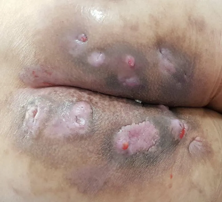
Figure 1 Clinical appearance of buttocks.
His vital signs were normal.He had several scars and nodules on the bilateral buttocks(Figure 1).Purulent discharge drained from one site on the left buttock.Other sites on the left buttock,which showed no drainage,were composed of granulation tissue.Several nodules with skin lesions on the right buttock were observed;however,the discharge was not purulent but serous.There were no lesions in the anus on digital rectal examination.The site of the skin nodule did not originate from the anus through physical examination.He had no symptoms indicative of other diseases.Finally,he was diagnosed with recurrent abscess due to HS at the first visit.
Laboratory examinations
The laboratory test included complete blood count,liver function,kidney function,and electrolyte and coagulation profiles,which were within normal limits.Carcinoembryonic antigen and CA19-9 levels were 4.93 ng(normal range,0-10 ng)and 39.69 U/mL(normal range,0-37 U/mL),respectively.
Imaging examinations
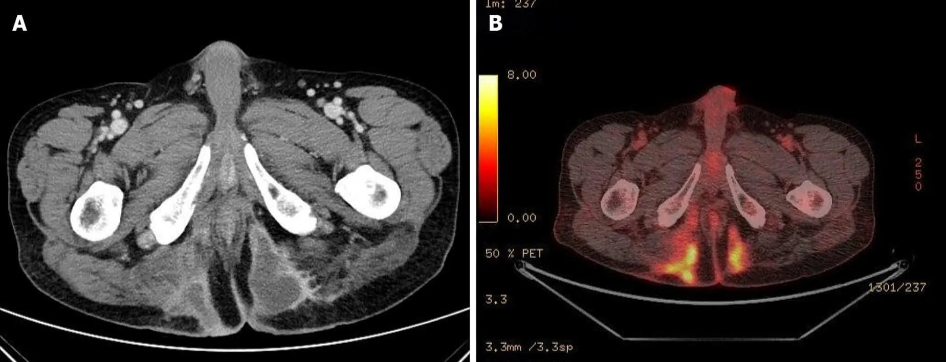
He underwent pelvic computed tomography(CT)before the operation because of the prolonged duration of symptoms suggesting the possibility of undiagnosed disease.CT showed an approximately 10 cm in length of abscess in the subcutaneous tissue of the bilateral buttocks(Figure 2A).There was no evidence of its relationship with the anus.After the first operation,he underwent gastroscopy,colonoscopy,transanal ultrasonography(TRUS),and positron emission tomography(PET)to determine the disease origin and condition,as he was diagnosed with mucinous adenocarcinoma.The results of all examinations other than PET were unremarkable.There was no evidence of anal fistula on TRUS.PET showed increased fluorodeoxyglucose uptake in the abscess cavity,buttock,and perianal wall(Figure 2B).It also showed increased fluorodeoxyglucose uptake in the prostate,which suggested inflammation or malignancy.The patient was diagnosed with inflammation of the prostate based on prostate biopsy findings.
Personal and family history
He had no significant personal or familial history.Moreover,there was no history of cancer in his family.
FINAL DIAGNOSIS
The histopathologic results after 2ndsurgery showed free resection margins on the leftside specimen and positive margins on the right-side specimen.The positive resection margin was close to the right ischium.The final diagnosis of the patient was mucinous adenocarcinoma without any evidence of anal fistulas.We performed additional immunochemistry tests,which yielded negative results for cytokeratin 7(CK7)and positive for CK20 andCDX2(Figure 3).A pathologist suggested that the disease originated from a chronic anal fistula.However,there was no evidence of anal fistulas.

Figure 2 Radiologic findings.A:Computed tomography findings before the first operation show a huge abscess cavity in bilateral buttocks;B:Positron emission tomography findings show 18F-fluorodeoxyglucose uptake along the abscess cavity.

Figure 3 Pathologic results of the buttock mass.A:There are variable-sized proliferated atypical glands and mucin pools(H&E,× 4);B:The atypical glands are positive for cytokeratin 20;C:The atypical glands are positive for CDX2.
TREATMENT
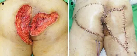
The patient underwent incision and drainage in the first operation.All nodules on each buttock were connected.The cavity of the abscess was deep,and it was not limited to the subcutaneous area.The discharge was purulent and subsequently mucinous.The patient was diagnosed with mucin-containing adenocarcinoma and treated with wide excision of the abscess cavity in the bilateral buttocks and V-Y advancement flap in the second operation(Figure 4).The surgeons resected the cavities carefully to prevent rupture.The abscess cavities in the bilateral buttocks were connected through the deep posterior anal space.The surgeons checked the anus during the second operation but did not find any indication of hidden disease.
OUTCOME AND FOLLOW-UP
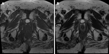
The patient has undergone clinical evaluation every 6 mo with a physical examination,laboratory test,and radiologic study.He has remained free of recurrence for 24 mo(Figure 5).
DISCUSSION
It is difficult to diagnose mucinous adenocarcinoma of the perineum.They are usually associated with chronic inflammation and manifest as persistent symptoms,regardless of the conventional treatment[8,9].Most patients present with perianal purulent discharge,a palpable peripheral mass,an ulcer or a recurrent abscess,which are nonspecific symptoms regardless of original disease[3,4,9,10].Patients with a tumor originating from a perianal fistula have a history of a fistula or persistent disease[3,11,12].Although diagnostic criteria have been developed,the diagnosis of adenocarcinoma from a perianal fistula is difficult as the symptoms are nonspecific[6].Therefore,mucinous discharge from the anal fistula provides evidence suggestive of carcinoma[13].T2-weighted magnetic resonance imaging shows a high signal intensity in some cases[3,14,15].However,many patients are diagnosed on the basis of official or operative biopsy[16,17].

Figure 4 Photographs after the second operation.A:Wide excision;B:V-Y flap operation.

Figure 5 Follow-up magnetic resonance imaging after 24 mo.There is no recurrence.A:T1 weighted image;B:T2 weighted image.
Patients with a tumor originating from HS also have a history of HS and symptoms similar to those of patients with a perianal fistula[4,18].Patients with a tumor originating from HS present with HS for at least 10 years[19,20].The tumor originating from HS may be located in the perianal region,perineum,and buttocks[4,20].The most common tumor type originating from HS is squamous adenocarcinoma,but it could be mucinous adenocarcinoma in some cases,in contrast to the tumor types originating from a perianal fistula[19,20].
Immunochemistry is commonly performed to determine the site of origin of the carcinoma[21,22].CKs are an epithelial class of intermediate-sized filament proteins in the cytoskeleton[23].CK7 is present in lung,breast,and female genital tract carcinomas[23].CK20 is found in the gastrointestinal,urothelium,and Merkel cell carcinomas[23,24].Adenocarcinomas of the small bowel,appendix,and colorectum express negative CK7 and positive CK20[23-25].Although a report of a case of mucinous adenocarcinoma from perianal abscesses stated that CK7 and CK20 were immunopositive,the immunochemistry results in this study indicated that the carcinoma had a colorectal origin[3].Another useful marker isCDX2,which indicates intestinal differentiation.CDX2is a caudal homeobox gene related to the proliferation and differentiation of intestinal epithelial cells[26].CDX2expression presents in at least 85% of overall colorectal adenocarcinomas[26,27].In this study,the first abscess of the patient developed in the left buttock,and the lesion persisted.The patient had no perianal symptoms.The initial diagnosis made in the outpatient clinic was recurrent HS.However,the immunohistochemistry results showed that the disease originated from the lower gastrointestinal tract.The authors suggested that this mucinous adenocarcinoma might be related to the anal fistulas that healed but remained as an abscess.
The treatment of mucinous adenocarcinomas of the buttock and perianal region depends on the location of the lesion and origin of the disease.Mostly,the surgical treatment is abdominoperineal resection(APR)with or without a wide local excision.Patients with mucinous adenocarcinoma involved with an anal fistula undergo APR,similar to patients with Crohn's anal fistula[5,11].Even if the disease does not involve the anus,most patients with associated HS undergo APR[4,19].Patients with wide excision underwent reconstructive operations[12,17,28].In a case report similar to this one,the patient underwent a local wide resection,and there was no gross evidence of anal canal involvement[10].The authors also considered APR as a curative treatment;however,the patient refused permanent colostomy,as there was no evidence of fistula.Moreover,many patients with perianal mucinous adenocarcinoma underwent further aggressive therapy,such as chemotherapy with or without radiotherapy[16,29,30].Neoadjuvant or adjuvant chemoradiotherapy showed better results,although the number of patients was small[16,29,30].The authors considered performing adjuvant chemoradiotherapy;however,the patient refused further treatment.
CONCLUSION
Mucinous adenocarcinomas of the buttock are rare and have uncertain etiologies and diagnostic criteria.They must be suspected in cases of continuous inflammation with an atypical course.Although the patient in the present case showed no evidence of fistula,immunochemistry was performed to detect the disease origin.Therefore,we need larger studies to confirm the etiology,diagnosis,and treatment of mucinous adenocarcinomas of the buttock.
杂志排行
World Journal of Clinical Cases的其它文章
- Special features of SARS-CoV-2 in daily practice
- Gastrointestinal insights during the COVID-19 epidemic
- From infections to autoimmunity:Diagnostic challenges in common variable immunodeficiency
- One disease,many faces-typical and atypical presentations of SARS-CoV-2 infection-related COVID-19 disease
- Application of artificial neural networks in detection and diagnosis of gastrointestinal and liver tumors
- Hepatic epithelioid hemangioendothelioma:Update on diagnosis and therapy
