Unilateral pleuroparenchymal fibroelastosis as a rare form of idiopathic interstitial pneumonia:A case report
2020-04-08JaeHaLeeHangJeaJangJinHanParkHyunKukKimSunggunLeeJiYeonKimSeongHoKim
Jae-Ha Lee,Hang-Jea Jang,Jin-Han Park,Hyun-Kuk Kim,Sunggun Lee,Ji-Yeon Kim,Seong-Ho Kim
Jae-Ha Lee,Hang-Jea Jang,Jin-Han Park,Hyun-Kuk Kim,Sunggun Lee,Seong-Ho Kim,Department of Internal Medicine,Haeundae Paik Hospital,Inje University College of Medicine,Busan 48108,South Korea
Ji-Yeon Kim,Department of Pathology,Haeundae Paik Hospital,Inje University College of Medicine,Busan 48108,South Korea
Abstract BACKGROUND Pleuroparenchymal fibroelastosis(PPFE)is a rare idiopathic interstitial pneumonia characterized by predominantly upper lobe involvement with pleural fibrosis and subjacent parenchymal fibrosis.Recently,there have been increasing reports of PPFE,and PPFE might coexist with other interstitial lung diseases in the lower lobe and upper lobe.However,cases of unilateral PPFE are scarce.CASE SUMMARY A 75-year-old Korean male presented to our hospital with chronic dry cough and exertional dyspnea.The patient’s symptoms started 6 mo previously and had been gradually worsening.At the time of presentation,he felt dyspnea when walking at his own pace.Radiologic findings suggested PPFE,but the lesion was localized in the upper lobe of the right lung.After multidisciplinary discussion,a transbronchial lung biopsy in the right upper lobe revealed collapsed alveoli with parenchymal fibroelastosis,and elastic van Gieson staining demonstrated septal elastosis with intra-alveolar collagenosis,which met the histopathologic criteria of definite PPFE.After multidisciplinary discussion in an experienced interstitial lung disease center,we confirmed the diagnosis of unilateral PPFE.Furthermore,we confirmed the progression of PPFE on radiologic findings during the followup period.CONCLUSION Clinicians should consider PPFE,even in cases with unilateral,predominantly upper lung involvement in interstitial lung disease patients through multidisciplinary discussion.
Key Words:Interstitial lung disease;Pleuroparenchymal fibroelastosis;Progression;Unilateral;Multidisciplinary discussion;Case report
INTRODUCTION
Pleuroparenchymal fibroelastosis(PPFE)is a rare idiopathic interstitial pneumonia(IIP)characterized by predominantly upper lobe involvement with pleural fibrosis and subjacent parenchymal fibrosis[1].After the first report in 2004 by Frankelet al[2],it was classified as the group of rare IIP in the international consensus classification of IIP in 2013.There was no consensus on the diagnostic criteria for PPFE,but the radiological and histopathological criteria suggested by Reddyet al[3]in 2012 has been generally accepted.Although PPFE is classified as a rare disease,recent reports increasingly reveal that PPFE may not be as rare as once thought,and various underlying conditions related with PPFE have been reported.
Chaet al[4]reported that among seven Korean patients with PPFE,three patients(42.9%)had a history of organ transplantation(two bone marrow and one liver).In our previous study,we also reported that among 26 patients with PPFE,16 patients(61.5%)had relevant underlying conditions(eleven infection,three chemotherapy and two organ transplantation)[5].In patients with PPFE,the coexistence of other interstitial lung diseases(ILDs)in the lower lobe has also been increasingly reported.The prevalence of coexistent ILD is relatively high(43.0%-91.7%),and among patients with ILDs,usual interstitial pneumonia was the most common radiologic and histopathologic finding[6-9].But unilateral lung involvement in patients with PPFE is very rare;therefore,we herein report a case of unilateral PPFE.
CASE PRESENTATION
Chief complaints
A 75-year-old Korean man presented to our hospital with chronic dry cough and exertional dyspnea.
History of present illness
The patient’s symptoms started 6 mo previously and had been gradually worsening.At the time of presentation,he felt dyspnea upon walking at his own pace.
History of past illness
He was an ex-smoker with a 35 pack-year history.He worked for a rubber-related product manufacturer for 30 years.He was not aware of any previous respiratory disease.
Physical examination
During the physical examination,auscultation with a stethoscope revealed inspiratory crackles in the right upper lung field.He appeared to have a slender habitus and flattened thoracic cage.His body mass index(BMI)was 18.4 kg/m².
Laboratory examinations
Laboratory findings showed that antinuclear antibody was positive(1:40,mixed and speckled pattern).Rheumatoid factor,anti-cyclic citrullinated peptide antibody and anti-neutrophil cytoplasmic antibody were unremarkable.
Further diagnostic work-up
Pulmonary function test showed amoderate restrictive ventilatory defect and low level of diffusion capacity of carbon monoxide.Forced vital capacity and total lung capacity(TLC)were 53% and 66% of the predicted level,respectively.Defect and low level of diffusion capacity of carbon monoxide was 47% of the predicted level,and residual volume(RV)was 73% of the predicted level.RV/TLC ratio was 48%.
Bronchoalveolar lavage,performed in the right upper lobe by flexible bronchoscopy,showed few lymphocytes(9%).The other results obtained for culture were unremarkable.
Imaging examinations
Initial chest X-ray showed a subpleural and irregularly shaped increased opacity in the right upper lung zone(Figure 1A).High-resolution computed tomography(HRCT)revealed subpleural thickening and consolidation with traction bronchiectasis in only the right upper lung field,which met the radiologic criteria of definite PPFE(Figure 2A).
Histological examination
Transbronchial lung biopsy was performed in the right upper lung field to make a histopathologic diagnosis.The histopathological findings of lung tissue obtained from transbronchial lung biopsy revealed collapsed alveoli with parenchymal fibroelastosis on higher magnification view,and elastic van Gieson stain demonstrated septal elastosis with intra-alveolar collagenosis,which met the histopathologic criteria of definite PPFE(Figure 3).
MULTIDISCIPLINARY EXPERT CONSULTATION
The clinical information and radiologic and histopathologic data were sent to The Kanagawa Cardiovascular and Respiratory Center in Yokohama,Japan for consultation of the multidisciplinary discussion(MDD).
Tae Iwasawa,Doctor,ILD specialist,Department of Radiology in Kanagawa Cardiovascular and Respiratory Center
HRCT showed mainly ground glass opacity and fibrosis with traction bronchiectasis,suggestive of PPFE.The lesion was located in the right upper lobe only with laterality.Radiologic finding was indeterminate for usual interstitial pneumonia,suggestive PPFE.
Tamiko Takemura,Doctor,ILD specialist,Department of Pathology in Kanagawa Cardiovascular and Respiratory Center
Hematoxylin and eosin-stained images revealed dense fragmentation and dilated alveolar space surrounding alveolar with elastosis.There was no granuloma.These findings suggested PPFE with high confidence.
Takashi Ogura,Doctor,ILD specialist,Department of Pulmonology in Kanagawa Cardiovascular and Respiratory Center
We diagnosed the patient with PPFE with laterality.We suggested that this abnormal presentation of PPFE may be due to occupational rubber exposure.
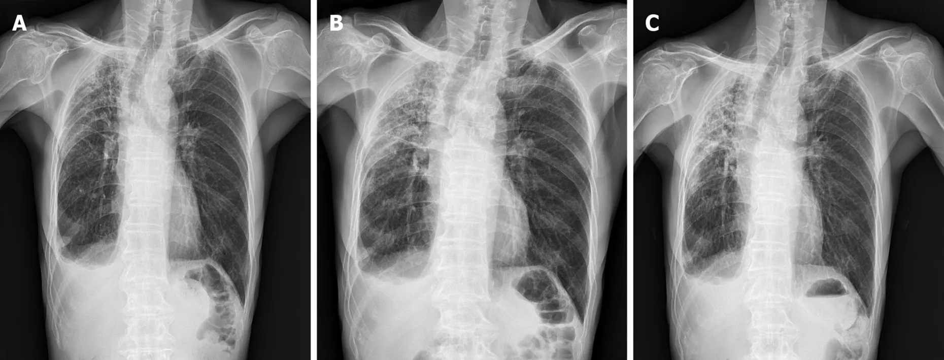
Figure 1 Chest X-ray showed subpleural and irregular shaped increased opacity in right upper lung zone.Sequential changes during 6 mo showed increased opacity in right upper lung zone.A:Initial visit of diagnosis;B:After 3 mo;C:After 6 mo.

Figure 2 High-resolution computed tomography revealed subpleural thickening and consolidation with traction bronchiectasis in only right upper lung field.Follow-up high-resolution computed tomography showed increased alveolar collapse with consolidation and volume loss compared to 6 mo ago.A:Right upper lobe at initial visit;B:Carina level at initial visit;C:Right lower lobe at initial visit;D:Right upper lobe after 6 mo;E:Carina level after 6 mo;F:Right lower lobe after 6 mo.
FINAL DIAGNOSIS
Through the MDDs in both our hospital and The Kanagawa Cardiovascular and Respiratory Center,the final diagnosis of the presented case was PPFE with laterality,unilateral PPFE.
TREATMENT
Based on the diagnosis,the patient has not been treated with medications.The patient was encouraged to undergo rehabilitation with nutritional support.

Figure 3 Transbronchial lung biopsy specimen in right upper lobe.A:On higher magnification view,hematoxylin and eosin-stained images revealed dense fragmentation and dilated alveolar space surrounding alveolar with elastosis;B:Elastic van Gieson stain demonstrated septal elastosis(orange arrows)with intra-alveolar collagenosis.
OUTCOME AND FOLLOW-UP
After diagnosis,the patient was scheduled to attend an outpatient clinic every 3 mo to estimate his clinical symptoms and disease progression.The respiratory symptoms have not changed for 6 mo,but chest X-ray and HRCT revealed progression of the PPFE(Figures 1 and 2).On HRCT,subpleural reticulation and traction bronchiectasis with a notable volume loss in the right upper lung was significantly increased when compared to the previous exam obtained 6 mo earlier.Therefore,we identified progression of PPFE.
DISCUSSION
PPFE is a new disease entity that is classified as a rare IIP.For nearly a decade,there have been an increasing number of reports that showed the heterogeneity of PPFE,including its incidence,etiology and prognosis.
The international guideline of the American Thoracic Society/European Respiratory Society recommended MDD as the diagnostic reference standard for ILD[10].The patient in the present case was diagnosed with unilateral PPFE after MDD involving a pulmonologist,rheumatologist,radiologist and pathologist in our hospital.However,because unilateral PPFE is so uncommon and we had never experienced it before,we also consulted with MDD in an experienced ILD center in Japan.The combination of the results from the two MDDs confirmed the diagnosis of unilateral PPFE.
In previous studies,a relatively lower BMI and a higher ratio of RV/TLC in PPFE patients compared to other IIP patients were reported as typical physiologic features of PPFE[5,6,8,11,12].Although the reason for the lower BMI in PPFE patients is not well understood,Suzukiet al[12]suggested that the characteristics of upper lobe predominant fibrosis and a more severe restrictive ventilatory defect might exacerbate the impaired energy and protein balance,resulting in low weight and muscle wasting[12].In terms of RV/TLC,Odaet al[7]suggested that despite upper lobe volume loss,RV/TLC was higher in PPFE patients because RV was relatively more increased than TLC owing to compensatory hyperinflation of the middle and lower lobes[7].The patient in the present case shared similar features with the aforementioned previous studies:A lower BMI of 18.4 kg/m² and a relatively increased ratio of RV/TLC(0.48).
Having also encountered cases of upper lung field fibrosis,we included apical caps,which is defined as a wedge and triangle-shaped opacity with broad pleural contact in the apex of the lung and is a sequelae of old tuberculous infection in the differential diagnosis[13].However,we confirmed the progression of the upper lung fibrosis over a period of 6 mo,a feature distinct from other diseases.
A number of studies report conflicting results regarding clinical outcomes in PPFE patients,and the prognosis of PPFE is not well defined.Recently,Katoet al[14]reported that among 36 patients who were clinically diagnosed with PPFE by HRCT,one-third died within 12 mo,and the median survival was 24 mo.Another previous study reported that PPFE patients had a poorer prognosis in the advanced stage than idiopathic pulmonary fibrosis patients[15].However,in cases of rapid progression of PPFE,no treatment has been shown to be effective.Only a few case reports suggested that pirfenidone reduced the decline in lung function[16].
PPFE might be a heterogeneous disease entity.PPFE might be related to relevant underlying conditions and coexist with other ILDs with lower lung involvement.As in the present case,PPFE can exist unilaterally[17].In the near future,PPFE must be investigated further,and clinicians need more information,including the pathogenesis,etiology,prognosis and in particular an effective treatment for PPFE.
CONCLUSION
PPFE is a rare IIP that is characterized by predominantly upper lobe involvement.However,coexistent interstitial involvement of the lower lobes has been described with increasing frequency in the literature.Herein,we reported a very rare case of unilateral PPFE.The diagnosis was confirmed by multidisciplinary discussion.In addition to fulfilling the diagnostic criteria,the patient also demonstrated the clinical features of PPFE.Physicians should consider PPFE in cases of unilateral upper lung predominant interstitial lung diseases.
杂志排行

World Journal of Clinical Cases的其它文章
- Lymphoplasmacyte-rich meningioma with atypical cystic-solid feature:A case report
- Left bundle branch pacing with optimization of cardiac resynchronization treatment:A case report
- Arterial embolism caused by a peripherally inserted central catheter in a very premature infant:A case report and literature review
- Gitelman syndrome caused by a rare homozygous mutation in the SLC12A3 gene:A case report
- Massive pulmonary haemorrhage due to severe trauma treated with repeated alveolar lavage combined with extracorporeal membrane oxygenation:A case report
- Successful treatment of encrusted cystitis:A case report and review of literature