Choriocarcinoma with lumbar muscle metastases: A case report
2020-04-08LiPangXiaoXinMa
Li Pang, Xiao-Xin Ma
Li Pang, Xiao-Xin Ma, Department of Obstetrics and Gynecology, Shengjing Hospital Affiliated to China Medical University, Shenyang 110004, Liaoning Province, China
Abstract BACKGROUND Choriocarcinoma is a highly malignant trophoblastic tumor that presents with early symptoms similar to those of an ectopic pregnancy. Here we present a patient with suspected ectopic pregnancy diagnosed by laparoscopic surgery in our hospital. The patient was found to have choriocarcinoma that had metastasized to the lumbar muscle and presented with symptoms similar to those of an ectopic pregnancy.CASE SUMMARY The patient was a 34-year-old female who complained of amenorrhea lasting 53 d,7 d of right lower back pain, and 3 d of right lower abdominal pain. Transvaginal ultrasonography revealed the absence of a gestational sac in the uterus and a mass in the left adnexa. After 6 d of re-examination, ultrasound and computed tomography (CT) examination were performed on the mass located in the left adnexa area. We also noted that the patient’s serum β-human chorionic gonadotropin (hCG) level was increased. Considering an ectopic pregnancy, we performed a laparoscopy and hysteroscopy. During the operation, a left ovarian mixed echogenic mass approximately 2.5 cm × 2.0 cm with no villous tissue was found. Postoperative levels of serum hCG continued to increase. Lung CT examination showed lung nodules. Both CT and magnetic resonance imaging showed a mixed echogenic mass in the lumbar muscle. Considering lumbar metastasis of choriocarcinoma, six courses of cisplatin, dactinomycin, and etoposide chemotherapy were given after surgery. The patient’s serum β-hCG level decreased to normal and the mixed echogenic mass in the lumbar muscle decreased in size after the fifth course of chemotherapy. All symptoms subsequently disappeared after treatment.CONCLUSION In summary, lumbar metastasis from choriocarcinoma is extremely rare.Appropriate chemotherapy can successfully treat these metastasized tumors.
Key Words: Choriocarcinoma; Ectopic pregnancy; Lumbar; Malignant trophoblastic tumor; Metastasis; Human chorionic gonadotropin; Case report
INTRODUCTION
Choriocarcinoma is a highly malignant tumor originating from trophoblastic tissue,which may be secondary to any type of pregnancy, a hydatidiform mole, or intrauterine or ectopic pregnancy[1,2]. Choriocarcinoma tends to metastasize to the lung,vagina, pelvis, or liver in more than 50% of patients[3-7]. Depending on the disease site,the clinical manifestations of choriocarcinoma are diverse and unique in each case,making diagnosis a challenge. For example, lung lesions can cause coughing and hemoptysis, brain lesions can cause headaches and visual impairment, metastasis to the uterus may cause vaginal bleeding, abdominal pain, intra-abdominal bleeding, and clinical symptoms similar to an ectopic pregnancy[8]. The most common clinical symptoms of choriocarcinoma are reported to be cardiopulmonary discomfort(20.66%), followed by symptoms of the gastrointestinal tract (18.43%), and the central nervous system (17.67%). In addition, 9.91%, 5.28%, and 4.95% of cases involved pregnancy bleeding, renal manifestations, and ocular manifestations, respectively[9]. In the current case presented here, the primary lesion was located in the lumbar region,which caused back and lower abdominal pain. This metastasis site is extremely rare.
The findings of this report demonstrate that if a patient’s serum human chorionic gonadotropin (hCG) levels are elevated, physicians should pay attention to patient complaints and symptoms. If these clinical manifestations are not due to pregnancy, it may lead to an early tumor diagnosis; this can help physicians provide treatment in a timely manner to improve clinical prognosis. With early diagnosis and appropriate chemotherapy treatment, tumors can be treated effectively. To our knowledge, this is the first report of choriocarcinoma metastasizing to the lumbar muscles.
CASE PRESENTATION
Chief complaints
A 34-year-old female was admitted to The Second Shengjing Hospital of China Medical University (Shenyang, China) complaining of amenorrhea lasting 53 d, 7 d of right lower back pain, and 3 d of right lower abdominal pain.
History of present illness
Transvaginal ultrasonography scans performed upon admission revealed a thickened endometrium of approximately 1.4 cm with an uneven echo. A 1.9 cm × 1.4 cm mass was seen in the left adnexa area, which was irregular in shape and had a mixed echo of low and medium. A circular blood flow signal was detected around the mass. No obvious space-occupying lesions were seen on the uterine wall (Figure 1A). The patient’s serum β-human chorionic gonadotropin (β-hCG) level was 33541 mIU/mL at admission.
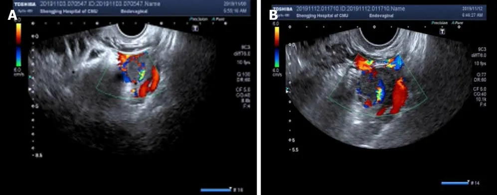
Figure 1 Ultrasonographic image of the lesion. The uterine cavity and cervical canal were empty. Ultrasonography revealed that the mass was heterogeneous with a mixture of cystic and solid echogenicity. A: The left adnexa mass on the day of hospital admission; B: The left adnexa mass six days after admission.
History of past illness
The patient had a history of three pregnancies. One was an ectopic pregnancy, which resulted in a laparoscopically resected right fallopian tube in 2016; the last pregnancy was aborted. Her menstrual history was unremarkable with her last menstrual period occurring 53 d before this hospital admission.
Physical examination
There were no abnormalities noted on physical examination.
Laboratory examinations
Her serum β-hCG level had increased to 35377 mIU/mL and 46553 mIU/mL on the second and sixth day after admission, respectively. However, all other blood test results were normal.
Imaging examinations
After 6 d, she was re-examined by transvaginal ultrasonography and computed tomography (CT). These images revealed that the mixed echogenic mass in the left adnexa area had increased to 2.5 cm × 2.1 cm (Figure 1B). CT examination also revealed a 2.9 cm × 3.1 cm mixed echogenic mass in front of the right lumbar muscle.Surgical consultation was not sought at that time. An ectopic pregnancy was suspected based on these findings and an emergency laparoscopy was performed under general anesthesia, along with a hysteroscopy adhesion separation and endometrial biopsy.The laparoscopy revealed a cystic projection measuring 2.5 cm × 2 cm located on the left ovary. No abnormalities were observed in the left fallopian tube and right ovary,and the right fallopian tube was absent. The uterus was slightly larger and smoother than average. A left ovary mass was excluded but no pregnancy tissues were visible to the naked eye. Hysteroscopy revealed an adhesion measuring 4 cm located on the anterior and posterior walls of the uterine cavity and the appearance of decidual changes in the uterine cavity. The adhesions and a small amount of intrauterine tissue were sent for pathological analysis and were reported as decidua. During the operation, the ultrasound director requested abdominal ultrasound of the pelvic cavity. However, no pelvic mass was found. Laparoscopy also did not reveal a mass in the pelvic or abdominal cavity. The patient’s serum β-hCG level was 44795 mIU/mL during the operation and persisted at a high-level following surgery, with changes in serum β-hCG before surgery and 5 d after surgery (Figure 2A).
Histopathological examination identified the uterine contents as decidual tissue,and the left ovarian surface mass was luteal tissue. CT examination demonstrated small nodules in the right upper lobe subpleural tip one day after surgery. Lung metastasis was detected on CT and enhanced pelvic magnetic resonance imaging(MRI) demonstrated a cystic solid mass in front of the right lumbar muscle approximately 2.5 cm × 2.1 cm in size. The parenchymal signal of the lesion was slightly mixed, showing a long T2 signal, an equal and slightly longer T1 signal. On enhancing the scan edge, the boundary between the lumbar muscles was not clear.There was no abnormal strengthening of the myometrium (Figure 3). Whole abdominal CT demonstrated a cystic solid mass approximately 2.9 cm × 3.1 cm in size,which was in the right lumbar muscle with an unclear border and thick wall. The wall of the enhanced scan was significantly strengthened.
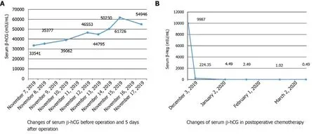
Figure 2 Patient serum β-human chorionic gonadotropin levels. A: Increasing levels of serum β-human chorionic gonadotropin (β-hCG) levels before surgery; B: β-hCG dropped to normal levels after cycle 2 of chemotherapy. β-hCG: β-human chorionic gonadotropin.
FINAL DIAGNOSIS
Based on these findings, the patient was diagnosed with choriocarcinoma (IV:9).
TREATMENT
The patient underwent six cycles of cisplatin, dactinomycin, and etoposide therapy.
OUTCOME AND FOLLOW-UP
Complete disappearance of the metastatic lesion in the lung was confirmed by CT after four cycles of chemotherapy; the lumbar muscle mass disappeared after five cycles.The patient’s serum β-hCG level was normal after the second cycle of chemotherapy(Figure 2B).
DISCUSSION
Gestational trophoblastic neoplasia is a group of malignant tumors related to pregnancy. Histologically derived from placental trophoblasts, the group is divided into either an invasive mole, choriocarcinoma, placental site trophoblastic tumor, or epithelioid trophoblastic tumor. Choriocarcinoma is a common gestational trophoblastic tumor with significant ethnic and regional differences[10]. In China, the incidence of choriocarcinoma is 1/2882 pregnancies, which makes this a high incidence area[11]. Due to the highly vascular nature of choriocarcinoma,hematogeneous metastasis can occur early and affect various organs and tissues throughout the body. Many patients only show symptoms of metastatic lesions. More than 60% of patients have pulmonary metastases at diagnosis[12]. It is the only gynecologic tumor recognized by the International Federation of Obstetrics and Gynecology and the International Gynecological Oncology Association that can be clinically diagnosed without histopathological evidence[12].

Figure 3 Magnetic resonance imaging of the lesion. Low signal intensity on T2-weighted image (T2WI) (A) and high signal intensity on T1-weighted image(T1WI) (B); C: T2WI, coronal plane; D: T1WI, coronal plane. Orange arrows indicate the lesion.
In this case report, we noted that the serum β-hCG levels in our patient continued to increase and her lower back pain symptoms persisted and gradually worsened. A color Doppler ultrasound was performed three times after admission, which showed that the left attachment area mass gradually increased. Concurrently, her β-hCG levels continued to increase which led to the diagnosis of ectopic pregnancy. CT showed a cystic solid mixed mass in the anterior psoas muscle before operation, although we were not concerned with this finding. During the operation, no villi were found in her pelvic or abdominal cavity. After careful examination of the lumbar muscles, a mass of approximately 3 cm × 2 cm was identified, although no treatment was performed considering the dense adhesion to blood vessels. The patient’s β-hCG level did not decrease during laparoscopy and hysteroscopy, but continued to increase on the first postoperative day. A lung CT examination revealed metastatic lung lesions and a full abdominal CT and pelvic MRI examination revealed a lumbar muscle mass associated with choriocarcinoma. The patient underwent six cycles of chemotherapy. After the fifth cycle of treatment, the patient’s symptoms were relieved and her β-hCG level returned to normal. Surprisingly, the lumbar muscle mass also disappeared. In this case, we did not have pathological evidence to confirm the diagnosis of choriocarcinoma. However, pathological evidence of choriocarcinoma is not a necessary condition for a final diagnosis[13]. The patient’s β-hCG levels and clinical symptoms were more important in this regard. To our knowledge, this is the first report of choriocarcinoma metastasizing to the lumbar muscle.
Choriocarcinoma misdiagnosed as an ectopic pregnancy is common. The main reason for this is because lung enhanced CT examinations are important for diagnosing the presence or absence of lung metastases. Misdiagnoses that delay the detection of metastasis is due to negative chest radiographs without lung CT at the first admission. It has been reported that if only chest radiography is performed,approximately 40% of patients with lung metastases may be missed[14]. Therefore, a craniocerebral and full abdominal MRI should be performed to understand metastatic lesions in cases such as the one presented here. This will potentially improve the diagnosis and cure rate.
When the clinical, pathological, and auxiliary imaging examinations do not match,we must carefully identify the cause, reflect on whether the diagnosis is correct, and closely follow up the patient. Choriocarcinoma is relatively uncommon in the clinic.The characteristics of choriocarcinoma are similar to those of an ectopic pregnancy,which easily leads to misdiagnosis. Therefore, in future clinical diagnoses, medical staff should understand the subtle differences between ectopic pregnancy and choriocarcinoma. Considerations should include the possibility of choriocarcinoma in order to reduce misdiagnosis. Although choriocarcinoma has a highly malignant biological behavior, it responds well to chemotherapy[15]. Chemotherapy should be administered immediately after choriocarcinoma diagnosis, which is crucial for a good prognosis. One study reported that chemotherapy could successfully treat choriocarcinoma, even in advanced stages. In the current case report, the pituitary metastasis of choriocarcinoma disappeared following several chemotherapy cycles.Notably, the lumbar muscle metastasis disappeared after conventional chemotherapy treatment.
CONCLUSION
In summary, lumbar metastasis from choriocarcinoma is extremely rare. Appropriate chemotherapy can successfully treat these metastasized tumors. In addition, the final diagnosis was not only based on imaging results, but also based on laboratory examinations and clinical symptoms.
杂志排行

World Journal of Clinical Cases的其它文章
- Effective administration of cranial drilling therapy in the treatment of fourth degree temporal, facial and upper limb burns at high altitude:A case report
- Rare imaging findings of hypersensitivity pneumonitis: A case report
- Successful management of a tooth with endodontic-periodontal lesion: A case report
- Primary chondrosarcoma of the liver: A case report
- Monocular posterior scleritis presenting as acute conjunctivitis: A case report
- Chest, pericardium, abdomen, and thigh penetrating injury by a steel rebar: A case report