Primary breast cancer patient with poliomyelitis: A case report
2020-04-07XingMiaoWangYiZiCongGuangDongQiaoSongZhangLiJuanWang
Xing-Miao Wang, Yi-Zi Cong, Guang-Dong Qiao, Song Zhang, Li-Juan Wang
Xing-Miao Wang, Yi-Zi Cong, Guang-Dong Qiao, Song Zhang, Department of Breast Surgery, The Affiliated Yantai Yuhuangding Hospital of Qingdao University, Yantai 264400, Shandong Province, China
Li-Juan Wang, Department of Radiology, The Affiliated Yantai Yuhuangding Hospital of Qingdao University, Yantai 264400, Shandong Province, China
Abstract BACKGROUND Poliomyelitis is an acute infection caused by an enterovirus, which primarily infects the human gastrointestinal tract. In general, patients with polio have no association with the occurrence of cancer. The present case study presents a rare case of poliomyelitis combined with primary breast cancer.CASE SUMMARY A 61-year-old woman who was diagnosed with poliomyelitis at 5 years old and confirmed invasive breast cancer by core needle biopsy (CNB) after hospitalization. The patient received a modified radical mastectomy and four cycles of chemotherapy with the TC (docetaxel and cyclophosphamide) regimen.The patient was also prescribed endocrine therapy without radiotherapy after chemotherapy. The patient had no evidence of lymphedema in the right upper extremities and no evidence of either regression or distant metastasis at the 1-year follow-up.CONCLUSION The pectoral muscles of patients with polio are easily damaged in traumatic procedures, such as CNB, local anesthesia for tumor excision, and general anesthesia for surgery. A CNB, modified radical mastectomy, and four cycles of TC chemotherapy were successfully completed for the present case and the adverse reactions were found to be tolerable. This case may indicate the relationship between breast cancer and polio, and the examination and treatment methods used could be used as a guide for similar cases in the future.
Key Words: Breast cancer; Diagnostic imaging; Follow-up; Poliomyelitis; Surgery; Case report
INTRODUCTION
Poliomyelitis is an acute infectious disease caused by a neurotropic poliovirus. It primarily invades the motor nerve cells of the central nervous system, most commonly the motor neurons in the anterior horn of the spinal cord[1]. The majority of the patients affected are 1-6 years old. The main symptoms of poliomyelitis are fever, general malaise, myalgia, and varying degrees of flaccid paralysis with irregular distribution.The clinical manifestations of poliomyelitis vary, but include mild non-specific lesions,aseptic meningitis (non-paralytic poliomyelitis), and flaccid weakness of various muscle groups (paralytic poliomyelitis). In patients with poliomyelitis, due to the damage of the motor neurons in the anterior horn of the spinal cord, the muscles associated with the neurons lose their control over nerve regulation and atrophy occurs. Concurrently, atrophy also occurs in the subcutaneous fat, tendons, and bone,making the whole limb thinner. Nevertheless, China has made significant achievements in eradicating polio following the generation of the oral live attenuated polio vaccine[2]. In general, patients with polio are not associated with the occurrence of cancers and poliomyelitis-associated malignancies are rarely reported. Breast cancer is the most common type of cancer for women worldwide[3]. However, the rate of early detection of breast cancer is increasing due to the widespread use of breast magnetic resonance imaging (MRI)[4]. The present paper reports a cases of primary breast cancer in a patient with previous poliomyelitis.
CASE PRESENTATION
Chief complaints
A post-menopausal woman, aged 61 years, visited The Affiliated Yantai Yuhuangding Hospital of Qingdao University after finding a painless lump in the right breast 1 wk previously.
History of present illness
The patient felt a painless lump in the right breast while taking a bath accidentally.
History of past illness
The patient suffered from a poliovirus infection over 50 years ago, which was diagnosed as poliomyelitis. She gave birth at the age of 25 but did not breastfeed, and she experienced irregular menstrual cycles and had no family history of cancer. The patient is right-handed and can write and walk around without crutches or a wheelchair.
Physical examination
She was diagnosed with invasive breast cancer by core needle biopsy (CNB) after hospitalization. A physical examination of the patient revealed a 1.0 cm × 1.0 cm, 1 cm from the nipple, non-tender mass in the upper outer quadrant of the right breast. No mass was palpable in the left breast and no tumescent lymph node was palpable in the bilateral axilla and supraclavicular fossae. A general physical examination revealed that her vision was clear, and both eyeballs moved freely without diplopia or nystagmus in any direction. Her left limb muscle strength, muscular tension, and tendon reflex were normal, whilst the right limb muscle strength was reduced, and she experienced muscle hypotonia and tendon areflexia. Her abdomen was slightly protruded and the size of her liver and spleen was normal, while both lower limbs were not swollen.
Laboratory examinations
Laboratory examinations were normal.
Imaging examinations
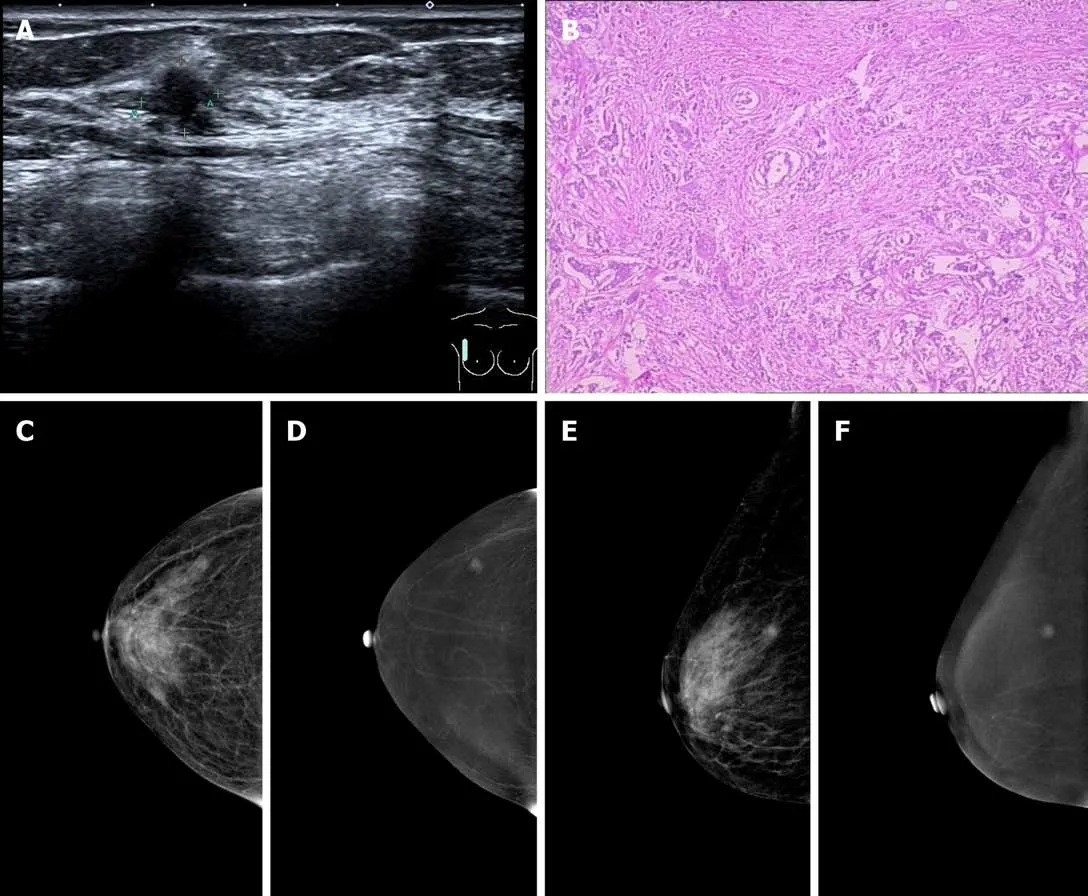
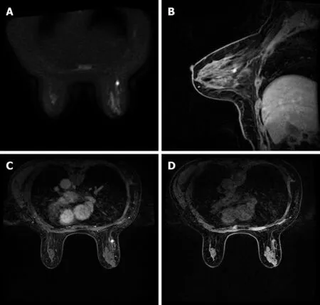
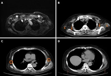
Breast ultrasound revealed a hypoechoic nodule approximately 0.8 cm × 0.8 cm in size in the upper outer quadrant of the right breast, with an irregular shape, obscure boundary, heterogenous internal echo, and punctate blood flow signals, which was classified as breast imaging reporting and data system (BI-RADS) 4b (Figure 1).However, no tumescent lymph node was detected in the right axillary or supraclavicular region. Preoperative contrast enhanced spectral mammography identified an irregular and hard-density mass of 1.0 cm × 1.0 cm in size in the upper outer quadrant of the right breast, which had an obscure boundary with no abnormal calcification, and was significantly enhanced and classified as BI-RADS 4b (Figure 1).A breast MRI scan revealed an ill-defined mass with uniform equal signal intensity.Axial diffusion weighted imaging (b = 800) identified the mass with high signal intensity. Axial and sagittal post-contrast fat-saturated T1-weighted MRI showed a 0.8× 0.9 cm enhancing irregular mass at the 9-10 o'clock position, approximately 6 cm from the nipple (Figure 2). Finally, a chest computed tomography scan was used to identify the atrophy occurring in the right pectoral muscles (pectoralis major,pectoralis minor, subclavius, and serratus anterior), teres major, subscapularis muscle,and latissimus dorsi muscle (Figure 3).
MULTIDISCIPLINARY EXPERT CONSULTATION
Multidisciplinary team (MDT) recommended that she need adjuvant chemotherapy combined with endocrine therapy after surgery but have no need for radiotherapy.
FINAL DIAGNOSIS
The final diagnosis was confirmed to be primary breast cancer combined with poliomyelitis.
TREATMENT
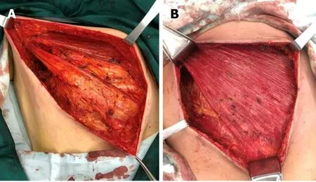
As the patient had no desire to conserve the breast tissue, a mastectomy and sentinel lymph node biopsy for the right breast were performed under general anesthesia. An axillary lymph node dissection was performed because the sentinel lymph node biopsy was also positive. The pectoralis major, pectoralis minor, serratus anterior, and external intercostal muscles were discovered to be atrophying to varying degrees during the operation (Figure 4). Due to the pectoralis major muscle of the patient being atrophied, with no clear boundary being observed between the fat and fascia in the posterior space of the breast, there was a higher probability of injury occurring during the operation compared with normal patients (Figure 4).
The postoperative pathology revealed a grade II right breast invasive ductal carcinoma, in which the size of the lump was 0.9 cm × 0.9 cm. Immunohistochemical staining indicated that the carcinoma was positive for estrogen receptor (90% strongly positive) and progesterone receptor (10% weakly positive) and negative for human epidermal growth factor receptor 2. In addition, positive Ki-67 staining was observed in 40% of the tumor cells. No cancer cells were observed in the nipple and deep resection plane, and axillary lymph node dissection revealed cancer metastasis in only one sentinel lymph node. The patient was ultimately diagnosed with breast cancer in the right breast of luminal B subtype, pT1N1M0, stage IIa. A TC (75 mg/m2i.v.docetaxel, and 600 mg/m2i.v.cyclophosphamide on day 1) regimen was administered every 3 wk for 4 cycles following the operation, and she received the formal advice from the MDT that there was no need for radiotherapy following surgery. The patient was prescribed endocrine therapy consisting of 2.5 mg letrozole and 600 mg vitamin D3 once a day for 1 year (The endocrine therapy was planned for 5 years).

Figure 1 Breast imaging. A: Ultrasound examination revealed an ill-defined hypoechoic mass in the right breast with an irregular shape; B: Microscopic examination revealed invasive ductal carcinoma cells invading the parenchyma in the right breast; C and E: On contrast enhanced spectral mammography (CESM)imaging, the low energy images showed the mass on the upper outer quadrant of the right breast with small spicules at the periphery; D and F: On CESM imaging,the subtraction images showed the significant enhancement of the lesions.
OUTCOME AND FOLLOW-UP
The patient exhibited no evidence of lymphedema in the right upper extremities and no signs of recurrence or distant metastasis at the 1-year follow-up. However, the functional abilities of the right upper extremities of the patient were significantly affected following the operation due to post-polio syndrome.
DISCUSSION
Polio (short for poliomyelitis) is an infectious disease caused by the poliovirus.Children under the age of five are the most infected age group compared with any other age group. According to the World Health Organization, 1/200 of people with polio experience permanent paralysis. However, as a result of the global polio eradication initiative of 1988, numerous countries and regions have now been certified polio-free. The polio vaccine was developed in 1953 and made available in 1957, which caused the cases of polio to significantly drop worldwide[5].

Figure 2 Brest imaging. A: Axial diffusion weighted imaging (b = 800) showed the mass with high signal intensity; B and C: Sagittal and axial post-contrast fatsaturated T1-weighted magnetic resonance imaging identified a 0.8 × 0.9 cm enhancing irregular mass at the 9-10 o'clock position, approximately 6 cm from the nipple; D: Transverse non-enhanced T1 weighted imaging revealed an ill-defined mass with uniform equal signal intensity.
Polio can affect the nervous system and lead to partial or complete paralysis, which is caused by the poliovirus inhabiting the throat and intestines of infected individuals[1]. It can be spread directly by person-to-person contact, by contact with infected nasal or oral mucus or sputum, or through contact with infected feces. The incidence of the disease has been significantly reduced since the development of the polio vaccine. There are three basic types of polio infection: Subclinical infection, nonparalytic infection, and paralytic infection[6]. In total, approximately 95% of infections are classified as subclinical infections, which may be asymptomatic. Clinical poliomyelitis affects the central nervous system (brain and spinal cord), and is divided into non-paralytic and paralytic types, which may occur following the recovery from a subclinical infection. Currently, the aim of poliomyelitis treatment is to control the symptoms while the infection runs its course. The prognosis depends on the subtype of the disease (subclinical, non-paralytic, or paralytic) and the site of infection; for example, if the spinal cord and brain are not involved, > 90% of individuals completely recover[1,7-9].
However, approximately 1% of polio cases can develop into the paralytic type.Paralytic polio leads to the paralysis of the spinal cord (spinal polio), brainstem(bulbar polio), or both (bulbospinal polio). The initial symptoms are similar to those of non-paralytic polio; however, after a week or so, more severe symptoms begin to appear. These symptoms include the loss of reflexes, severe spasms, muscle pain, limb relaxation, temporary or permanent paralysis, and the deformation of the limbs. In addition, polio may recur following recovery, which can even happen 15 to 40 years later. The common symptoms of post-polio syndrome (PPS) include muscle and joint weakness, severe muscle pain, muscle atrophy, dyspnea, and dysphagia[9,10]; it is estimated that 25%-50% of polio survivors will experience PPS. Currently, there is no cure for polio, only treatment options aimed at alleviating the symptoms are available[10]. Heat and physical therapy are used to stimulate the muscles and antispasmodic drugs are provided to relax the muscles. While this can improve mobility, it cannot reverse permanent polio paralysis[7,10,11]. Thus, the most effective way to treat polio is through preventative measures such as vaccinations.
To date, there has been no direct association identified between cancer and poliomyelitis. Previous studies have reported that the incidence rate of tumors of the central nervous system in patients with poliomyelitis may increase, and the incidence of breast cancer and skin cancer was higher in female patients with poliomyelitis compared with the general population[12]. Another previous study suggested that poliovirus infection in colon cells may induce resistance to the development of colon cancer to some extent, which subsequently reduces the mortality of colon cancer[13].However, further investigations are required to determine the relationship between poliomyelitis and cancer.

Figure 3 Chest imaging. A: Axial magnetic resonance imaging; B, C, and D: CT scan of the chest revealed atrophy of the pectoralis major (star), teres major(orange triangle), serratus anterior (orange circle), subscapularis muscle (solid arrow), and latissimus dorsi muscle (hollow arrow).

Figure 4 Breast surgery. A: Pectoralis major, pectoralis minor, and serratus anterior muscles were atrophied, the muscle bundles were thin, and the boundary between the pectoralis major and fat and fascia of the posterior space of breast was ill-defined; B: Normal pectoralis major, pectoralis minor, and serratus anterior.
Although breast cancer is the most common type of malignancy diagnosed in women, to date, no previous studies have reported any cases of breast cancer due to poliomyelitis complications. In addition, there are no reports investigating the chemotherapy risk assessment in patients with polio. The pectoralis major and minor muscles during poliomyelitis usually atrophy to differing degrees, which causes the thinning of the chest. Therefore, traumatic procedures, such as CNB, local anesthesia for tumor excision, and general anesthesia for surgery should be performed with caution to avoid damaging the chest organs. Breast MRI can predict the features of tumor, pectoral muscle and axillary lymph node status, and the risk of recurrence after breast conserving surgery[14,15].
CONCLUSION
In conclusion, the present paper reports a case of breast cancer following poliovirus infection, and to the best of our knowledge, this is the first report demonstrating this occurrence. In addition, we did not find precedent of chemotherapy in almost all instances of reported poliomyelitis. The patient received a chemotherapy regimen of TC (75 mg/m2i.v.docetaxel and 600 mg/m2i.v.cyclophosphamide on 1stday) every 3 wk for 4 cycles following the operation and did not receive radiotherapy following the chemotherapy. The patient was also prescribed endocrine therapy consisting of 2.5 mg letrozole and 600 mg vitamin D3 once a day. After 1 year of follow-up, local recurrence and distant metastasis were not present, and the patient will continue to be monitored in the future.
杂志排行
World Journal of Clinical Cases的其它文章
- Novel triple therapy for hemorrhagic ascites caused by endometriosis: A case report
- Discontinuous polyostotic fibrous dysplasia with multiple systemic disorders and unique genetic mutations: A case report
- Symptomatic and optimal supportive care of critical COVID-19: A case report and literature review
- Multiple ectopic goiter in the retroperitoneum, abdominal wall, liver,and diaphragm: A case report and review of literature
- Acute celiac artery occlusion secondary to blunt trauma: Two case reports
- Uncontrolled central hyperthermia by standard dose of bromocriptine: A case report
