Rare narrow QRS tachycardia with atrioventricular dissociation: A case report
2020-04-07ChaoZhuMingXingChenGuiJianZhou
Chao Zhu, Ming-Xing Chen, Gui-Jian Zhou
Chao Zhu, Ming-Xing Chen, Gui-Jian Zhou, Department of Cardiology, The Affiliated Hospital of Yangzhou University, Yangzhou University, Yangzhou 225009, Jiangsu Province, China
Abstract BACKGROUND Most Mahaim fibers are right free-wall atriofascicular accessory pathways with only antegrade conduction. Concealed Mahaim fiber is not very rare; however,concealed nodoventricular fiber is a very rare kind of retrograde accessory pathway in supraventricular tachycardia with atrioventricular (AV) dissociation.Only a few cases about successful ablation of the nodoventricular accessory pathway have been reported. We describe the case of a 32-year-old woman who underwent an electrophysiology study and radiofrequency (RF) ablation of a rare narrow QRS tachycardia with AV dissociation.CASE SUMMARY A 32-year-old woman with a history of paroxysmal palpitation was admitted to our hospital for RF ablation. Electrocardiography revealed a narrow QRS complex tachycardia with the same morphology in sinus rhythm. Echocardiography showed no structural heart disease. A right-sided concealed AV accessory pathway and a right-sided concealed nodoventricular accessory pathway were involved in the orthodromic atrioventricular reciprocating tachycardia. His bundle-ventricular interval during tachycardia was the same as that in sinus rhythm. The tachycardia could be initiated and entrained by ventricular pacing.Premature right ventricular stimulus introduced during the His-bundle refractory period when tachycardia occurred was able to advance the next atrial potential.The earliest atrial activation was mapped near the proximal slow AV nodal pathway. RF ablation of both accessary pathways was successfully performed under the guidance of a three-dimensional mapping system by recording the earliest retrograde atrial potential, and tachycardia could no longer be induced.CONCLUSION Narrow QRS tachycardia with AV dissociation is inducible by concealed nodoventricular fiber and ablated by recording the earliest retrograde atrial potential.
Key Words: Atrioventricular dissociation; Nodoventricular pathways; Ablation; Narrow QRS wave tachycardia; Mahaim fiber; Case report
INTRODUCTION
Most Mahaim fibers are right free-wall atriofascicular accessory pathways with only antegrade conduction[1]. Recent research revealed that concealed Mahaim fiber is not very rare[1]. However, the atrioventricular (AV) accessary pathway as a kind of atypical Mahaim fiber is very uncommon. AV dissociated tachycardia with a faster ventricular rate can be easily attributed to ventricular tachycardia with the exception of supraventricular tachycardia as atrioventricular node reentrant tachycardia (AVNRT)or nodofascicular and nodoventricular tachycardias[2]. Concealed nodoventricular Mahaim fibers connecting the AV node and proximal ventricle appear to be scarcely observed in clinical practice. Only a few cases about successfully ablating the nodoventricular accessory pathway have been reported[3]. We describe the case of a 32-year-old woman who underwent an electrophysiology (EP) study and radiofrequency(RF) ablation of a rare narrow QRS tachycardia with AV dissociation.
CASE PRESENTATION
Chief complaints
A 32-year-old woman with a history of paroxysmal palpitation was admitted to our hospital for RF ablation.
History of present illness
In July 2018, a 32-year-old woman with a history of paroxysmal palpitation was admitted to our hospital for RF ablation. Electrocardiography (commonly known as ECG) revealed a narrow QRS complex tachycardia with the same morphology in sinus rhythm. Hotter recordings demonstrated 13093 atrial premature beats in 24 h.Echocardiography showed no structural heart disease.
History of past illness
The patient had no previous medical history.
Physical examination
The patient’s temperature was 37.7 °C, heart rate was 75 beats per min, respiratory rate was 16 breaths per min, blood pressure was 128/85 mmHg, and oxygen saturation in room air was 98%. Clinical physical examination revealed no abnormalities.
Laboratory examinations
The results of routine blood tests, routine urine tests, urinary sediment examination,routine fecal tests, occult blood test, blood biochemistry, immune indexes, and infection indexes were in the normal range. Electrocardiogram, chest X-ray, and arterial blood gas were also normal. Preoperative blood urea nitrogen and serum creatinine value were also normal.
Imaging examinations
Imaging examination revealed no abnormalities.
Electrophysiology study
EP study was performed later in our EP lab. After successfully puncturing both femoral veins, standard 4-polar electrode catheters were introduced to the His bundle and right ventricular (RV) apex through 7-French sheaths. A steerable decapolar electrode catheter was placed in the coronary sinus with the proximal electrode at the ostium. A 4-mm tip electrode was introduced into the cardiac chamber to map the earliest atrial activation during tachycardia. The basic heart rhythm was sinus rhythm,with an atrial-His interval of 70 ms and His bundle-ventricular (HV) interval of 50 ms. The HV interval in atrial S1S1 340 ms overdrive pacing was the same as that in sinus rhythm, which indicated no overt pre-excitation.
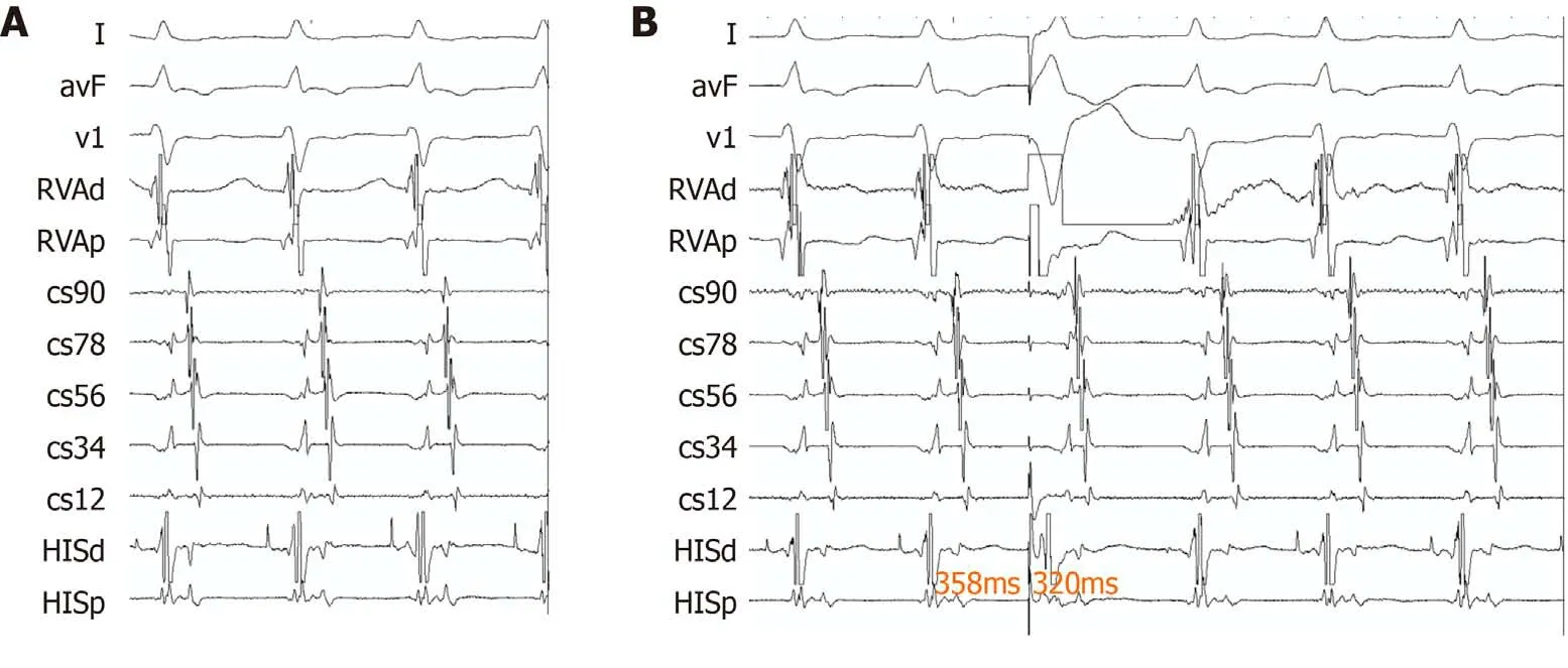
Tachycardia with a cycle length of 360 ms occurred after atrial S1 S2 stimulation,with 1:1 ventriculoarterial conduction (Figure 1A). Premature ventricular beats during a His refractory period advanced the following atrial potential (Figure 1B). The threedimensional mapping system revealed the earliest atrial activation in tachycardia at 7 o’clock of the tricuspid annulus.
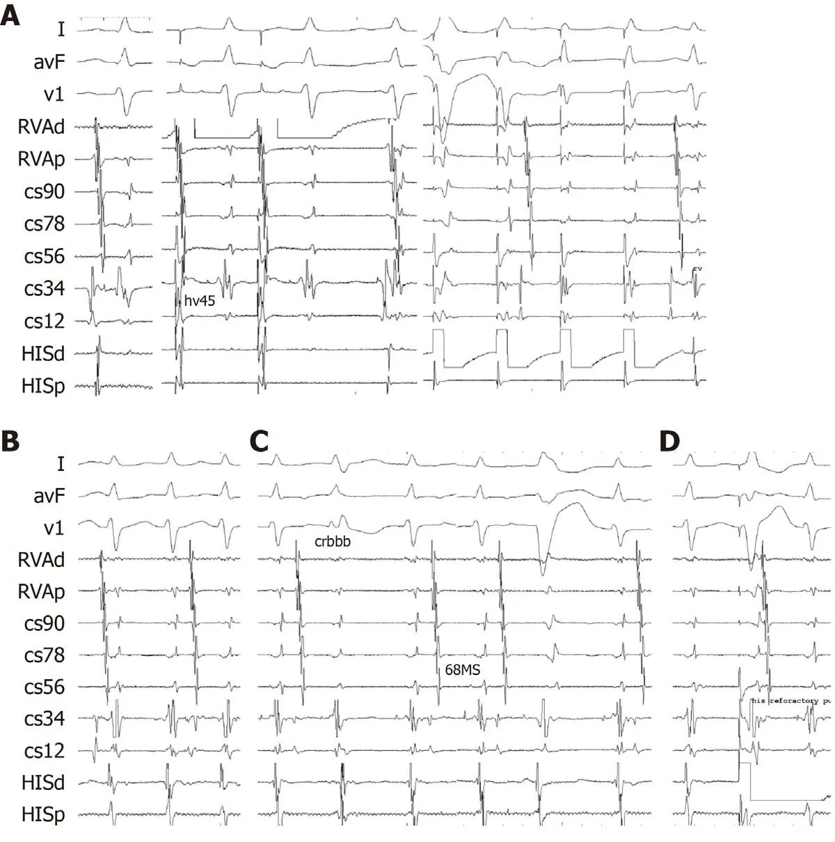
After successfully ablating this AV accessory pathway, the atrial-His interval was 75 ms, and the HV interval was 45 ms. Another narrow QRS complex tachycardia with ventriculoarterial dissociation was induced by both atrial stimulation and ventricular stimulation, which manifested the same surface ECG QRS waves as tachycardia;subsequently, atrial and ventricular stimulation was performed (Figure 2A and B).
The second episode of tachycardia had many electrophysiologic characteristics: (1)The P wave could be positive or negative during tachycardia; (2) This tachycardia could be induced by both atrial and ventricular stimulation; (3) The HV interval during tachycardia was the same as that during sinus rhythm and during the first episode of tachycardia; and (4) This tachycardia could be terminated by an adenosine triphosphate injection.
The second tachycardia had the same HV interval and surface ECG morphology as the first tachycardia. Both right bundle branch block and left bundle branch block could be detected during tachycardia. The HV interval was prolonged for about 20 ms during left bundle branch block; however, the HV interval remained the same during right bundle branch block as that without branch bundle block, which indicated nonventricular tachycardia (Figure 2C). RV stimulus in the His refractory period advanced the next H wave, and it could be recognized as an accessory pathway (Figure 2D).
Another electrophysiologic phenomenon we found was that the ventricularventricular interval shortened from about 10 ms to 30 ms when there was an A wave between the two ventricular waves. Regardless of this phenomenon, stimulus to the H interval with an A wave between them was still 20 ms less than the HH interval in tachycardia with the A wave between them and about 50 ms less than the HH interval in tachycardia without the A wave between them. After the His refractory period stimulation, the next A wave always appeared before the next H wave and V wave when AV dissociation existed. Atrial activation mapping showed earliest atrial activation just in front of the coronary sinus ostium, around the area of the slow pathway of the AV node.
FINAL DIAGNOSIS
The final diagnosis of the presented case was a rare narrow QRS tachycardia with AV dissociation.
TREATMENT
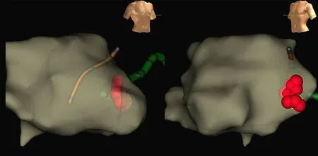
After successful ablation (application, 30 W; temperature, 55 °C; time, 120 s) of the slow pathway of the AV node, tachycardia could no longer be induced (Figure 3). The patient was well tolerated without any complication.

Figure 1 The first tachycardia and premature ventricular beats. A: The first tachycardia, the cycle length was 360 ms, with VA 1: 1 conduction; B:Premature ventricular beats during His refractory period advanced the following atrial potential.

Figure 2 Electrophysiology study. A: In sinus rhythm, AH interval was 75, His bundle-ventricular (HV) interval was 45, tachycardia could be induced both by stimulating atrium and ventricle; B: This tachycardia showed complete atrioventricular dissociation with ventricular rate faster than atrial rate; C: During tachycardia,HV interval remained the same in right bundle branch block pattern as in sinus rhythm, HV interval prolonged about 20 ms in left bundle branch block pattern; D: Intra His refractory period stimulation advanced the next H wave.
OUTCOME AND FOLLOW-UP
At follow-up visit3 mo after the ablation procedure, the patient was asymptomatic.
DISCUSSION

Figure 3 The ablation target was just before the ostium of coronary sinus around the area of slow pathway of the atrial ventricular node.
Nodoventricular fibers are usually recognized as traditional Mahaim fibers connecting the AV node and base of the ventricle and related atriofascicular pathways[4]. Usually nodoventricular fibers originate from a slow pathway region of the AV node itself and then adjacent to the para-Hisian regionviadirect insertion into the ventricular myocardium[5]. Mahaim fibers were thought to be only antegrade in reentrant tachycardia. In clinical observation of true nodoventricular Mahaim fibers, it is rare for them to have only retrograde AV connection. These pathways could be ablated at any region of their length, but it is more common to ablate them at the AV node of their origins, as it is inappropriate and difficult to find the ventricular insertion site of nodoventricular pathways[6]. In our case, it was obvious to distinguish these two kinds of fibers according to complete AV dissociation with a faster ventricular rate. This orthodromic tachycardia utilized the His-Purkinje fiber as its antegrade limb and concealed the nodoventricular accessory pathway as its retrograde limb.
Another interesting finding in our case was that the P wave could be positive or negative during tachycardia, as illustrated during different origination of atrial activation. The negative P wave indicated atrial activation originating from the bottom of the right atrium or AV node area, whereas the positive P wave indicated retrograde atrial activation blocking the AV node and atrial activation originating from the sinus node. Because of the presence of concealed conduction, the cycle length of tachycardia was shortened when there was an atrial wave between two ventricular waves and prolonged when there was no atrial wave.
The differential diagnoses of nodoventricular accessory pathway are as follows: (1)Nodofascicular accessory pathway; (2) Intra-Hisian reentry; (3) AVNRT with retrograde block at the upper common pathway; (4) Ventricular tachycardia; (5)Fasciculoventricular fiber-induced tachycardia; and (6) Junctional tachycardia. The tachycardia could be initiated and entrained by ventricular pacing, revealing a reentrant mechanism, which excluded junctional tachycardia. It could be differentiated from intra-Hisian reentrant tachycardia and fasciculoventricular reentrant tachycardia based on HV interval stability in tachycardia and sinus rhythm[7]. Premature RV stimulus introduced during the His-bundle refractory period when tachycardia occurred was able to advance the next atrial potential, which could rule out AVNRT[8].The existence of different types of branch bundle block alterations in tachycardia with alteration of the HV interval excluded ventricular tachycardia as well.
It was not easy to differentiate the nodofascicular accessory pathway from the nodoventricular accessory pathway. In nodofascicular reentry, the V-H interval in supraventricular tachycardia is less than the V-H interval during RV pacing;however, in nodoventricular reentry, the V-H interval in tachycardia is slightly more than that in RV pacing[6]. Therefore, premature ventricular beats induced in the His refractory period demonstrated concealed nodoventricular pathway if atrial activation was in front of the following His and ventricular activation when AV dissociation was present. In our study, in the His refractory period stimulation, the next A wave always appeared before the next H wave and V wave were observed, which indicated that this tachycardia was nodoventricular reentry.
There were also some limitations in our study. It might have been better to use high density mapping to manifest the detail of the reentrant circle.
CONCLUSION
A narrow QRS tachycardia with AV dissociation can be induced by concealed nodoventricular fiber, and it can be ablated by recording earliest retrograde atrial potential.
杂志排行
World Journal of Clinical Cases的其它文章
- Strategies and challenges in the treatment of chronic venous leg ulcers
- Peripheral nerve tumors of the hand: Clinical features, diagnosis,and treatment
- Treatment strategies for gastric cancer during the COVID-19 pandemic
- Oncological impact of different distal ureter managements during radical nephroureterectomy for primary upper urinary tract urothelial carcinoma
- Clinical characteristics and survival of patients with normal-sized ovarian carcinoma syndrome: Retrospective analysis of a single institution 10-year experiment
- Assessment of load-sharing thoracolumbar injury: A modified scoring system
