Candidal periprosthetic joint infection after primary total knee arthroplasty combined with ipsilateral intertrochanteric fracture: A case report
2020-04-07JunXinQingShanGuoHuaYuZhangZhiYangZhangTomerTalmyYuZhuoHanYuXieQiuZhongSiRuZhouYangLi
Jun Xin, Qing-Shan Guo, Hua-Yu Zhang, Zhi-Yang Zhang, Tomer Talmy, Yu-Zhuo Han, Yu Xie, Qiu Zhong,Si-Ru Zhou, Yang Li
Jun Xin, Qing-Shan Guo, Hua-Yu Zhang, Zhi-Yang Zhang, Yu-Zhuo Han, Yu Xie, Si-Ru Zhou, Yang Li, Department of Trauma Surgery, State Key Laboratory of Trauma, Burns and Combined Injuries, Medical Center of Trauma and War Injury, Daping Hospital, Army Medical University of PLA, Chongqing 400042, China
Tomer Talmy, The Institute of Research in Military Medicine, The Hebrew University of Jerusalem, Hadassah Medical Center, Jerusalem 91120, Israel
Qiu Zhong, Department of Clinical Laboratory, Daping Hospital, Army Medical University of PLA, Chongqing 400042, China
Abstract BACKGROUND Candidal periprosthetic joint infection is a rare and difficult to diagnose complication of total knee arthroplasty. The treatment of such complications is inconclusive and may include prosthesis removal, debridement, arthrodesis, and extensive antifungal therapy to control the infection.CASE SUMMARY A 62-year-old male with a history of total knee arthroplasty (TKA) in his left knee presented with ipsilateral knee pain and a sinus discharge 20 mo after TKA. The patient was previously evaluated for left knee pain, swelling, and a transient fever one month postoperatively. Prothesis removal and insertion of a cement spacer were performed in a local hospital six months prior to the current presentation.Medical therapy included rifampicin and amphotericin which were administered for 4 wk following prosthesis removal. A second debridement was performed in our hospital and Candida parapsilosis was detected in the knee joint. Fourteen weeks following the latter debridement, the patient suffered a left intertrochanteric fracture and received closed reduction and internal fixation with proximal femoral nail anterotation. Two weeks after fracture surgery, a knee arthrodesis with autograft was performed using a double-plate fixation. The patient recovered adequately and was subsequently discharged. At the two-year follow-up, the patient has a stable gait with a pain-free, fused knee.CONCLUSION Fungal periprosthetic joint infection following TKA may be successfully and safely treated with prosthesis removal, exhaustive debridement, and arthrodesis after effective antifungal therapy. Ipsilateral intertrochanteric fractures of the affected knee can be safely fixated with internal fixation if the existing infection is clinically excluded and aided by the investigation of serum inflammatory markers.
Key Words: Infection; Periprosthetic joint infection; Intertrochanteric fracture; Fungal;Arthrodesis; Case report
INTRODUCTION
Periprosthetic joint infection (PJI) is one of the most serious complications of total knee arthroplasty (TKA)[1]. With the improvement of surgical techniques and conditions, the incidence of periprosthetic infection is decreasing. However, with the rapid increase in the total number of arthroplasties, the absolute number of PJI has also increased.Fungal PJIs are rarely reported, accounting for less than 1% of all PJIs[2]. However,fungal infections may result in disastrous consequences for the patient. Many factors may increase the risk for fungal PJIs following arthroplasties, including impaired host immunity, malignant tumors, excessive or inappropriate use of antibiotics, and catheter retention (urinary or extra-intestinal hypertrophic tubes). Due to atypical early clinical signs and lack of specificity in laboratory examinations, the early diagnosis of fungal PJIs is difficult. In addition, due to the lack of high-level evidence,no standard management has been determined. Although a two-stage exchange arthroplasty is preferred by most surgeons[3], controversies still exist with regard to the ideal interval between implant removal and reimplantation, the usefulness of antifungal-loaded cement spacers and the duration of systemic antifungal treatment. If other fractures occur in the interval between implant removal and reimplantation, the case would be more complicated.
Here, we report a case of candidal PJI following TKA combined with ipsilateral intertrochanteric fracture after the first stage of implant removal and cement spacer insertion.
CASE PRESENTATION
Chief complaints
A 62-year-old male presented with pain and a sinus effusion in the left replaced knee for 3 mo.
History of present illness
A TKA of the left knee was performed 20 mo prior to the current admission. One month postoperatively,the patient experienced pain and swelling of the left knee with transient fever. The patient was administered amoxicillin for one week by a local community clinic without abatement of the symptoms. Three months after the TKA,he was referred to the operating hospital where a knee aspiration was performed with no indicative findings. Radiography of the left replaced knee did not reveal any notable abnormalities (Figure 1). Gentamicin and vancomycin were given empirically without a clinically significant response. The knee was re-aspirated without any indicative findings. The patient was then discharged without a definitive diagnosis.Fourteen months after TKA, the patient experienced left knee pain with further exacerbation of joint swelling and an inability to walk. Thus, the hospital chose to remove the implant and insert an antibiotic-impregnated (vancomycin) acrylic bone cement spacer (Figure 2). Bacterial cultures were performed on the intraoperative excised tissue and the result remained negative. Rifampicin was administered for another four weeks post-operatively. Three months later, a draining sinus with effusion appeared on the left knee and the patient was referred to our hospital.
History of past illness
The patient had no history of cardiovascular disease, diabetes, immune deficiency, or long-term steroid use.
Physical examination
Upon examination, a drainage sinus tract with effusion was observed superomedial to the patellar tendon. The local temperature was slightly elevated, with a terminally restricted range of motion upon flexion and pain associated with both passive and active motion of the knee. The patient was neurologically intact and distal pulsations were well felt.
Laboratory examinations
Laboratory evaluation including complete blood count, procalcitonin, interleukin-6,erythrocyte sedimentation rate, and urine microscopy revealed no significant abnormalities besides an elevated C-reactive protein (31.1 mg/L). Knee joint aspiration was sent for analysis. Gram staining of the bacterial culture was negative, but fungal culture was positive forCandida parapsilosis (C. parapsilosis).
Imaging examinations
No abnormalities were found in the anteroposterior lateral radiographs of the knee after primary TKA.
FINAL DIAGNOSIS
Candidal periprosthetic joint infection after primary total knee arthroplasty.
TREATMENT
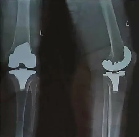
Figure 1 Anteroposterior and lateral radiographs after primary total knee arthroplasty. The surgery was performed 20 mo prior to current presentation.
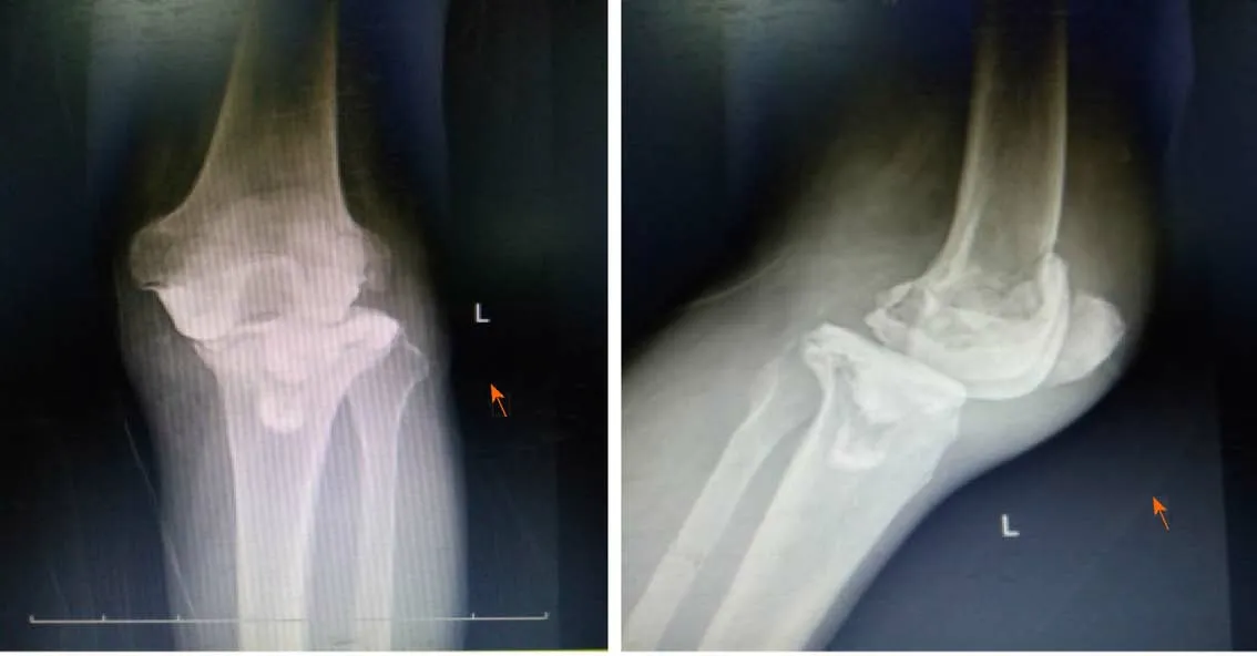
Figure 2 Radiograph of the patient’s left knee after prosthesis removal and insertion of an antibiotic (vancomycin) impregnated cement spacer.
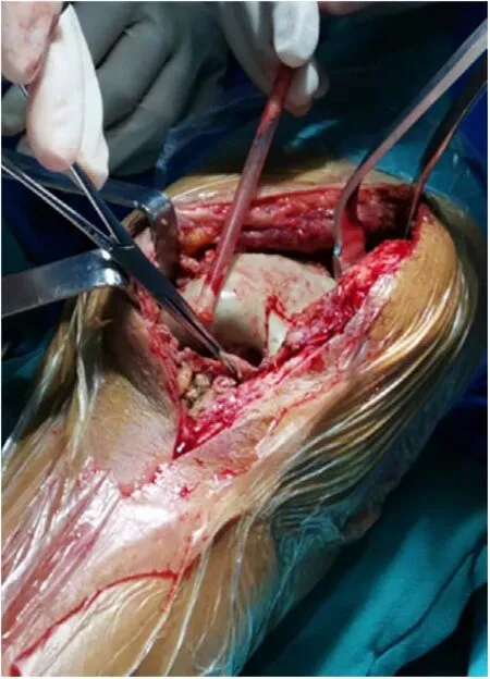
Figure 3 Cheesy like necrotic tissue was observed on the surface of the spacer which had an unpleasant odor during the second debridement procedure.
Considering the recurrent failure in obtaining a definitive diagnosis or clinically significant findings from cultured samples, we decided to perform an additional operation to replace the cement spacer. During the operation, necrotic tissue with a white cheesy appearance was observed on the surface of the spacer which had an unpleasant odor (Figure 3). The intraoperative specimen was sent for aerobic,anaerobic,acid-fast bacilli, andfungalcultures. After removal of the spacer, exhaustive debridement was performed. After the debridement, examination revealed mediolateral knee instability. Therefore, a cross-knee external fixator was applied to optimize stabilization, followed by the insertion of a new cement spacer (Figure 4).Fungalculture was positive forC. parapsilosissensitive to voriconazole and itraconazole with bacterial cultures remaining negative. Post-operative medical therapy included intravenous voriconazole for 4 wk followed by oral itraconazole for 12 wk. Unfortunately, 14 wk postoperatively (2 wk before completion of the antifungal treatment course), the patient suffered a fall resulting in a left intertrochanteric fracture (AO classification: 31A2.2) (Figure 5). Upon examination of the left knee, we found that it was free of any sinuses or signs of surgical wound infection, with an intact range of motion. The patient’s C-reactive protein, procalcitonin, and erythrocyte sedimentation rate were all within the normal reference ranges. The patient underwent a closed reduction and internal fixation with proximal femoral nail anterotation for the fracture and was able to walk on crutches the following day (Figure 6). Two weeks after the latter operation, no signs of infection were observed, and inflammatory markers all declined or were within the normal range. Considering the extensive softtissue defect around the knee joint, a knee arthrodesis with autograft was performed using a double-plate fixation (Figure 7). The patient recovered adequately and was subsequently discharged.
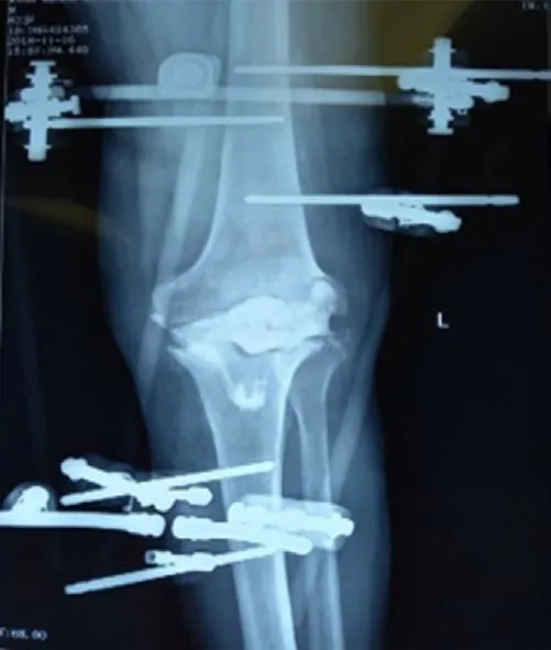
Figure 4 Radiograph of the patient’s left knee after the second debridement.
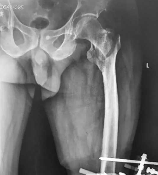
Figure 5 Radiograph of the left intertrochanteric fracture (31A2.2) after the second debridement.
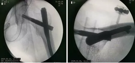
Figure 6 Ipsilateral intertrochanteric fracture fixed by proximal femoral nail anterotation.
OUTCOME AND FOLLOW-UP
At the two-year follow-up, the patient is able to walk without crutches for more than 3 km with a pain-free fused knee. However, radiography shows significantly worsened contralateral knee degeneration (Figure 8).
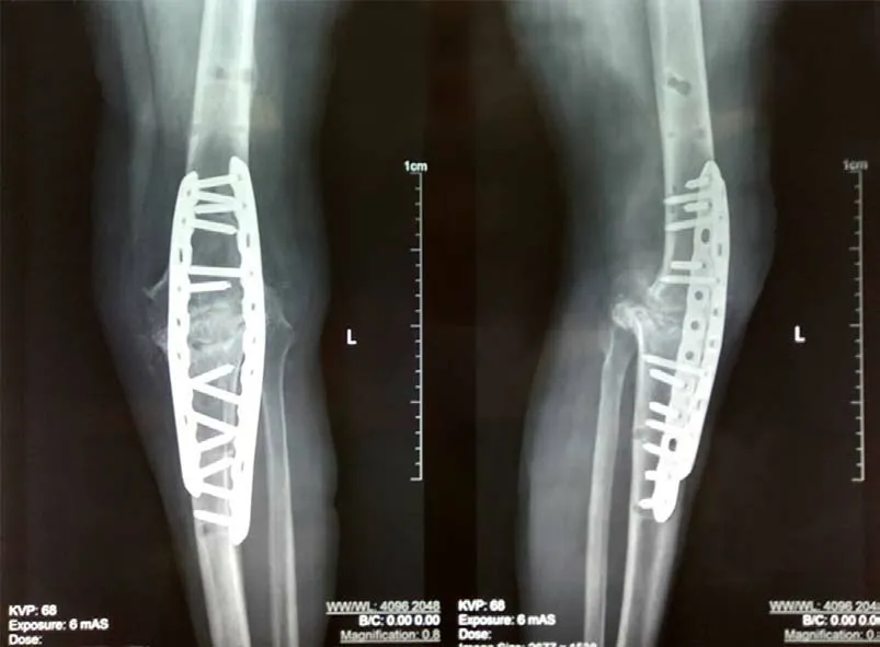
Figure 7 Knee arthrodesis with autograft using a double-plate fixation.
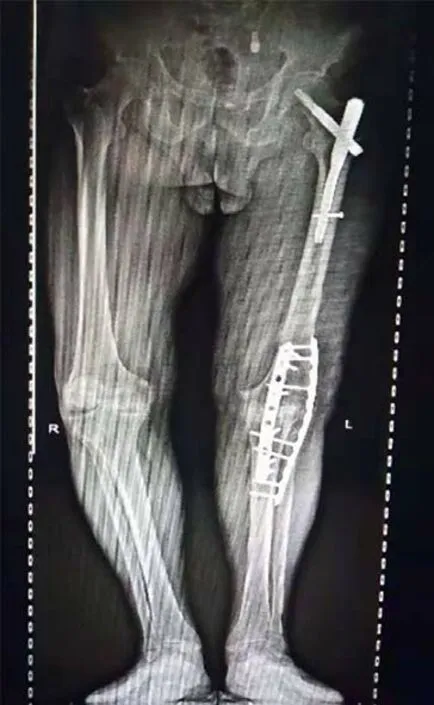
Figure 8 Radiography at the two-year follow-up.
DISCUSSION
Fungal PJIs are rarely reported worldwide. Thus, evidence-based guidelines on the treatment of fungal PJIs have yet to be established[4]. The presence of a persistent fungal infection is thought to be a result of an often delayed diagnosis[5,6]. Several different treatment methods, including antifungal drugs, debridement with retained prosthesis, resection arthroplasty, one-stage or two-stage exchange arthroplasty and arthrodesis have been reported, with variable outcomes[3,5].
Currently, the trigger of fungal infection remains unclear. Patients who are immunocompromised, have an underlying systemic illness or have been subject to prolonged antibiotic regimens may have a higher risk of fungal infection. Prolonged wound drainage has also recently been identified as a potential risk factor[7]. The most likely cause of fungal infection in the reported case is the extensive use of antibiotics before identifying a definitve cause of the prosthetic joint infection.
In previous studies,Candida albicanswas reported to be the most common pathogen in PJIs followed byC. parapsilosis[8]. Candidal PJI was reported in patients with preoperative cutaneous candidiasis[9]. However, No primary source of candidal infection was identified in our case. It should be noted thatC. parapsilosisinfections are characterized by the creation of highly resolute biofilms, making eradication of the mycotic infection a significant obstacle[5,6].
Fungal infections are characterized by a subtle onset of symptoms, and typical local and systemic signs of infection such as swelling, fever, erythema and pain are often absent. The progression of symptoms is often slow, usually manifesting weeks to years after arthroplasty[10]. In this case, the patient’s initial symptoms were not obvious and without systemic symptoms, further obscuring early diagnosis.
Fungal culture is not considered a routine test in many hospitals. Negative bacterial culture results of synovial fluid aspiration cannot exclude infection. For patients with potential risk factors such as long-term use of antibiotics, immunosuppressants,catheterization, and cutaneous fungal infections, one should expand beyond the spectrum of bacterial cultures to consider the possibility of a fungal etiology[11].
In the presented case, the fracture occurred during the late stage of the anti-fungal course after the second debridement. The fracture was managed under the assumption that the fungal infection had resolved. After weighing the risks of the complication of the fracture and infection, we decided to fixate the fracture with minimally invasive surgery to avoid the complication of long-term immobilization.
Due to exhaustive debridement, a considerable amount of affected tissue was resected and mediolateral instability of the knee was present intraoperatively. Thus,reimplantation was unsuitable for this patient. Considering the high non-fusion rate,we used a double-plate fixation arthrodesis with autograft. Follow-up radiography showed successful fusion after one year.
The choice and the duration of antifungal therapy remains unclear. Amphotericin B and fluconazole are the most commonly used antifungal drugs, but their side effects are greater while voriconazole and itraconazole are less toxic[12]. Keuninget al[6]recently reported a case ofC. parapsilosisPJI following revision of TKA, successfully treated with a combination of voriconazole and micafungin. The lack of evidence regarding the choice of antifungals warrants further research in order to optimize treatment protocols for mycotic PJI.
Another controversial issue is the duration of antifungal treatment. A long period of oral antifungal treatment has been recognized as being an essential factor for the success of staged reimplantation after a fungal peri-prosthetic joint infection.However, no evidence was found for the routine use of arthrodesis. Theoretically,arthrodesis is much safer than reimplantation. Therefore, a protocol of intravenous voriconazole for 4 wk followed by oral itraconazole for 12 wk was selected for this case. As of the writing of this report, the patient did not experience recurrence of symptoms.
CONCLUSION
Fungal PJI following TKA can be successfully and safely treated by prosthesis removal, exhaustive debridement, and arthrodesis after effective antifungal therapy.Ipsilateral intertrochanteric fractures of the affected knee can be safely fixated using internal fixation if resolution of the existing fungal infection can be establishedviaclinical and laboratory investigations.
杂志排行
World Journal of Clinical Cases的其它文章
- Strategies and challenges in the treatment of chronic venous leg ulcers
- Peripheral nerve tumors of the hand: Clinical features, diagnosis,and treatment
- Treatment strategies for gastric cancer during the COVID-19 pandemic
- Oncological impact of different distal ureter managements during radical nephroureterectomy for primary upper urinary tract urothelial carcinoma
- Clinical characteristics and survival of patients with normal-sized ovarian carcinoma syndrome: Retrospective analysis of a single institution 10-year experiment
- Assessment of load-sharing thoracolumbar injury: A modified scoring system
