Seven-year follow-up of the nonsurgical expansion of maxillary and mandibular arches in a young adult: A case report
2020-04-07TingTingYuJingLiDaWeiLiu
Ting-Ting Yu, Jing Li, Da-Wei Liu
Ting-Ting Yu, Jing Li, Da-Wei Liu, Department of Orthodontics, Peking University School and Hospital of Stomatology, National Engineering Laboratory for Digital and Material Technology of Stomatology, Beijing Key Laboratory of Digital Stomatology, Center for Craniofacial Stem Cell Research and Regeneration, Peking University School and Hospital of Stomatology,Beijing 100081, China
Abstract BACKGROUND Palatal expansion treatment has been used to expand the constricted maxillary arch and has become a routine procedure in orthodontic practice over the past decades. However, the long-term stability of expansion in the permanent dentition without a surgical approach is uncertain.CASE SUMMARY We present the case of a 15-year-old boy with Class II malocclusion and constricted arches. The patient was treated with rapid palatal expansion (RPE)followed by a fixed orthodontic appliance. A 7-year follow-up evaluation was performed by analyzing cephalometric radiographs, plaster models, and photographs. The patient’s constricted maxillary and mandibular arches were relived after the expansion treatment. A Class I occlusion and normal arch form were established and maintained in the long-term.CONCLUSION RPE treatment is successful in solving constricted dental arch in the permanent dentition without a surgical approach. Permanent retention and even occlusal contact help prevent long-term relapse.
Key Words: Rapid palatal expansion; Transverse maxillary deficiency; Stability;Orthodontics; Case report
INTRODUCTION
The goal of orthodontic treatment is to achieve favorable aesthetics, ideal function, and permanent maintenance. Relapse after orthodontic treatment or age-related remodeling will lead to instability of the dental arch[1,2].
The constricted arch form can cause a range of problems, such as, crowding, jaw discrepancy, or incisor protrusion[3]. Palatal expansion can be used to correct the transverse dimension discrepancy and create space for alignment and front teeth retrusion[4,5]. However, longitudinal studies demonstrated that the arch width reduction continues throughout adult life[6,7]. Therefore, expansion of the dental arch beyond the original dental status will lead to potential high risk relapse posttreatment[8], especially in patients who have past the peak of growth stage[9,10]. Only a few studies have determined the systemic long-term stability in expansion patients[11,12].
Stability is one of the key elements in orthodontic treatment, and the argument of long-term stability for maxillary and mandibular arches expansion has been ongoing for decades. This case report describes the procedure in a 15-year-old boy with Class II malocclusion and constricted maxillary and mandibular arches. The outcome of the expanded arches with rapid palatal expansion (RPE) has been followed for 7 years post-treatment.
CASE PRESENTATION
Chief complaints
A boy aged 15 years and 4 mo was referred for an evaluation of orthodontic treatment at the Orthodontic Department, Peking University School of Stomatology, with the chief complaint of tooth crowding.
History of present illness
His medical history was unremarkable, but his dental care was irregular and he had a bad habit of thumb sucking. The patient had passed his growth peak period, his height was 183 cm and weight was 77 kg at his first visit.
History of past illness
His immediate family members have no similar malocclusions.
Physical examination
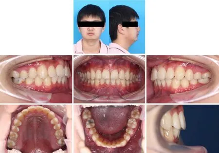
The extra-oral examination (Figure 1, Photographic machine: EOS 60D, Canon, Oita Prefecture, Japan) showed that the patient had a lower anterior facial height and a convex profile with a retruded mandible. His lip looked competent with a slightly acute nasolabial angle and a deep labiomental sulcus. There was no significant facial asymmetry.

Figure 1 Pretreatment photographs.
Intraoral clinical examination (Figure 1) revealed cusp to cusp class II molar relationships on both sides, and a full class II relationship on the right canine as well as cusp to cusp class II relationships on the left canine. His maxillary anterior teeth were in protrusion with an 11-mm overjet and an 80% overbite, which led to a traumatic bite on his left incisors. The upper and lower arch form was both V shaped and symmetric. He had moderate crowding in both arches and a deep curve of Spee in his mandibular arch. He also had cross-bite on his right second molars. His maxillary dental midline was coincident with his facial midline, and the mandibular dental midline deviated 0.5 mm to the left.
Imaging examinations
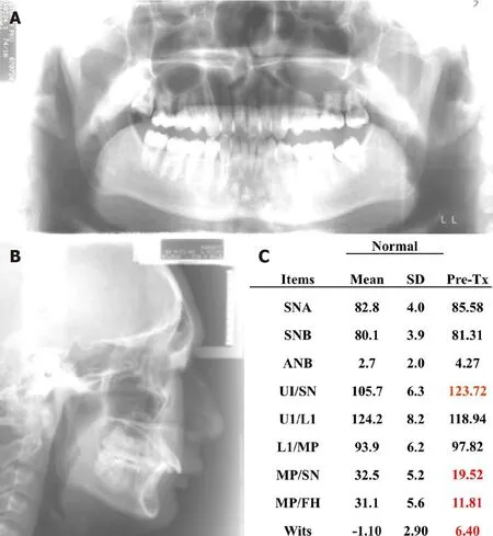
The lateral cephalometric analysis (Figure 2, Software used for cephalometric measurements: Huazheng software, developed by the Craniofacial Growth and Development Center, School of Stomatology, Peking University, and School of Computer Science, Peking University, Beijing, China) indicated a skeletal class I pattern based on the subspinale-nasion-supramental (ANB) angle (4.27°), but skeletal class II pattern according to the Wits value (6.40). It also showed protruding upper incisors [upper first incisor (U1): Sella-nasion (SN) = 123.72], well-positioned lower incisors [U1: Mandibular plane (MP) = 97.82], and a low mandibular plane angle(MP:SN = 19.52).
FINAL DIAGNOSIS
Based on the clinical and imaging outcomes, the patient was diagnosed with Class II malocclusion and constricted maxillary and mandibular arches.

Figure 2 Pretreatment radiographs. A: Panoramic; B: Lateral cephalometric; C: Cephalometric measurements. ANB: Subspinale-nasion-supramental; U1:Upper first incisor; L1: Lower first incisor; MP: Mandibular plane; SN: Sella-nasion; FH: Frankfort horizontal plane; Wits: Witwatersrand.
TREATMENT
The treatment objectives were to:(1)Reduce gingival inflammation;(2)Correct upper and lower arch constriction; (3) Relieve the crowding in both arches; (4) Establish the class I molar and canine relationship. Correct overjet, overbite and dental midline; and(5) Improve the aesthetic harmony of the profile.
Before orthodontic treatment, the patient was referred to a periodontist for scaling and oral hygiene instruction treatment in order to reduce gingival inflammation and establish a healthy oral condition. In the first phase of orthodontic treatment, we performed non-extraction and used a rapid palatal expansion (RPE) device (Leone,Italy) to expand his upper arch to relieve his severe arch width discrepancies. The activation protocol was one-quarter of a turn (0.2 mm) per 12 h, the overall activation period was 20 d followed by a 3-mo retention period[13]. In case of relapse, after the RPE device was removed, Quad Helix (Laboratory of the Department of Orthodontics,Peking University Hospital of Stomatology, Beijing, China) was used to maintain the arch width during the alignment and leveling process. As expansion provided spaces to relieve crowding, we bonded 0.018 × 0.025-inch pre-adjusted brackets (Formula-R,Tomy Inc., Tokyo, Japan) on the mandibular and maxillary arches, and the alignment and leveling phase proceeded with a sequence of 0.012-in, 0.014-in, 0.016-in Nickel-Titanium (NiTi) and 0.016 X 0.022-in NiTi sectional and continuous wires (SMART,Beijing, China) for both arches. The first phase of treatment lasted 20 mo in total.
After the first phase of orthodontic treatment, the cast analysis (Figure 3) showed that the width between the upper first premolar and first molar had increased by 7 mm and 6 mm, respectively, the width between the lower canines was expanded by 5.5 mm along with the upper arch. The severe overjet and Class II malocclusion were corrected after the expansion. Crowding in both arches was relieved, and an acceptable overbite and overjet were also achieved. The first phase of treatment established a Class I molar relationship, and the canine relationship was improved with a canine-protected occlusion. On the mid-term evaluation, the patient still had decreased lower facial height and a convex profile; and low mandibular plane angle.The second phase of orthodontic treatment mainly focused on the control of gingivitis and improvement of the profile esthetic harmony.

Figure 3 Posttreatment photographs.
To improve patient profile esthetic harmony, the convex profile with retruded mandible, could be solved by surgical-orthodontic treatment with the extraction of two lower premolars, and finished with a class III molar relationship. After communicating with the patient and his family regarding the second phase treatment plan, they declined the surgical options, as the patient was satisfied with his current facial profile and his chief complaint was crooked front teeth. Thus, we continued leveling the teeth to relieve the deep overbite using 0.016 X 0.022-in NiTi, and ended with a 0.018 X 0.025-in stainless steel archwire (SMART, Beijing, China). The patient used a vacuum-formed retainer for at least 22 h per day over the first year and only at night during sleep to the present. We also provided the patient with Hawley retainers in order to secure the stability of both arches.
OUTCOME AND FOLLOW-UP
The treatment resulted in significant improvements in dental alignment and occlusal stability. The posttreatment analysis indicated that the treatment objectives were almost achieved. The facial photographs (Figure 3) show the slight improvement in the patient’s profile esthetics, especially his retruded mandible after the non-surgical orthodontic treatment. An ideal occlusion with a Class I molar relationship, normal overbite and overjet were established by the treatment regimen. The patient’s constricted maxillary and mandibular arches were improved after the expansion treatment. The increased angle of L1/MP from 97.82° to 109.26° indicated that the arches expansion effect included protrusion of the lower incisors. Anterior and lateral occlusion were also checked at the end of treatment. Moreover, the 7-year follow-up evaluation showed a stable arch with a Class I occlusion and excellent interdigitation.
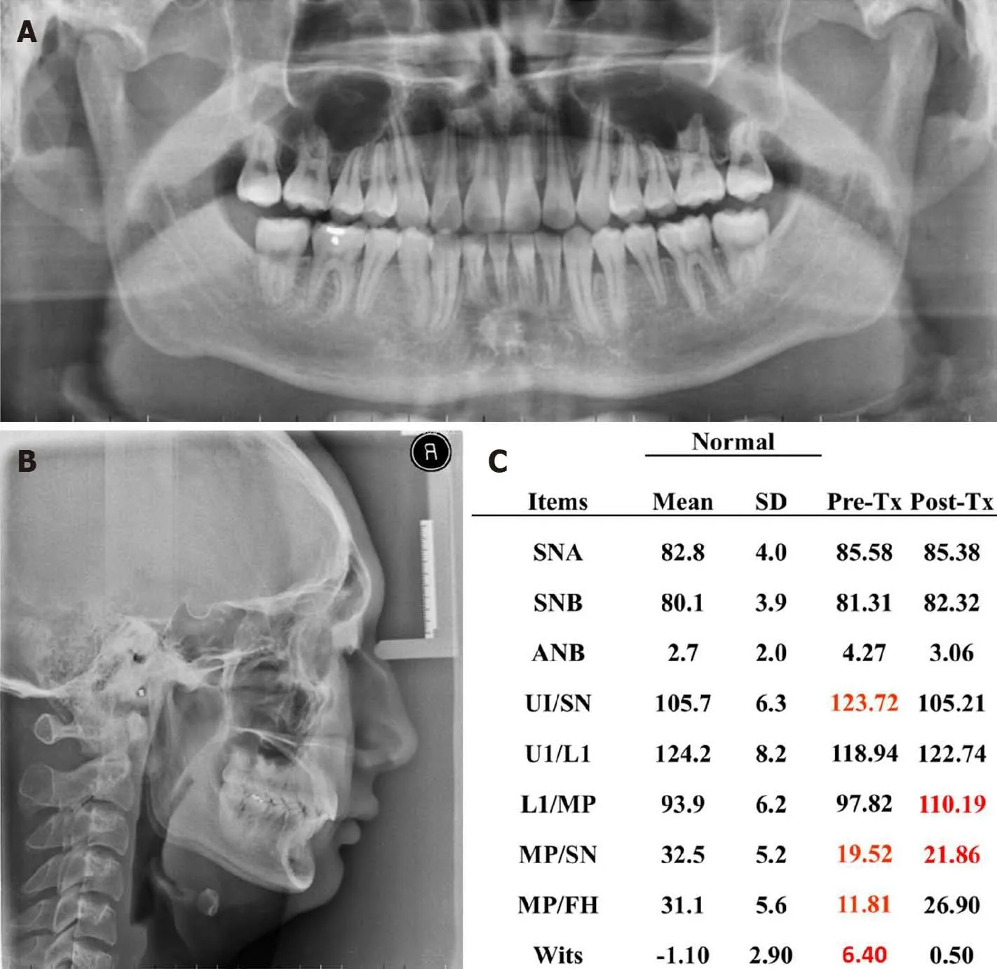
Post-treatment panoramic radiography showed acceptable root parallelism and no significant signs of bone or root resorption (Figure 4A). The post-treatment lateral cephalometric analysis (Figure 4B and C) showed a slight change in skeletal movement, including an increase of the mandibular plane angle [MP/SN, 21.12;MP/Frankfort horizontal plane (FH), 13.42]. The 7-year post-treatment follow-up(Figure 4) showed that there was no significant relapse in the width of both arches,and only a slight movement of lower anterior teeth was found, and the patient had a stable occlusion.

Figure 4 Posttreatment radiographs. A: Panoramic; B: Lateral cephalometric; C: Cephalometric measurements. ANB: Subspinale-nasion-supramental; U1:Upper first incisor; L1: Lower first incisor; MP: Mandibular plane; SN: Sella-nasion; FH: Frankfort horizontal plane; Wits: Witwatersrand.
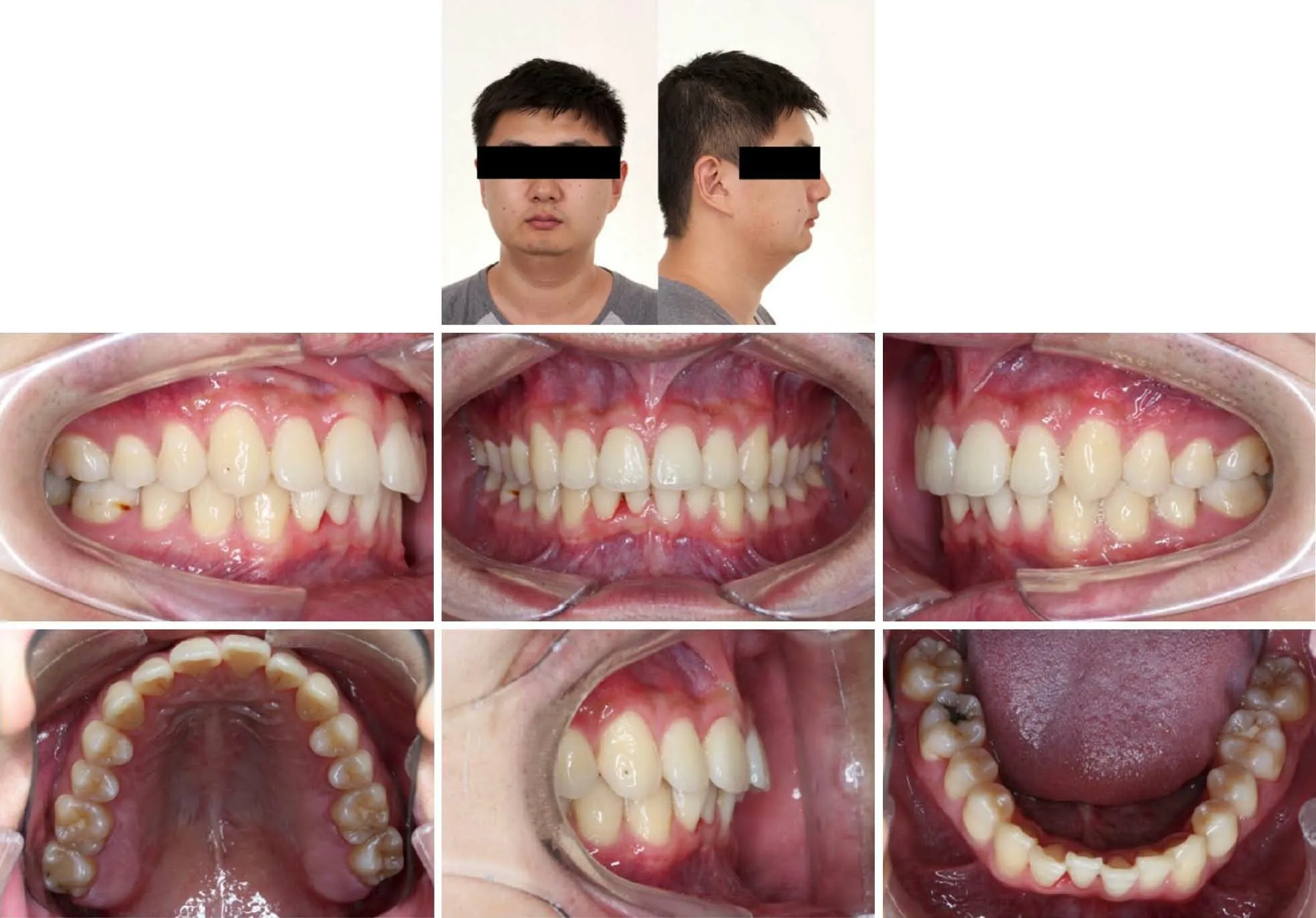
The 7-year posttreatment examination (Figure 5) showed that the width of both arches had no significant relapse, only a slight movement of the upper and lower frontal teeth was found, the patient maintained a stable occlusion.
DISCUSSION
This case report describes the long-term outcome after maxillary and mandibular arches expansion using nonsurgical rapid maxillary expansion (RPE) treatment followed by a fixed appliance on permanent dentition. The 7-year follow-up evaluation of the patient’s occlusion and expansion stability make this issue interesting and valuable.
RPE treatment has been used to expand constricted maxillary arches and has become a routine procedure in orthodontic practice over the decades. However, a relapse tendency has been found in patients who were treated with these appliances for maxillary expansion after long-term evaluation[12]. A number of researchers have investigated the stability of RPE treatment, and factors such as age of the patient[11,14],length of retention period[9], rate of expansion, design of the device[15,16], and biological response of the maxillary suture[17]have been discussed in detail.
Our case report showed that maxillary and mandibular arch expansion followed by a fixed orthodontic appliance led to increases in arch widths of 5 to 7 mm in a young adult patient. Similar RPE treatment outcomes in adult patients were found in previous investigations[11,18], even though not all had used the Hyrax type of expander.Some reports have indicated that the Haas type of palatal expander may result in a better therapeutic outcome due to the acrylic pad supported against the palate[19].However, a more recent report has shown no functional differences between the Haas and Hyrax expansion appliances[20]. A treatment alternative for RPE could be carried out using titanium[21]or stainless steel[22]orthodontic miniscrews. These devices present good clinical reliability[23]and excellent mechanical properties and with small diameters[24]. However, the miniscrew-supported RPE technique is more invasive and less desirable for patients.

Figure 5 Seven-year posttreatment photographs.
Age and maturation are important factors when considering the RPE treatment[25].The rigidity of the craniofacial skeleton increased with maturity, which will significantly affect long-term stability. Many researchers have demonstrated that RPE treatment is more stable in growing individuals than in young adults and adults. In our case report, we challenged the current understanding of RPE treatment in a young adult, and the 7-year posttreatment records showed that the patient maintained the arches width with no significant relapse after RPE. Based on this case, we provide some points in using RPE to prevent relapse. The first point is to accomplish a proper occlusion. It was previously reported that an interdigitated dentition with even occlusal contacts and correct occlusal loading of teeth, is more likely to be stable[2]. In this case, anterior and lateral occlusal guidance was checked at the end of treatment, to ensure an even occlusal contact and correct occlusal loading of teeth, which helped to prevent a relapse. The second point is periodontal and gingival remodeling. When teeth are moved, the periodontal ligament and gingival tissue will control the moved teeth tending to their original position. Normally, the fibers will take 8 mo or more to remodel[26], and long-term retention is needed to minimize the risk. In this case, after 20 days of activation using RPE, a 3-mo retention period was observed to maintain the arch width. Moreover, in case of relapse, after removal of the RPE device, Quad Helix was used to help maintain the arch width during the alignment and leveling process.After finishing the orthodontic treatment, a vacuum-formed retainer was provided for the patient and he was asked to wear the retainer throughout the day over the first year and only at night during sleep to the present.
In this case report, we conducted a 7-year follow-up evaluation by cephalometric radiographs, plaster models and photos. The limitation of this study is that we were unable to analyze the relationship between teeth roots and surrounding bone. To solve this problem, clinicians can perform a 3D evaluation of the bone surrounding the upper and lower teeth in future follow-up studies using CBCT.
CONCLUSION
In this patient, maxillary and mandibular dental arch expansion followed by the use of fixed orthodontic appliances increased the arch width by 5 to 7 mm and resulted in a long-term clinically proper stable occlusion. Long-term stability and a balanced occlusion prevented relapse after 7 years.
杂志排行
World Journal of Clinical Cases的其它文章
- Strategies and challenges in the treatment of chronic venous leg ulcers
- Peripheral nerve tumors of the hand: Clinical features, diagnosis,and treatment
- Treatment strategies for gastric cancer during the COVID-19 pandemic
- Oncological impact of different distal ureter managements during radical nephroureterectomy for primary upper urinary tract urothelial carcinoma
- Clinical characteristics and survival of patients with normal-sized ovarian carcinoma syndrome: Retrospective analysis of a single institution 10-year experiment
- Assessment of load-sharing thoracolumbar injury: A modified scoring system
