Norwegian Scabies Mimicking Seborrheic Scalp Dermatitis in a Patient with Chronic Hepatitis C
2020-04-03LvYaZhangXiaoYuJuanJuanZouYouHongHuLinJieYiWenWuMuPingFang
Lv-Ya Zhang, Xiao Yu, Juan-Juan Zou, You-Hong Hu, Lin Jie, Yi-Wen Wu, Mu-Ping Fang∗
Department of Dermatology, Xiaogan Central Hospital, Xiaogan, Hubei 432100, China.
Introduction
Human scabies has coexisted with humans for more than 2,500 years, and affects people of all ages, races, and socioeconomic classes.1Human scabies is caused by an ectoparasite mite called S. scabiei var. hominids that belongs to the Stigmata order.S.scabiei mites burrow into the skin and lay eggs in the tunnels,which then hatch into the nymph stage, and after a few days, the nymphs leave the tunnels and appear on the skin surface.2Norwegian scabies(NS)(crusted scabies)is an acute skin disorder seen in immunocompromised patients.
Case report
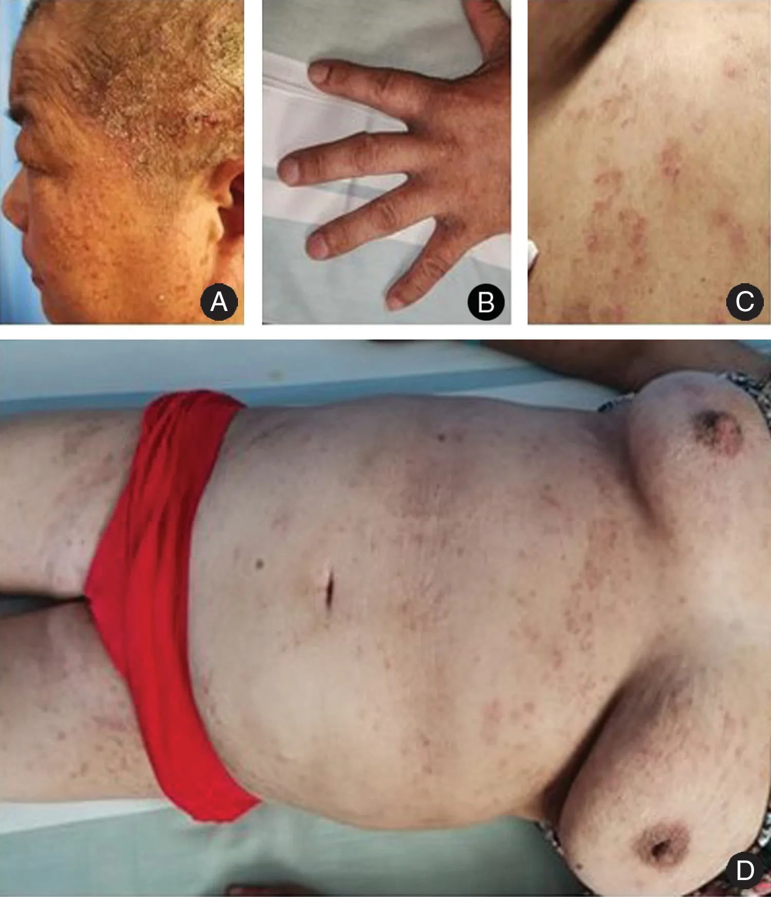
A 55-year-old woman presented with dermatitis, skin lesions,pruritus with infiltrated erythema,a large amount of greasy scales, and crusting on the scalp for 5 months,and erythema and papules on the body and extremities for 1 week (Fig. 1). The patient also had hypertension and coronary disease.The scalp lesions had been misdiagnosed as seborrheic dermatitis, and the symptoms had not improved after intermittent outpatient treatment comprising oral antihistamines and topical administration of 0.05% fluticasone propionate cream and halcinonide solution for 5 months. New rashes had appeared on the trunk and extremities in the past week. The patient was first seen on August 26, 2019. Laboratory examinations revealed several abnormalities. Complete blood count revealed an elevated platelet count of 89×109/L. Biochemistry analysis revealed an elevated glutamic-pyruvic transaminase concentration of 242U/L(reference range 7-40U/L), elevated glutamic oxaloacetic transaminase concentration of 105U/L (reference range 13-15U/L),decreased albumin concentration of 36.7g/L (reference range 40-55g/L),and decreased serum potassium concentration of 3.35mmol/L (reference range 3.5-5.3mmol/L).The patient tested positive for hepatitis B e-antibody,hepatitis B core antibody,anti-HCV IgG,anti-SSA/Ro-52 antibody, and antinuclear antibody. The primary karyotype titer for antinuclear antibody was 1:320. Color Doppler echocardiography showed mild enlargement of the left atrium and decreased left ventricular diastolic function. There were no abnormalities detected on chest radiography, ultrasonographic examination of the liver,gallbladder,pancreas,and spleen,urinalysis,routine stool testing, and thromboelastography. Histopathological examination revealed S. scabiei mites in the epidermis and inflammatory cell infiltration in the superficial dermis(Fig. 2).
The scabies infestation was eradicated after a 3-week course of medical therapy comprising both sulfur ointment and lindane cream. There was no recurrence during 5 weeks of follow-up.
Discussion
NS is a rare skin infestation caused by the S. scabiei that presents as pruritic dermatitis. NS has an extremely high mite burden within the epidermis and is therefore exceedingly contagious.Scabies is reported in many parts of the world,especially in developing countries.The main factors affecting the distribution of scabies are overcrowding, poor personal hygiene, and living in rural areas.3NS is an acute form of scabies that is seen in immunocompromised patients, including patients with immune system suppression, autoimmune diseases, malnutrition, malignancy, organ transplantation, and HIV.4Immunosuppression allows the S.scabiei mites to rapidly spread throughout the skin of the whole body and create a generalized infestation. Furthermore, patients with T-cell leukemia/lymphoma, human T-cell leukemia/lymphotropic retroviruses-1 infection,or neurological pathologies with altered sensitivity and reduced sensation (e.g.,leprosy, syringomyelia, Parkinson’s disease, Down’s syndrome, dementia, and mental retardation) who contract NS often do not scratch; this means that the S.scabiei tunnels are not mechanically destroyed, which enables them to proliferate. NS is also more prevalent in patients with defective T-cell responses(such as those with AIDS), organ transplantation, physical debilitation, critical illness, and those using potent topical corticosteroids.On presentation, patients often display extensive hyperkeratotic plaques with yellow-green crusts,most commonly located on the torso,extremities,face, and scalp.5This presentation is often misdiagnosed by clinicians who are inexperienced or unfamiliar with S. scabiei infection;common misdiagnoses include pityriasis, systemic lupus erythematous, bullous pemphigoid, eczema, or adverse drug reactions.6
The pathological manifestations of scabies are induced by the infiltration of inflammation cells (including eosinophils, lymphocytes, and histocytes), epidermal hyperproliferation, increased permeability of skin vessels,and secretion of several cytokines by T and B lymphocytes.Th1 responses are predominant in ordinary scabies,while Th2 responses are active in NS.7
The prevention and treatment of human scabies requires good personal hygiene, including frequent bathing,changing of clothes, and washing and drying of bedding.Patients with scabies should avoid cohabitation and shaking hands and should wash their clothes separately.The patient’s clothes should be boiled to kill the insects;if the clothes cannot be boiled,they should be wrapped in a plastic bag for 1 week and cleaned after the mites have died from starvation.

Figure 1. The clinical presentation of the patient with Norwegian scabies.(A)The scalp displays infiltrated erythema,a large amount of greasy scales, and crusts. The extremities (B) and trunk (C and D) display erythema, papules, and canals.
Traditional treatments have been successfully applied in the management of NS, with slight alterations. The purpose of treatment is to kill the scabies mites, relieve pruritus,and treat complications.Outcomes are improved by early detection, diagnosis, and treatment. All patients with scabies who are living in one home or in collective units should be treated at the same time.Medications that are commonly used to treat scabies include 10% sulfur and 3% salicylic acid ointment (the dosage is halved for use in children); 1% r-666 cream, which is contraindicated during pregnancy, lactation, and in infants younger than 2 years, but may be used with caution in children older than 2 years; 10%-25% benzyl benzoate lotion or emulsion; permethrin cream; application of 40%sodium thiosulfate solution twice before drying,and 4%dilute hydrochloric acid solution twice every morning and evening for 3-4 days;and 10%clomiphene emulsion or liniment.Treatments for scabies nodules include tar gel application once a night for 2-3 weeks, injection of prednisone,triamcinolone,or triamcinolone into the skin lesions, and topical application of triamcinolone acetonide neomycin.8
A review of the present patient’s family history revealed that all family members had recurrent scabies infestations and their symptoms were successfully relieved via conventional therapy with sulfur ointment application from the neck to the feet.However,the scalp lesions on the present patient persisted after several treatments. The treatment failure was considered to be due to her immunocompromised condition and a failure to apply the ointments to the scalp.In severe infestations,surgical debridement has been used as a successful adjunct procedure to debulk the lesions and intensify the ability of topical medications to penetrate the dermal layers and eliminate the mites.9

Figure 2. Histopathological results of lesions(H&E staining).Irregular acanthosis,inflammatory cell infiltration can be seen around the vessels in the superficial dermis (A, ×40). Scabies can be seen in the tunnel of the stratum corneum (B, ×100; C, ×400) even through the stratum spinosum to the superficial dermis (D, ×400).
杂志排行
国际皮肤性病学杂志的其它文章
- Instructions for Authors
- Bowenoid Papulosis
- Co-Infection of Chlamydia trachomatis and Neisseria gonorrhoeae on Adult Conjunctivitis:A Case Report
- Mycobacterium Chelonae/Abscessus Co-infection of the Limbs: A Challenging Case
- Lipoid Proteinosis Due to Homozygous Deletion Mutation (c.735delTG) in the ECM1 Gene Presents with Seizures and Hoarseness but No Skin Involvement
- Pathogenesis of Photoaging in Human Dermal Fibroblasts
