18氟-氟代脱氧葡萄糖PET/CT对多系统结节病和淋巴瘤的鉴别诊断
2019-12-16徐冰柯淑君黄海栋
徐冰 柯淑君 黄海栋


[摘要]目的 探讨18氟-氟代脱氧葡萄糖(18F-FDG)PET/CT检查在多系统结节病和淋巴瘤鉴别诊断中的价值。方法 回顾性分析2009年6月~2018年5月我院收治的15例结节病患者和20例淋巴瘤患者的18F-FDG PET/CT图像,所有病例均经病理证实。记录两组患者的最大淋巴结大小、淋巴结的最大SUV值、两侧肺门淋巴结是否对称性肿大及肿大淋巴结的融合、坏死、钙化情况。结果 两组的淋巴结最大SUV值、淋巴结钙化率、淋巴结坏死率比较,差异无统计学意义(P>0.05);结节病组的最大淋巴结直径小于淋巴瘤组,差异有统计学意义(P=0.001);结节病组的两侧肺门淋巴结对称性肿大发生率高于淋巴瘤组,差异有统计学意义(P=0.006);结节病组的淋巴结融合率低于淋巴瘤组,差异有统计学意义(P=0.007)。结论 结节病和淋巴瘤在肿大淋巴结的大小、两侧肺门淋巴结是否对称性增大、淋巴结的融合倾向方面有差异,PET/CT作为全身性的检查方法,对其鉴别诊断有较高的价值。
[关键词]结节病;淋巴瘤;体层摄影术;正电子发射断层显影术;最大摄取值
[中图分类号] R445 [文献标识码] A [文章编号] 1674-4721(2019)10(c)-0116-04
[Abstract] Objective To explore the value of 18F-fluorodeoxyglucose (18F-FDG) PET/CT examination in the differential diagnosis of the multisystem sarcoidosis and lymphoma. Methods 18F-FDG PET/CT images of 15 sarcoidosis patients and 20 lymphoma patients admitted to our hospital from June 2009 to May 2018 were retrospectively analyzed. All cases were confirmed by pathology. The size of the largest lymph node, mean maximum standard uptake value (SUVmax) of the lymph nodes, whether the bilateral hilar lymph nodes were symmetrically enlarged, and presence of fusion, necrosis, calcification of the enlarged lymph nodes were recorded in the two groups. Results There were no significant differences in the SUVmax, calcification of the lymph node, and necrosis of the lymph node between the two groups (all P>0.05). The size of the largest lymph node in the sarcoidosis group was significantly smaller than that in the lymphoma group, the difference was statistically significant (P=0.001). The symmetric enlargement in the bilateral hilar lymph nodes in the sarcoidosis group was higher than that in the lymphoma group, the difference was statistically significant (P=0.006). Lymph node fusion in the sarcoidosis group was lower than in the lymphoma group, the difference was statistically significant (P=0.007). Conclusion There are differences between sarcoidosis and lymphoma in the size of enlarged lymph nodes, symmetry of bilateral hilar lymph nodes and the tendency of lymph node fusion. As a general examination method, PET/CT is of great value in the differential diagnosis of sarcoidosis and lymphoma.
[Key words] Sarcoidosis; Lymphoma; Tomography; Positron-emission tomograpgy; Maximum standard uptake value
結节病是一种全身性、系统性的类上皮肉芽肿性疾病,病变主要累及淋巴结,尤其是胸内淋巴结,特征性表现为双侧肺门淋巴结对称性肿大,但身体的任何部位均可受累及,如肺、脾、骨头、眼睛、心脏、神经系统等[1-2]。恶性淋巴瘤是体内淋巴样器官的恶性增生性疾病,淋巴样器官遍及全身,包括骨髓、胸腺、淋巴结、脾脏、呼吸道和胃肠道的淋巴滤泡等,分布广泛,全身各部位均可发病,且发病率和病死率均较高[3]。结节病和淋巴瘤均是全身性疾病,均以累及淋巴系统为主,而且多侵犯胸内淋巴结,因此两者在鉴别诊断上存在着很大困难[4-6]。本研究回顾性分析35例以全身多发淋巴结肿大为主要表现的患者的18氟-氟代脱氧葡萄糖(18F-FDG)PET/CT图像,旨在提高两者的诊断准确性。
1资料与方法
1.1一般资料
回顾性分析我院2009年6月~2018年5月经穿刺病理或手术病理证实的35例以全身多发淋巴结肿大为主要表现的患者的临床及影像学资料,其中结节病15例,淋巴瘤20例。纳入标准:所有病例均经病理证实;所有患者均在病理取材前行18F-FDG PET/CT检查,且间隔时间不超过1周;无其他任何恶性肿瘤史;均为初治患者,未经任何治疗。排除标准:孤立单个的淋巴结肿大;就诊时1个月内有感染性疾病史;既往曾有肺结核病史。15例结节病患者中,男6例,女9例;年龄34~77岁,平均(54±12)岁;临床症状主要为咳嗽、咳痰、乏力、体重减轻等,其中6例体检发现,无任何临床症状;组织病理学均为非干酪样坏死性肉芽肿。20例淋巴瘤患者中,男性11例,女性9例;年龄25~84岁,平均(57±14)岁;临床症状主要为乏力、纳差、消瘦、发热、体表淋巴结肿大等;其中弥漫大B淋巴瘤12例,NK/T细胞淋巴瘤2例,间变性大细胞淋巴瘤1例,非霍奇金淋巴瘤5例。本研究获上海市浦东新区浦南医院医学伦理委员会批准,所有患者均签署书面知情同意书。
1.2 18F-FDG PET/CT成像方法
所有患者均行全身18F-FDG PET/CT扫描,示踪剂18F-FDG由原子科兴公司提供,显像仪为西门子Biograph 64。患者检查前需要禁食4~6 h,并监测其血糖水平,判断血糖是否符合要求,一般要求其血糖水平在11 mmol/L以下。药物的注射量根据患者的体重计算(0.15~0.20 mci/kg),采用静脉注射,于60~90 min后开始扫描。先行投射采集,CT扫描参数为120 KV、100 mAs、0.5 s/r、3 mm层厚,螺距(pitch)1.05 mm的进床速度,20~30 s扫描完毕,然后自动切入PET数据采集,一般从腹股沟开始向上采集,矩阵为128×128。应用CT数据进行衰减校正,重建算法采用滤波反投影法,融合图像为麦迪克斯(MedEx)工作站软件处理获得。
1.3图像分析
由2名高年资PET-CT诊断医生分别阅片,共同确定病变范围和程度,依次记录以下内容。①淋巴结的大小:用同机CT测量最大淋巴结最大截面的平均直径;②淋巴结的最大SUV值(SUVmax):取至少5个不同部位的病灶分别测量,取其平均值;③双侧肺门淋巴结是否对称性肿大;④肿大淋巴结是否有融合、坏死、钙化。
1.4统计学方法
采用SPSS 20.0统计软件对数据结果进行分析,计量资料以均数±标准差(x±s)表示,采用t检验;计数资料以率表示,采用χ2检验,如样本量过小χ2检验近似无效时,采用Fisher精确概率法统计,以P<0.05为差异有统计学意义。
2结果
2.1结节病和淋巴瘤最大淋巴结大小和SUVmax的比较
结节病组最大淋巴结的直径为(1.91±0.51)cm,小于淋巴瘤组的(3.07±0.81)cm,差异有统计学意义(t= 11.296,P<0.05)。结节病组的SUVmax为11.34±3.94,淋巴瘤组的SUVmax为12.89±5.32,两组数据差异无统计学意义(t=1.088,P>0.05)。
2.2结节病和淋巴瘤受累淋巴结的影像学特征
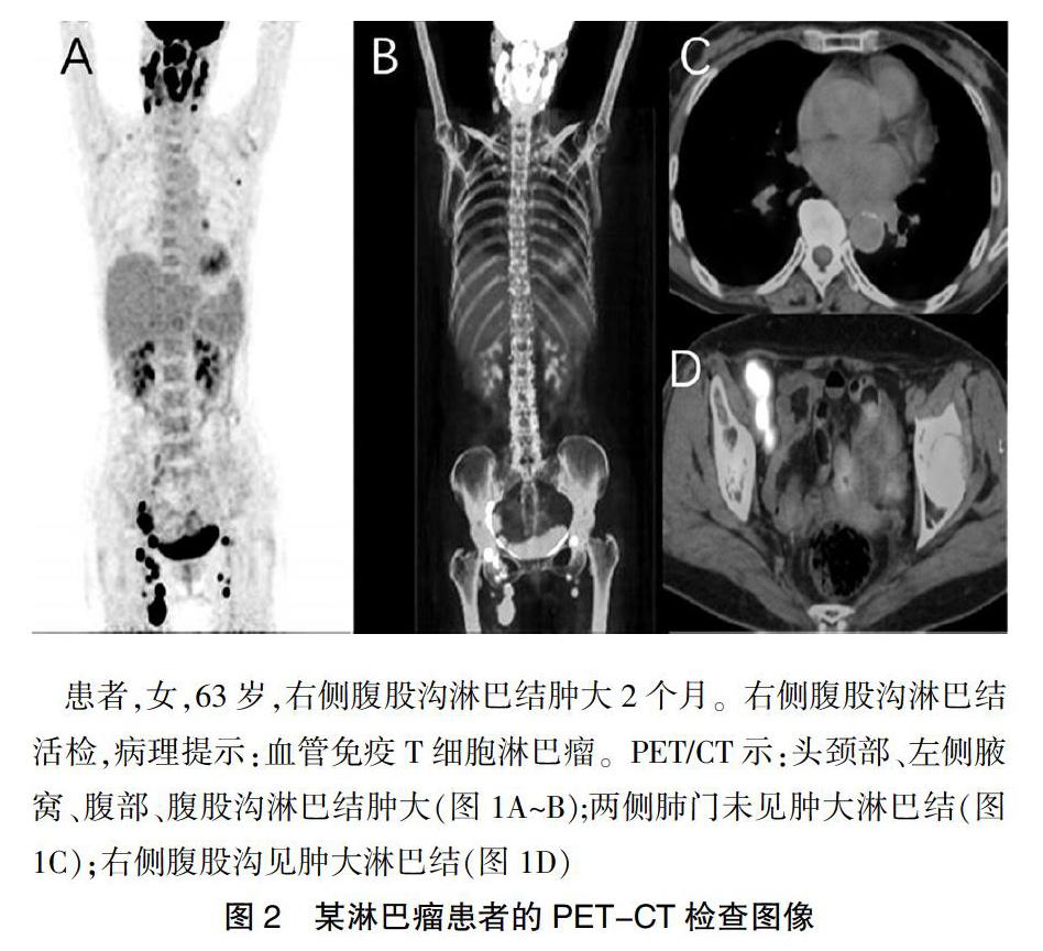
结节病组的两侧肺门淋巴结对称性肿大发生率高于淋巴瘤组,差异有统计学意义(P<0.05);结节病组的淋巴结融合率低于淋巴瘤组,差异有统计学意义(P<0.05);两组的淋巴结坏死率和钙化率比较,差异无统计学意义(P均>0.05)(表1)。具体影像学图片如图1、2所示。
3讨论
目前,18F-FDG PET/CT是检测肿瘤的重要工具,淋巴瘤是淋巴系统的恶性肿瘤,18F-FDG PET/CT对大多数淋巴瘤的灵敏度能达到90%以上[7]。结节病是一种全身肉芽肿性疾病,大量的研究显示,18F-FDG PET/CT对结节病同样具有较高的敏感性[8-9]。18F-FDG PET/CT作为一种全身性的检查工具,不仅能显示病变的部位、累及的范围,而且能显示出病變部位组织的代谢情况,对结节病和淋巴瘤的鉴别诊断有重要的参考价值[10]。
结节病和淋巴瘤在肿大淋巴结的大小上有差异,这可能与这两种疾病的性质及病程有关[11]。结节病是一种良性肉芽肿性疾病,病程多数较慢,而且具有自愈倾向,肿大淋巴结会逐渐吸收变小甚至消失[12];而淋巴瘤作为一种恶性肿瘤,其生长速度较快,在短期内就会迅速增殖[13],所以往往发现时其受累淋巴结就已经明显肿大,部分可能会出现融合现象,这也解释了为什么淋巴瘤组中淋巴结的融合现象较结节病组多见[14]。
SUV为FDG的标准化摄取值,用来描述葡萄糖的代谢情况,一般作为肿瘤良恶性的鉴别诊断及疗效评价的重要指标[15-16]。因为恶性肿瘤细胞内糖酵解作用明显增强,其FDG的摄取明显高于正常组织和良性病变,而结节病虽然是良性病变,但其病变组织中炎细胞(如淋巴细胞、巨噬细胞等)的葡萄糖代谢也会增加,因此两者都有FDG的摄取增加[7,17]。本研究中,两组的SUVmax比较,差异无统计学意义(P>0.05),提示SUVmax对两者鉴别意义不大。
结节病和淋巴瘤肿大的淋巴结一般密度较均匀,但也可以发生钙化[18]。钙化多为纤维组织营养不良造成,和结核或者高钙无关,大多数钙化发生在肺门或者气管旁淋巴结,钙化的形态无特异性,可为针尖样、颗粒状或爆米花样,有报道称部分可以见到蛋壳样的钙化,比较有特异性,仅见于结节病和矽肺或煤工尘肺中[19]。未治疗的淋巴瘤的钙化比较少见,一般见于个案报道[20],但放疗或者化疗后,钙化比较常见。
两侧肺门淋巴结对称性肿大是结节病的典型表现,这种情况不光表现在肺门,而且在其他部位如颈部、腹部等如有淋巴结受累,也会出现相应的对称性分布的特点[21-23]。本研究中,结节病组的两侧肺门淋巴结对称性肿大发生率明显高于淋巴瘤组(P<0.05),与既往文献报道一致,由此可以看出,两侧肺门淋巴结对称性肿大是结节病的特征性表现。
综上所述,结节病和淋巴瘤在淋巴结的大小、两侧肺门淋巴结是否对称性肿大、肿大淋巴结是否融合等方面有差异,可以作为两者的鉴别要点,这对临床诊断工作具有指导意义。
[参考文献]
[1]关志伟,姚树林,王瑞民,等.22例结节病18F-FDG PET/CT影像学特征分析[J].中华核医学杂志,2011,31(5):334-338.
[2]Colden DG,Busaba NY.Sarcoidosis presenting as recurrent nasal polyps[J].Otolaryngol Head Neck Surg,2000,123(4):519-521.
[3]Delsol G.The 2008 WHO lymphoma classification[J].Ann Pathol,2008,28(1):S20-S24.
[4]Maayan H,Ashkenazi Y,Nagler A,et al.Sarcoidosis and lymphoma:case series and literature review[J].Sarcoidosis Vasc Diffuse Lung Dis,2011,28(2):146-152.
[5]Oskuei A,Hicks L,Ghaffar H,et al.Sarcoidosis-lymphoma syndrome:a diagnostic dilemma[J].BMJ Case Rep,2017,2017:bcr2017220065.
[6]Papanikolaou IC,Sharma OP.The relationship between sarcoidosis and lymphoma[J].Eur Respir J,2010,36(5):1207-1209.
[7]Nishii R,Higashi T,Kagawa S,et al.Diagnostic usefulness of an amino acid tracer,α-[N-methyl-11C]-methylaminoisobutyric acid (11C-MeAIB),in the PET diagnosis of chest malignancies[J].Ann Nucl Med,2013,27(9):808-821.
[8]Kaira K,Oriuchi N,Otani Y,et al.Diagnostic usefulness of fluorine-18-alpha-methyltyrosine positron emission tomography in combination with 18F-fluorodeoxyglucose in sarcoidosis patients[J].Chest,2007,131(4):1019-1027.
[9]Koo HJ,Kim MY,Shin SY,et al.Evaluation of mediastinal lymph nodes in sarcoidosis,sarcoid reaction,and malignant lymph nodes using CT and FDG-PET/CT[J].Medicine (Baltimore),2015,94(27):e1095.
[10]Shiozaki T,Sadato N,Senda M,et al.Noninvasive estimation of FDG input function for quantification of cerebral metabolic rate of glucose:optimization and multicenter evaluation[J].J Nucl Med,2000,41(10):1612-1618.
[11]张健雄,李云霄,王浩彦.66例结节病首发表现与首诊科室回顾分析[J].心肺血管病杂志,2018,37(1):29-32.
[12]Soto-Gomez N,Peters JI,Nambiar AM.Diagnosis and management of sarcoidosis[J].Am Fam Physician,2016,93(10):840-848.
[13]Impivaara S,Makela L,Hernberg M,et al.Cutaneous sarcoidosis after Hodgkin lymphoma[J].Int J Dermatol,2018, 58:e1-e21.
[14]Cozzi D,Bargagli E,Calabrò AG,et al.Atypical HRCT manifestations of pulmonary sarcoidosis[J].Radiol Med,2018,123(3):174-184.
[15]Yu C,Xia X,Qin C,et al.Is SUVmax helpful in the differential diagnosis of enlarged mediastinal lymph nodes?A pilot study[J].Contrast Media Mol Imaging,2018,2018:3417 190.
[16]Dhillon T,Palmieri C,Sebire NJ,et al.Value of whole body 18FDG-PET to identify the active site of gestational trophoblastic neoplasia[J].J Reprod Med,2006,51(11):879-887.
[17]Bois JP,Muser D,Chareonthaitawee P.PET/CT evaluation of cardiac sarcoidosis[J].PET Clin,2019,14(2):223-232.
[18]Tortorich J,Woods M,Shintaku W,et al.Diagnostic considerations of calcified lymph nodes[J].J Tenn Dent Assoc,2013,93(2):8-10,quiz 11-2.
[19]Voortman M,CMR H,Elfferich P,et al.The burden of sarcoidosis symptoms from a patient perspective[J].Lung,2019, 197(2):155-161.
[20]Alobeidy ST,Ilowite J,Donovan V,et al.Calcification in untreated mediastinal Hodgkin′s lymphoma[J].J Thorac Imaging,2001,16(4):304-306.
[21]柯淑君,肖湘生.肺結节病的临床与影像研究[J].国际医学放射学志,2015,38(4):331-334.
[22]王旭华,聂斌,汤宇,等.胸部结节病的CT表现[J].中国CT和MRI杂志,2005,3(2):29-30,34.
[23]Polverosi R,Russo R,Coran A,et al.Typical and atypical pattern of pulmonary sarcoidosis at high-resolution CT:relation to clinical evolution and therapeutic procedures[J].Radiol Med,2014,119(6):384-392.
(收稿日期:2019-03-14 本文编辑:祁海文)
