Observation on Cardiac Opening of the Inferior Vena Cava in Goat Fetuses
2019-09-10JieZHANGJinlongZHANG
Jie ZHANG Jinlong ZHANG
Abstract [Objectives] This study was conducted to better understand the characteristics of the fetal blood circulation.
[Methods] The cardiac opening of the inferior vena cava was observed in goat fetuses. There are two cardiac openings of the inferior vena cava, namely, the foramen ovale with the valves and the right atrium opening of the inferior vena cava. The blood from the inferior vena cava moves easily into the right atrium via the right atrium opening because the right atrium opening without the valves is located in the lower right of the foramen ovale, and is bigger than the interatrial foramen.
[Results] The blood may flow into the left atrium through the foramen ovale when the blood volume into the right atrium exceeds the limit of the right atrium. The fetal inferior vena cava compared with after birth receives oxygenated blood in the umbilical vein from the placenta besides the normal blood. Accordingly the blood volume into the right atrium exceeds the limit of the right atrium. Extra oxygenated blood is diverted directly into the left atrium through the foramen ovale.
[Conclusions] This study provides a reference for studying the blood circulation of fetuses and even adults.
Key words Fetal circulation; Inferior vena cava; Foramen ovale; Interatrial foramen
Received: August 12, 2019Accepted: October 23, 2019
Supported by the Priority Academic Program Development of Jiangsu Higher Education Institutions (PAPD); Scientific Research Fund of Nanjing General Hospital of Nanjing Military Command, China (2016033).
Jie ZHANG (1991-), female, P. R. China, resident doctor, master, devoted to research about Cardiology.
*Corresponding author. Email: zjl@yzu.edu.cn.
The fetal umbilical cord is an important link between the fetus and the mother, and includes therein two umbilical arteries and an umbilical vein, in which the blood flow is relatively large[1-2]. There is still much controversy about the blood circulation of fetuses. It is now widely believed that the umbilical vein blood from the placenta with rich nutrients and high oxygen content is injected into the inferior vena cava (called the caudal cava vein in animals) through the liver and ductus venosus, and then flows into the right atrium[3]. Because the cardiac opening of the inferior vena cava in human fetuses faces the foramen ovale, plus the effect of the inferior vena cava valve[4-5], most of the blood from the inferior vena cava is directly injected into the left atrium through the foramen ovale, and then leaves leaf ventricle and enters the aorta to supply nutrition to the head and neck, forelimb and heart of the fetus. The blood from the superior vena cava (called the cranial cava vein in animals) enters the right atrium, then flows into the right ventricle and enters the pulmonary artery through the orifice of pulmonary trunk. Since the lungs in fetuses still have no respiratory function, only a small amount of the blood enters the lung, and most of the blood enters the descending aorta via the arterial duct[5-7]. When using goats to carry out related experiments, we found that due to the difference in body position, the cardiac opening of the inferior vena cava of goat fetuses is different from that of human fetuses. Therefore, we dissected goat fetuses and observed from another angle to better help us understand the fetal blood circulation.
Materials and Methods
Thirteen aborted goat fetuses without malformation were collected from a sheep farm (because the goats were pregnant for about 5 months, so aborted fetuses of 4 to 5 months old were chosen, 5 rams, 8 ewes). The chests and abdomens were opened, and the fetuses were soaked in formalin for more than 2 weeks, and then dissected. The cardiac opening of inferior vena cava of the fetuses was observed and photographed.
Results and Analysis
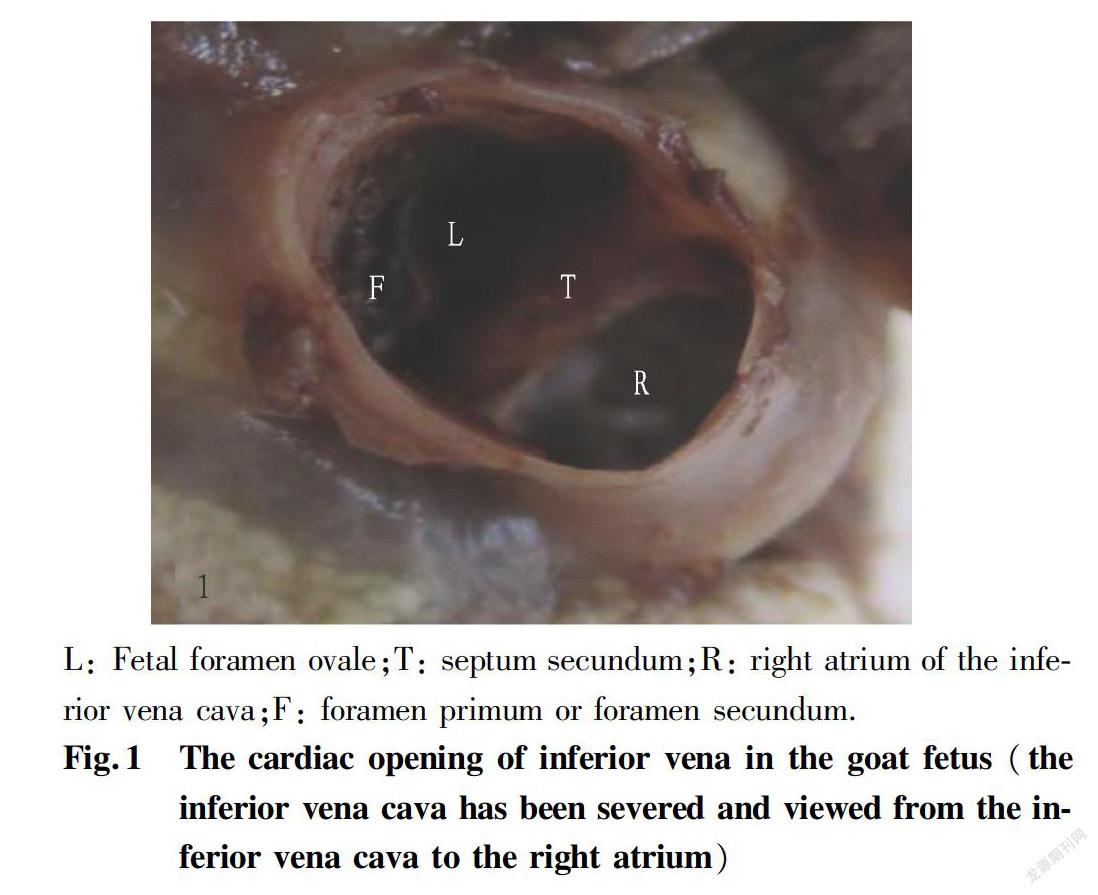
The inferior vena cava of goat fetuses has no venous valves at cardiac opening, and there are two openings in the inferior vena cava after entering the heart. One is the foramen ovale with the valves (Fig. 1L). The fetal foramen ovale is a hole formed between the septum secundum (Fig. 1T) and the left wall of the inferior vena cava, generally oval, and has valves formed by the septum primum on the left atrium side. The foramen primum or the foramen secundum (Fig. 1F) can be observed on the valves. The other opening is the right atrium of the inferior vena cava (Fig. 1R), which is slightly larger than the foramen ovale and is formed between the septum secundum (Fig. 1T) and the right wall of the inferior vena cava (or right atrium wall). It is located at the lower right of the foramen ovale, and has no valves on the right atrium. The right atrium of the inferior vena cava is much larger than the foramen primum or the foramen secundum.
Conclusions and Discussion
Human fetuses have inferior vena cava valves[4-5]. Related literature suggests that the valves have a diversion effect, so that most of the fetal inferior vena cava blood enters the left atrium through the foramen ovale. In this study, it was observed that there are no inferior vena cava valves at the cardiac opening of the inferior vena cava in goat fetuses, and there is no diversion effect of the inferior vena cava valves. Generally, the venous valve has the effect of preventing blood backflow[7]. What is the role of the human inferior vena cava valves?Whether the human inferior vena cava valves make the inferior vena cava blood enter the left atrium under its diversion effect or prevent the blood from flowing backwards remains to be discussed.
L: Fetal foramen ovale;T: septum secundum;R: right atrium of the inferior vena cava;F: foramen primum or foramen secundum.
In goat fetuses, there are two cardiac openings of the inferior vena cava, namely, the foramen ovale and the right atrium opening of the inferior vena cava. The blood from the inferior vena cava moves easily into the right atrium via the right atrium opening because the right atrium opening is bigger than the interatrial foramen and is located in the lower right of the foramen ovale, which means that the blood from the inferior vena cava gives priority to the right atrium. Then under what circumstances will the inferior vena cava blood be injected into the left atrium through the foramen on the foramen ovale? We believe that it should be dependent on the amount of blood received in the right atrium (the volume of the right atrium) and the amount of venous blood injected into the right atrium. When the amount of venous blood injected into the right atrium exceeds the limit of the right atrium, the excessive blood enters the left atrium through the foramen on the foramen ovale. Compared with after birth, the fetal inferior vena cava receives oxygenated blood in the umbilical vein from the placenta besides the normal blood. Accordingly the blood volume into the right atrium exceeds the limit of the right atrium. Extra oxygenated blood is diverted directly into the left atrium through the foramen ovale. For human fetuses, it is generally believed that the cardiac opening of the inferior vena cava in human fetuses faces the foramen ovale, and there is a diversion effect of the inferior vena cava valves, so the nutrientrich blood of the inferior vena cava is more likely to pass directly through the foramen ovale and enter the left atrium[5-7]. If this is the case, then the cardiac opening of the inferior vena cava in the newborn also faces the foramen ovale, and also has the diversion effect of the inferior vena cava valves, so the inferior vena cava blood should be equally easy to move into the left atrium through the foramen ovale, but in fact it is not the case. In fact, the human cardiac opening of the inferior vena cava is similar to the goat cardiac opening of the inferior vena cava, and the differences lies in their different orientations, and lies in that the cardiac opening of the inferior vena cava in human fetuses has inferior vena cava valves besides the valves on the foramen ovale[6]. For human, the right atrium opening of the inferior vena cava is also larger than the foramen. Similarly, the left atrium also has a limit for receiving blood (the volume of the left atrium). Since the fetuses have blood injected into the left atrium through the foramen ovale, they reduce the blood entering the left atrium through the pulmonary vein from the pulmonary circulation by regulating the pulmonary circulation (resistance of the lung), so that the blood in the pulmonary artery beyond the limit of the left atrium can be injected into the descending aorta through the arterial duct, thereby satisfying the blood supply of the umbilical artery in addition to ensuring that the descending aorta supplies normal organs normally. In addition, we believes that the blood flow into the left atrium through the foramen ovale, or the blood flow of the fetal pulmonary circulation, or the blood flow into the descending aorta via the arterial duct are related to the blood flow of the umbilical artery and umbilical vein. When the blood flow of the umbilical artery and the umbilical vein becomes larger, the blood injected into the right atrium of the inferior vena cava increases, the blood flow into the left atrium through the foramen ovale increases accordingly, the blood flow of the pulmonary circulation into the left atrium through the lung becomes smaller, and the blood flow in the arterial duct increases; and when the blood flow of the umbilical artery or umbilical vein becomes small (for example, the umbilical artery or umbilical vein is compressed), the blood flow into the left atrium through the foramen ovale becomes smaller, the blood volume of the pulmonary circulation becomes larger, and the blood flow in the arterial ducts also gets smaller. After birth, the umbilical artery and umbilical vein are shut, and the blood flow to the foramen ovale and the arterial duct through the interatrial septum is reduced to zero, at which the pulmonary circulation blood flow reaches its maximum. Of course, whether this is the case needs further study.
References
[1] TRUDINGER BJ, GILES WB, COOK CM, et al. Fetal umbilical artery flow velocity waveforms and placental resistance: clinical significance[J]. Br J Obstet Gynaecol, 1985(92): 23-30.
[2] BELLOTTI M, PENNATI G, DE GASPERI C, et al. Simultaneous measurements of umbilical venous, fetal hepatic, and ductus venosus blood flow in growthrestricted human fetuses[J]. Am J Obstet Gynecol, 2004(190): 1347-1358.
[3] KISERUD TORVID, ACHARYA GANESH. The fetal circulation[J]. Prenatal Diagnosis, 2004, 24(13): 1049-1059.
[4] WANG Y, SHEN B, GUO ZK. Observation and measurement of the aging changes in right atriums main structure in fetal heart[J]. Journal of Xinxiang Medical College, 2006, 23(6): 587-589. (in Chinese)
[5] KISERUD T, RASMUSSEN S, SKULSTAD S. Blood flow and the degree of shunting through the ductus venosus in the human fetus[J]. American Journal of Obstetrics and Gynecology, 2000(182): 147-153.
[6] CUI GY. Fetal blood circulation and changes after birth[J]. Bulletin of Biology, 1990(5): 20-22. (in Chinese)
[7] GU XS. Human anatomy[M]. Beijing: Science Press, 2004. (in Chinese)
杂志排行

农业生物技术(英文版)的其它文章
- Evaluation on Application and Spraying Effect of AirAssisted Sprayer in Apple Orchard with Dwarfing Rootstocks
- Problems in the Development of Traditional Chinese Medicinal Materials Planting Industry in Shiyan City and Countermeasures
- Study on Practical Mature Age of Individual Pinus thunbergii×P. densiflora
- Effects of Different Densityreducing Methods on Canopy Microenvironment, Tree Growth and Fruit Quality in Closed Apple Orchard
- Development of Whole Potato Flour Fish Noodles
- Preparation of Alkalisoluble Pachymaran Rice Wine