结直肠癌组织CD3免疫组织化学评分与肿瘤分化程度及预后关系分析
2019-08-20张畅胡阿锦龚苗子王跃王淑芳张雨王云帆缪琦张晋夏
张畅,胡阿锦,龚苗子,王跃,王淑芳,张雨,王云帆,缪琦,张晋夏
结直肠癌组织CD3免疫组织化学评分与肿瘤分化程度及预后关系分析
张畅,胡阿锦,龚苗子,王跃,王淑芳,张雨,王云帆,缪琦,张晋夏
100144 北京大学首钢医院病理科
分析结直肠癌组织 CD3 免疫组织化学评分与患者性别、年龄、肿瘤分化程度、TNM 分期、组织抗原表达及血清肿瘤抗原关系,评估 CD3 免疫组织化学评分在结直肠癌免疫状态评估中的作用。
采用免疫组织化学方法对 116 例结直肠癌患者肿瘤标本进行 CD3 免疫组织化学染色并评分,将患者分成 CD3 低表达组与 CD3 高表达组。比较分析两组患者的性别和年龄组成、肿瘤分化程度及 pTNM 分期。同时比较两组肿瘤组织样本 CEA、EGFR、MUC1、HER2、mesothelin、PD-1、PD-L1 及 FOXP3 免疫组织化学评分,并且比较两组患者血清中 CEA、CA125 及 CA199 水平。
174 例组织标本 CD3 免疫组织化学染色均可见阳性细胞,评分均在1 ~ 12 分之间。CD3 低表达组年龄
< 30 岁患者比例(9.20%)高于 CD3 高表达组(0%),> 65 岁患者比例(33.33%)低于高表达组(57.47%)。CD3 低表达组高分化癌(3.45%)及中分化癌(60.92%)比例低于 CD3 高表达组(分别为 9.20% 和 74.71%),而低分化癌(27.59%)及复发/转移癌(8.05%)比例高于 CD3 高表达组(分别为 9.20% 和 6.90%)。pTNM 分期越高预后越差,CD3 免疫组织化学评分越低(< 0.05)。CD3 低表达组患者组织样本中 PD-1(< 0.001)、PD-L1(< 0.001)及 FOXP3(< 0.01)水平低于 CD3 高表达组,而两组患者血清肿瘤标志物(CEA、CA125 和 CA199)水平无明显差异。
CD3 免疫组织化学评分可广泛应用于结直肠癌肿瘤组织,并反映患者的肿瘤相关免疫状态与预后。
抗原,CD3; 预后; 抗原,肿瘤; 结直肠肿瘤; 肿瘤免疫
T 细胞是肿瘤免疫中重要的功能细胞,在肿瘤相关免疫过程中发挥重要作用。CD3 是T 细胞表面的特异性标志物,CD3 免疫组织化学染色可很好地评估肿瘤组织中 T 细胞的浸润水平。因此 CD3免疫组织化学染色常被用来评估患者的免疫状态。本研究分析了肿瘤组织 CD3 免疫组织化学评分与患者性别和年龄分布、肿瘤组织分化程度、肿瘤组织抗原水平及血清肿瘤标志物水平关系。希望通过分析评估 CD3 免疫组织化学评分对肿瘤免疫水平进行评估。
本研究除对标本进行 CD3 免疫组织化学评分以外,还对标本进行了癌胚抗原(carcinoembryonic antigen,CEA)、表皮生长因子受体(epidermal growth factor receptor,EGFR)、间皮素(mesothelin)、黏蛋白 1(mucin 1,MUC1)、表皮生长因子受体 2(epidermal growth factor receptor 2,HER2)、程序性细胞死亡受体-1(programmed death 1,PD-1)、程序性死亡配体-1(programmed death-ligand 1,PD-L1)和叉状头型转录因子 3(forkhead box protein 3,FOXP3)的免疫组织化学评分。
1 资料与方法
1.1 资料
1.1.1 临床资料 纳入 2017 年 5 – 12 月在我院经组织病理学诊断确诊的结直肠癌患者 174 例,男 103 例,女 71 例,年龄 24 ~ 93 岁,平均61 岁,所有患者均经过手术治疗,肿瘤完整切除后,对手术标本进行病理分期、病理学诊断及免疫组织化学检测。所有病例在手术前均未经过放射治疗或化学药物治疗。所有样本中结直肠原发癌 156 例,复发/转移癌 18 例。
1.1.2 试剂与仪器 ZM-0061(CEA)、ZA-0579(mesothelin)、ZA-0505(EGFR)、ZM-0503(CD3)、ZM-0381(PD-1)抗体均购自北京中杉金桥生物技术有限公司;ab134182(HER2)、ab228415(PD-L1)、ab99963(FOXP3)抗体购自美国 Abcam 公司。使用 Dako AutostainerLink 48 自动染色仪一步法染色。
1.2 方法
1.2.1 样品采集与处理 新鲜手术切除标本经 10% 福尔马林固定液固定 48 h 后,常规制作石蜡切片,HE 及免疫组织化学染色。
1.2.2 免疫组织化学评分 组织切片免疫组织化学评分= 着色强度分 × 阳性细胞着色比例分。着色强度分:不着色 0 分,弱着色 1 分,中着色2 分,强着色 3 分。阳性细胞比例分:0 ~ 5% 阳性 0 分,6% ~ 25% 阳性 1 分,26% ~ 50% 阳性2 分,51% ~ 74% 阳性 3 分,> 75% 阳性 4 分。
1.2.3 肿瘤病理分期 采用国际抗癌联盟(UICC)恶性肿瘤 TNM 分期(第八版),结直肠恶性肿瘤病理分期(pTNM 分期)作为分期标准。
1.2.4 血清肿瘤标志物检测 患者血清肿瘤标志物检测结果为患者病理学诊断确诊后最近一次检测结果,数据由本院电子病例系统提供。
1.3 统计学处理
检验方法进行统计学分析。GraphPad Prism 7.0 软件(GraphPad 软件公司)用于统计学分析及统计图表制作。
2 结果
2.1 CD3 免疫组织化学染色
174 例组织标本 CD3 免疫组织化学染色均可见阳性细胞(图 1),评分 1 ~ 12 分。根据 CD3免疫组织化学评分将病例分为 CD3 低表达组(CD3 免疫组织化学染色评分≤ 6 分)与 CD3 高表达组(CD3 免疫组织化学染色评分 > 6 分)。CD3 低表达组患者 87 例,男 50 例(57.47%),女 37 例(42.53%);CD3 高表达组患者87 例,男 54 例(62.07%),女 33 例(37.93%),两组性别构成比均无明显差异。CD3 低表达组患者年龄 24 ~ 93 岁,平均 60.78 岁,< 30 岁8 例(9.20%),30 ~ 65 岁 50 例(57.47%),> 65 岁29 例(33.33%);CD3 高表达组患者年龄32 ~86 岁,平均 63.40 岁,< 30 岁0 例,30 ~ 65 岁 50 例(57.47%),> 65 岁 37 例(42.53%)。两组间不同年龄患者比例存在差异(< 0.05)(图 2A)。比较不同年龄患者肿瘤组织 CD3 免疫组织化学评分,发现年龄 < 30 岁患者 CD3 免疫组织化学评分明显偏低(< 0.01)(图 2B)。

图 1 CD3 免疫组织化学染色(A:CD3 免疫组织化学染色弱着色,着色强度分 1 分;B:CD3 免疫组织化学染色中着色,着色强度分 2 分;C:CD3 免疫组织化学染色强着色,着色强度分 3 分)
Figure 1 CD3 immunohistochemical staining (A: CD3 immunohistochemical staining weakly colored, with a color intensity score of 1; B: CD3 immunohistochemical staining for medium coloration with a color intensity score of 2; C: Strong staining of CD3 immunohistochemical staining with a color intensity score of 3)

病例数(例)Number of patients60 40 20 0CD3 低表达CD3 low expressionCD3 高表达CD3 high expressionCD3 免疫组织化学评分CD3 immunohistochemicalscores10 8 6 4 2 0 < 30 30 ~ 65 > 65 < 30 30 ~ 65 > 65 年龄AgeA 年龄AgeB
Figure 2 Sex, age and tumor tissue CD3 immunohistochemical scores in patients with different CD3 expression (A: CD3 low expression group and CD3 high expression group number of patients; B: CD3 immunohistochemical scores of tumor tissues of patients of different ages)
2.2 不同组织分化程度肿瘤组织 CD3 免疫组织化学评分
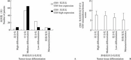
CD3 低表达组患者组织病理诊断为高分化癌者 3 例(3.45%),中分化 53 例(60.92%),低分化 24 例(27.59%),转移/复发 7 例(8.05%);CD3 高表达组患者高分化 8 例(9.20%),中分化 65 例(74.71%),低分化 8 例(9.20%),转移/复发 6 例(6.90%)。两组间不同肿瘤分化程度患者比例存在差异(< 0.05)(图 3A)。将所有病例按组织分化程度分为高分化组、中分化组、低分化组及转移/复发组,并比较各组 CD3 免疫组织化学染色评分。结果显示,CD3免疫组织化学评分与肿瘤组织分化程度成正相关(< 0.05)(图 3B)。
2.3 不同 pTNM 分期患者肿瘤组织 CD3 免疫组织化学评分
CD3 低表达组患者 pTNM 分期情况,I 期9 例(10.34%),II 期 34 例(39.08%),III 期27 例(31.03%),IV 期 17 例(19.54%);CD3 高表达组 pTNM 分期情况,I 期 14 例(16.19%),II 期 42 例(48.28%),III 期 23 例(26.44%),IV 期 8 例(9.20%)。两组间不同 pTNM 分期患者比例存在差异(< 0.05)(图 4A)。比较不同 pTNM 分期患者肿瘤组织CD3 免疫组织化学染色评分。结果显示,pTNM 分期越高预后越差,CD3 免疫组织化学评分越低(< 0.05)(图 4B)。

病例数(例)Number of patients80 60 40 20 0CD3 低表达CD3 low expressionCD3 高表达CD3 high expressionCD3 免疫组织化学评分CD3 immunohistochemicalscores15 10 5 0 高分化High differentiation 中分化Medium differentiation 低分化Low differentiation 转移/复发Metastasis/recurrence 高分化High differentiation 中分化Medium differentiation 低分化Low differentiation 转移/复发Metastasis/recurrence 肿瘤组织分化程度Tumor tissue differentiationA 肿瘤组织分化程度Tumor tissue differentiationB
Figure 3 CD3 immunohistochemical scores of tumor tissues with different degrees of tissue differentiation (A: Number of different degrees of tissue differentiation in CD3 low-expression group and CD3 high-expression group; B: CD3 immunohistochemical scores of tumor tissues with different degrees of tissue differentiation)

病例数(例)Number of patients50 40 30 20 10 0CD3 低表达CD3 low expressionCD3 高表达CD3 high expressionCD3 免疫组织化学评分CD3 immunohistochemical scores10 8 6 4 2 0 I II III IV I II III IV pTNM 分期pTNM stagesA pTNM 分期pTNM stagesB
Figure 4 CD3 immunohistochemical scores of tumor tissues in patients with different pTNM stages (A: Number of different pTNM staging cases in the CD3 low expression group and the CD3 high expression group; B: Different pTNM staging cases CD3 immunohistochemical score of patient tumor tissue)

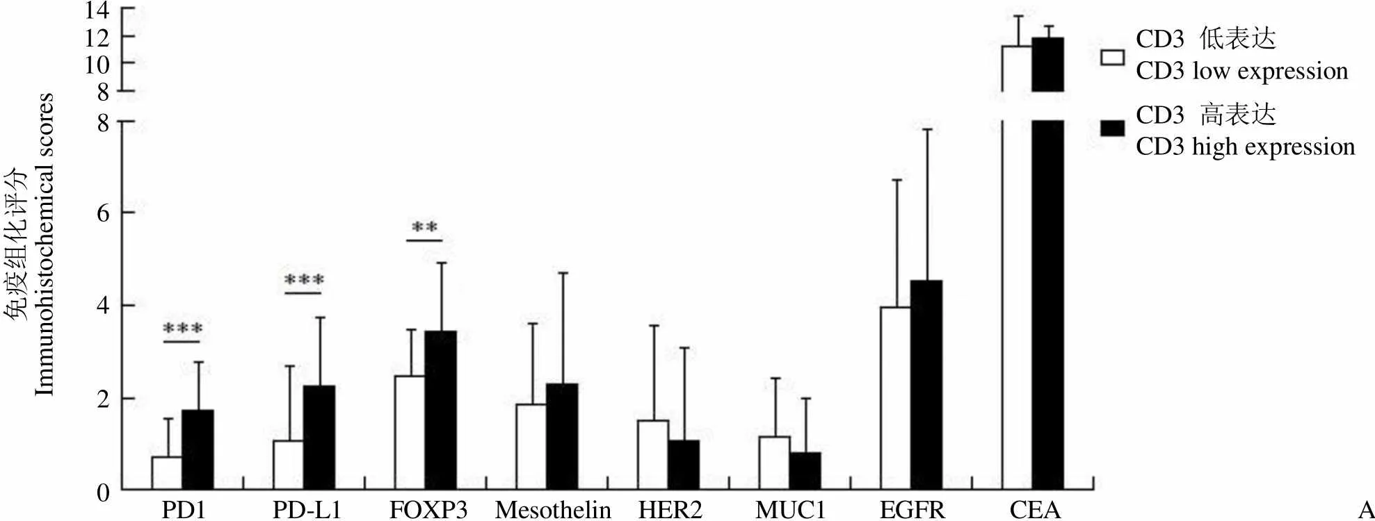
免疫组化评分Immunohistochemical scores14121088 6 4 2 0CD3 低表达CD3 low expressionCD3 高表达CD3 high expression PD1 PD-L1 FOXP3 Mesothelin HER2 MUC1 EGFR CEAA
Figure 5 Immunohistochemical staining scores of various antigens in tumor tissues (A) and serum tumor marker concentration in patients (B) with CD3 low expression and high expression (**< 0.01,***< 0.001)
2.4 不同 CD3 表达的肿瘤组织各种抗原免疫组织化学染色评分
CD3 低表达组 PD-1(< 0.001)、PD-L1(< 0.001)及 FOXP3(< 0.01)免疫组织化学评分低于 CD3 高表达组(图 5A)。CD3 低表达及高表达组mesothelin、HER2、MUC1、EGFR 及 CEA 免疫组织化学评分无明显差异。两组患者血清肿瘤标志物(CEA、CA125和 CA199)水平无明显差异(图 5B)。
3 讨论
CD3 分子是成熟 T 细胞表面重要的分子标志物,约 95% 成熟 T 细胞表面表达 CD3 分子。CD3 分子与 T 细胞受体(TCR)一同构成 TCR/CD3 复合物,在 T 细胞刺激信号转导与 T 细胞活化过程中发挥关键作用[1]。因此免疫组织化学染色检测中 CD3 分子被认为是 T 细胞的特异性标记。研究中所有组织标本 CD3 免疫组织化学染色均可见阳性细胞,说明在结直肠癌患者肿瘤组织中普遍存在 T 淋巴细胞浸润,因此 CD3 免疫组织化学评分在结直肠癌样本中可广泛应用。
CD3 低表达组与高表达组性别构成比例无明显差异,可见肿瘤组织中 CD3 表达水平与性别无明显相关性。两组年龄构成存在一定差异,其中 CD3 低表达组年龄较低患者(≤ 65 岁)比例低于高表达组。肿瘤组织 CD3 表达水平可能与患者免疫反应水平正相关。肿瘤组织 CD3 低表达可能与患者 T 细胞功能不良、免疫监视及免疫清除功能不全相关。免疫监视及免疫清除功能不全导致患者发病年龄偏低。免疫功能较低同样可能导致肿瘤分化程度较低,肿瘤恶性程度增高。
PD-1/PD-L1 及 FOXP3 均为 T 细胞功能抑制相关分子。肿瘤细胞表面表达 PD-L1 与淋巴细胞表面 PD-1 分子结合,形成 PD-1/PD-L1 复合体,激活抑制性信号通路,抑制淋巴细胞增殖及细胞因子分泌[2]。FOXP3 为叉状头转录因子家族成员,主要表达于 CD4+CD25+调节性 T 细胞(Treg),是其发育和功能的关键性分子。Treg 可抑制肿瘤浸润淋巴细胞的功能及活性,是肿瘤免疫微环境中的免疫抑制性因素[3]。肿瘤免疫微环境中的免疫抑制性因素可以抑制 T 淋巴细胞的功能与活性,促进肿瘤组织的恶化和增长,是肿瘤组织对机体免疫压力的反击性反应[4]。CD3 高表达组 PD-1/PD-L1 及 FOXP3 水平较 CD3 低表达组增高,可能是肿瘤组织对 T 细胞免疫作用的抑制性反应。说明在免疫反应较强的肿瘤组织局部,肿瘤组织对机体的免疫抑制反应也会相应增强。
总之,肿瘤组织 CD3 免疫组织化学评分可以反映机体的肿瘤免疫水平及预后情况,是良好的免疫评估手段。
[1] Smith-Garvin JE, Koretzky GA, Jordan MS. T cell activation. Annu Rev Immunol, 2009, 27:591-619.
[2] Acosta-Gonzalez G, Ouseph M, Lombardo K, et al. Immune environment in serrated lesions of the colon: intraepithelial lymphocyte density, PD-1, and PD-L1 expression correlate with serrated neoplasia pathway progression. Hum Pathol, 2019, 83:115- 123.
[3] Bennett CL, Christie J, Ramsdell F, et al. The immune dysregulation, polyendocrinopathy, enteropathy, X-linked syndrome (IPEX) is caused by mutations of FOXP3. Nat Genet, 2001, 27(1):20-21.
[4] Nirschl CJ, Drake CG. Molecular pathways: coexpression of immune checkpoint molecules: signaling pathways and implications for cancer immunotherapy. Clin Cancer Res, 2013, 19(18):4917-4924.
Analysis of relationship between CD3 immunohistochemical scores and tumor differentiation degrees and prognosis in colorectal cancer
ZHANG Chang, HU A-jin, GONG Miao-zi, WANG Yue, WANG Shu-fang, ZHANG Yu, WANG Yun-fan, MIAO Qi,ZHANG Jin-xia
Department of Pathology, Shougang Hospital, Peking University, Beijing 100144, China
The role of CD3 immunohistochemistry in the immune status assessment of colorectal cancer was evaluated by analyzing the relationship between CD3 immunohistochemistry scores and patients' gender, age, tumor differentiation degree, TNM stage, tissue antigen expression and serum tumor antigen levels.
CD3 immunohistochemical staining was performed on tumor specimens of 116 patients with colorectal cancer. The patients were then divided into groups with low CD3 expression and high CD3 expression according to CD3 immunohistochemistry scores. The gender and age composition, TNM stage and tumor differentiation degree of the two groups were compared and analyzed. Meanwhile, the immunohistochemical scores of CEA, EGFR, MUC1, HER2, mesothelin, PD-1, PD-L1 and FOXP3 in tumor tissue samples of the two groups were compared, and the serum levels of CEA, CA125 and CA199 were compared between the two groups.
All the 116 tissue specimens showed positive cells by immunohistochemical staining of CD3, and the scores were all between 1 and 12 points. The proportion of patients younger than 30 years old (9.20%) in the CD3 low-expression group was higher than that in the CD3 high-expression group (0%). However, the proportion of patients over 65 years old (33.33%) in the CD3 low-expression group was lower than that in the high-expression group (57.47%). The proportion of hyperdifferentiated cancer (3.45%) and medium-differentiated cancer (60.92%) in the CD3 low-expression group was lower than that in the CD3 high-expression group (9.20% and 74.71%, respectively), but the proportion of low-differentiated cancer (27.59%) and recurrent/metastatic cancer (8.05%) was higher than that in the CD3 high-expression group (9.20% and 6.90%, respectively). Patients with higher TNM stage had worse prognosis and lower CD3 immunohistochemical scores. The levels of PD-1 (< 0.001), PD-L1(< 0.001) and FOXP3 (< 0.01) in tissue samples of patients with low CD3 expression were lower than those of patients with high CD3 expression. There was no significant difference in serum tumor markers (CEA, CA125 and CA199) between the two groups.
CD3 immunohistochemistry scores can be widely used in tumor tissues of colorectal cancer and reflect the tumor-related immune status and prognosis of patients.
Antigens, CD3; Prognosis; Antigens, neoplasm; Colorectal neoplasm; Tumor-related immune
ZHANG Jin-xia, Email: kjfsa87@yeah.net
10.3969/j.issn.1673-713X.2019.04.010
北京大学首钢医院临床重点项目(1-138-2)
张晋夏,Email:kjfsa87@yeah.net
2019-03-22
