Role of behavioral training in reducing functional impairments after stroke
2019-07-17MahiraMoftah,NafisaM.Jadavji
Stroke is the leading cause of long-term disability worldwide.There are two main types of stroke, hemorrhagic and ischemic.A hemorrhagic stroke is when there a bleed in the brain, whereas an ischemic stroke is the result of blockage of blood flow to the brain, which leads to degeneration, neurotoxicity, inflammation,and apoptosis. This damage not only affects the ischemic core, but also neuronal, astrocyte, and synaptic survival in the peri-infarct region and connected areas (Kerr et al., 2011; Jadavji et al., 2018).The prevalence of ischemic stroke is predicted to increase as the global population ages (Mukherjee and Patil, 2011). Between 1970 and 2008 there has been a 100% increase in stroke incidence in low income countries. For example, the estimated losses in gross domestic product, as the result of vascular diseases, including stroke,have ranged from $20 million in Ethiopia to $1 billion in China and India (Mukherjee and Patil, 2011).
Currently, intravenous fibrinolytic therapy and endovascular clot disruption are the only treatments that provide benefit to stroke patients (Ploughman et al., 2014). A stroke often leads to functional impairments, and spontaneous recovery is limited after stroke,making it a leading source of disability in patients (Cramer, 2008;Ploughman et al., 2014). Furthermore, only 65% of stroke patients return to full functional independence (Austin et al., 2014). It is also important to note that the aging brain responds to stroke differently, when compared to younger brains (Kerr et al., 2011). With a predicted increase in prevalence of stroke in the aging population and reduced mortality after stroke, better post-stroke care is needed to decrease the burden worldwide.
Behavioral experience can facilitate functional outcome and support post-stroke plasticity (Kerr et al., 2011). For example, physical exercise after stroke may be a catalyst to promote neuroplasticity in the brain and functional recovery (Gordon et al., 2004; Ploughman et al., 2014). In addition, exercise improves attention and cognitive function, which may enhance re-learning of motor skills after stroke (Ploughman et al., 2007). In terms of mechanisms of action, exercise increases plasticity-promoting factors, such as brain derived neurotrophic factor (BDNF), synaptogenesis, as well as maintains brain volume in healthy adults (Ploughman et al., 2007).BDNF is a secreted protein that mediates activity-dependent processes in the brain, such as promoting neuronal growth, dendrite and synaptic plasticity.
Interventions based on task practice and repetition are important to the repair processes after stroke for two reasons. Firstly, the accompanying behavioral experience can influence the effectiveness of many potential restorative therapies. Task-oriented and repetitive training-based intervention may have significant value as the main therapy of interest. For example, constraint induced movement therapy, which is based on the idea of overcoming learned-nonuse of the affected hand after stroke (Cramer, 2008). Learning to use the good limb after a stroke worsens the disuse of the paretic limb and reduces the beneficial effect of later rehabilitative training focused on the paretic limb. It also decreases neuronal activation and results in a further reduction in the forelimb movement representations in the motor cortex of the injured hemisphere (Kerr et al., 2011).The rodent skilled reaching task is an example of motor task that requires daily practice and training (Jadavji et al., 2018).
Preclinical research has facilitated advancement of our understanding of stroke. For instance, ischemic stroke in model systems has demonstrated spreading depolarizations of electric activity,which leads to the expansion of the ischemic core into the penumbra (Leo, 1944). The penumbra has been regarded to be an attractive therapeutic target for stroke affected patients. When animal models have been used to understand the impact of exercise after stroke, they have reported increased levels of neurotrophic factors,such as BDNF, within the first 4 weeks after stroke (Ploughman et al., 2014). This is similar to what has been reported in human stroke patients (Ploughman et al., 2014). Increased BDNF levels create a cascade of events including elevated cyclic adenosine monophosphate response elements protein and synapsin-I. In addition to increased BDNF levels, insulin-like growth factor-1, and nerve growth factor are increased within the first 4 weeks after stroke.This creates an ideal environment in the brain for neuroplastic changes (Ploughman et al., 2014).
After stroke, behavioral experience such as training and intensity,can impact neuroplasticity within the damaged cortex (Kerr et al.,2011). Additionally, behavioral training after stroke has resulted in the re-organization of the motor map (Ploughman et al., 2007). This increase in plasticity seems to occur within the first month after the stroke event, peaking at 1 week, then shifting into a ‘maintenance'phase (Kerr et al., 2011). A study in stroke patients with a moderate-severe and severe stroke, classified by the Barthal index of daily living, showed the largest partial recovery of motor function within 30 days, whereas mildly impaired individuals experienced little to no benefit (Duncan et al., 1992). When a rehabilitative program is put into place, in both animal models and humans that suffer a moderate-severe focal ischemic stroke, they have a better chance to recover compared to those who suffer a mild stroke (Duncan et al.,1992; Kerr et al., 2011).
There are several factors that may increase risk of stroke, one of them being nutrition. Homocysteine is a non-protein amino acid and increased concentrations are associated with a higher risk for stroke (Jadavji et al., 2018). Folic acid plays an important role in reducing levels of homocysteine through one-carbon metabolism.The enzyme methylenetethrahydrofolate reductase (MTHFR) catalyzes the irreversible conversion of 5,10-methylenetetrahydrofolate to 5-methyltetrahydrofolate. The methyl group from 5-methyltetrahydrofolate is a substrate in the vitamin-B12-dependent methylation of homocysteine to form methionine by methionine synthase.In the human population there is a polymorphism in MTHFR(677C→T) that has been described and individuals with this polymorphism are at risk for vascular disease, such as stroke (Frosst et al., 1995). To study the role of MTHFR deficiency in vivo, a mouse model has been developed and the heterozygote mice mimic aspects of the polymorphism observed in the human population (Jadavji et al., 2018).
In our recent publication, using 1.5 year-old male mice, we demonstrate that Mthfr+/-mice are more vulnerable to ischemic stroke induced by photothrombosis within the sensorimotor cortex compared to wild-type animals (Jadavji et al., 2018). Additionally,we demonstrate that testing the Mthfr+/-mice daily on the skilled reaching task for 5 weeks after ischemic damage promotes functional recovery on the forepaw asymmetry and skilled walking tasks when compared to wild-type animals. Testing on the skilled reaching task began 2 days after ischemic damage and continued to 60 days, this maybe a critical time window for therapeutic benefit(Austin et al., 2014). The animals were euthanized approximately 6 weeks after damage. We did not observe any changes in lesion volume between wild-types and heterozygote mice, these maybe a result of the daily training on the skilled reaching task. Interestingly, at the lesion site within the sensorimotor cortex, we observed increased levels of neuronal apoptosis and reduced proliferation in Mthfr+/-mice. In primary astrocyte cultures we observed increased cell death, as well. Understanding the mechanism through which apoptosis occurs in neurons and astrocytes requires further investigation. In the study we used aged animals, and from previous reports we know that the levels of neurogenesis, synaptogenesis,and axon sprouting occurs more slowly in older animals (Kerr et al., 2011). But the aging brain does respond to experience, which was evident in the present study. We did not assess whether there were changes in plasticity between the genotype groups after stroke,however, this would be an interesting follow-up study.

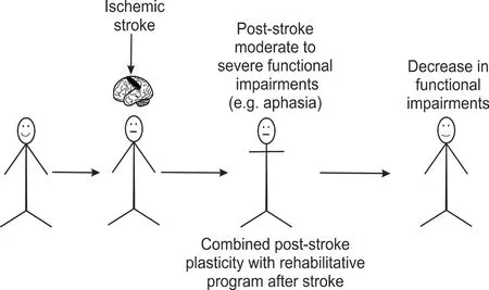
Figure 1 Summary of literature of stroke affected patients and model systems who experience moderate to severe functional impairments, including aphasia or motor impairments.Participation in rehabilitative programs of stroke combined with intrinsic neuroplasticity may reduce functional impairments.
Our experimental data, along with the literature suggests that a neurologic rehabilitation program after stroke should maximize a patients' physical, communicative, and cognitive functioning(Figure 1). A better understanding of the mechanisms involved in how rehabilitation after stroke changes the brain are also needed, so that these factors can be monitored during rehabilitation programs.Technological developments (e.g. tablets or robots) and advancements in imaging may help with stroke therapeutic development(Dobkin and Dorsch, 2013).
The work was funded by the Natural Sciences and Engineering Research Council (NSERC) Canada (to NMJ).
Mahira Moftah, Nafisa M. Jadavji*
Department of Neuroscience, Carleton University, Ottawa, ON,Canada
*Correspondence to: Nafisa M. Jadavji, PhD,nafisa.jadavji@mail.mcgill.ca.
orcid: 0000-0002-3557-7307 (Nafisa M. Jadavji)
Received: January 5, 2019
Accepted: March 18, 2019
doi: 10.4103/1673-5374.255967
Copyright license agreement: The Copyright License Agreement has been signed by both authors before publication.
Plagiarism check: Checked twice by iThenticate.
Peer review: Externally peer reviewed.
Open access statement:This is an open access journal, and articles are distributed under the terms of the Creative Commons Attribution-NonCommercial-ShareAlike 4.0 License, which allows others to remix, tweak, and build upon the work non-commercially, as long as appropriate credit is given and the new creations are licensed under the identical terms.
Open peer reviewer: Noela Rodriguez-Losada, University of Malaga, Spain.
杂志排行
中国神经再生研究(英文版)的其它文章
- Novel miRNA, miR-sc14, promotes Schwann cell proliferation and migration
- Neuromodulation and ablation with focused ultrasound - toward the future of noninvasive brain therapy
- Remodeling dendritic spines for treatment of traumatic brain injury
- Acute drivers of neuroinflammation in traumatic brain injury
- More than anti-malarial agents: therapeutic potential of artemisinins in neurodegeneration
- Why microglia kill neurons after neural disorders?The friendly fire hypothesis
