More than anti-malarial agents: therapeutic potential of artemisinins in neurodegeneration
2019-07-17BingWenLuLarryBaumKwokFaiSoKinChiuLiKeXie
Bing-Wen Lu , Larry Baum , Kwok-Fai So, , Kin Chiu, , Li-Ke Xie
1 Department of Ophthalmology, Eye Hospital, China Academy of Chinese Medical Sciences, Beijing, China
2 Department of Ophthalmology, Li Ka Shing Faculty of Medicine, The University of Hong Kong, Hong Kong Special Administration Region,China
3 State Key Laboratory of Brain and Cognitive Sciences, The University of Hong Kong, Hong Kong Special Administration Region, China
4 Center for Genomic Sciences, Li Ka Shing Faculty of Medicine, The University of Hong Kong, Hong Kong Li Ka Shing Faculty of Medicine, Hong Kong Special Administration Region, China
5 Department of Psychiatry, Li Ka Shing Faculty of Medicine, The University of Hong Kong, Hong Kong Li Ka Shing Faculty of Medicine, Hong Kong Special Administration Region, China
6 GHM Institute of CNS Regeneration, Jinan University, Guangzhou, Guangdong Province, China
Abstract Artemisinin, also called qinghaosu, is originally derived from the sweet wormwood plant (Artemisia annua),which is used in traditional Chinese medicine. Artemisinin and its derivatives (artemisinins) have been widely used for many years as anti-malarial agents, with few adverse side effects. Interestingly, evidence has recently shown that artemisinins might have a therapeutic value for several other diseases beyond malaria,including cancers, inflammatory diseases, and autoimmune disorders. Neurodegeneration is a challenging age-associated neurological disorder characterized by deterioration of neuronal structures as well as functions, whereas neuroinflammation has been considered to be an underlying factor in the development of various neurodegenerative disorders, including Alzheimer's disease. Recently discovered properties of artemisinins suggested that they might be used to treat neurodegenerative disorders by decreasing oxidation,inflammation, and amyloid beta protein (Aβ). In this review, we will introduce artemisinins and highlight the possible mechanisms of their neuroprotective activities, suggesting that artemisinins might have therapeutic potential in neurodegenerative disorders.
Key Words: artemisinin; inflammation; neuroinflammation; neurodegeneration; Alzheimer's disease;Parkinson's disease; Aβ; anti-oxidation; neuroprotection; neural regeneration
Introduction
Artemisinin and its derivatives from the sweet wormwood plant (Artemisia annua, Asteraceae) are called artemisinins;they have been used in traditional Chinese medicine for a long time (Tu, 2011; Guo, 2016; Wong et al., 2017). The plant was first recognized by the Chinese physician Hong Ge (born in the year 283) for its fever-reducing property (Efferth,2017). Youyou Tu, at the China Academy of Traditional Chinese Medicine, isolated artemisinin (Artemisinin structure research collaboration group, 1977; Tu, 2011; Guo, 2016)and tested it in clinical trials (Tu, 2016). Studies conducted in humans during 1980s-1990s led to the designation of artemisinins as a first-line treatment for malaria and helped Youyou Tu win the 2015 Nobel Prize in Physiology or Medicine (Chen, 2016). Artemisinin-based combination therapies have joined the currently established standard treatments of malarial parasites around the world (Qin et al., 2017).Interestingly, abundant evidence has recently accumulated demonstrating that artemisinins might also be useful for many other diseases beyond malaria, including cancers, inflammatory diseases, and autoimmune disorders (Raffetin et al., 2018).
Neurodegeneration is a type of neurological disorder that mainly occurs in the aging population and is characterized by deterioration of neuronal structures as well as functions(Kreiner, 2018). Neurodegenerative diseases, including Alzheimer's disease (AD) and Parkinson's disease, are currently major challenges around the world (Olanow et al., 2009).The causes for neurodegenerative disorders are still not fully known, but might include excitotoxicity, oxidative stress,inflammation, and apoptosis (Alexander, 2017; Feldmann et al., 2019; Toosi et al., 2019). Signs of neuroinflammation have been proven in many AD mouse models before Aβ accumulation (Camara and De-Souza, 2018). Thus, neuroinflammation is now widely regarded as an important pathogenic factor of neurodegenerative disorders (Braidy and Grant, 2017; McCauley and Baloh, 2018; Niranjan, 2018).
Newly discovered properties of artemisinins suggest that they might be able to treat neurodegenerative diseases(Okorji et al., 2016; Zheng et al., 2016; Yan et al., 2017; Zeng et al., 2017). This review will explore this evidence.
A literature review was conducted in February 2019 using the PubMed. Using the key word “artemisinins”, 632 relevant publications published from 1956 to 2019 were retrieved.Among them, around 60 publications were examined using the key words “inflammation” OR “neuro-” OR “oxidation”.
Chemical Structures and Pharmacological Actions of Artemisinins
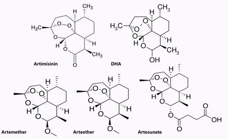
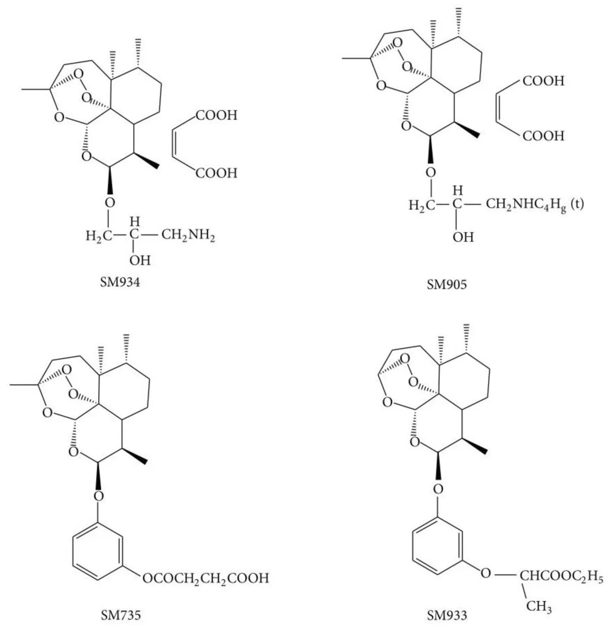
It was Youyou Tu who first deduced the structure of artemisinin. Using mass spectroscopy, spectrophotometry and X-ray crystallography, she discovered that artemisinin is a sesquiterpene lactone endoperoxide (Tu, 2016). The clinically important artemisinins include artesunate, artemether, and dihydroartemisin (DHA) (Figure 1), which were discovered and developed in 1986 (Nair et al., 1986). Today, these derivatives are more commonly used than artemisinin itself to treat malaria because of the minimal adverse effects, as well as their better efficacy and tolerability (Pinheiro et al., 2018).Artesunate is the most important analog, displaying a more favorable pharmacological profile than artemisinin because of its greater water-solubility and higher oral bioavailability that enable it to act more rapidly (Pinheiro et al., 2018). Recently, novel artemisinin derivatives have also been designed.These are named SM, each with a specific number to identify it (Figure 2).
Neuroprotective Activity of Artemisinins
Artemisinins have been shown to have neuroprotective effects, and therefore they are potential candidates for the treatment of neurodegenerative disorders (Okorji et al.,2016; Zheng et al., 2016; Yan et al., 2017; Zeng et al., 2017).Other factors in their favor as potential treatments are their abilities to cross the blood-brain barrier as small lipophilic molecules and to protect against oxidative stress (Zheng et al., 2016).
Anti-oxidation
Artemisinin has been shown to have a neuroprotective effect on sodium nitroprusside-induced oxidative insult to PC12 cells and brain primary cortical neurons (Zheng et al., 2016).PC12 cells were pretreated with artemisinin of different concentrations (3.1-100 μM) before their exposure to sodium nitroprusside (800 μM). Cell viability was protected in a concentration-dependent manner with artemisinin pretreatment, as measured by MTT assay, caspase 3/7 activities, and by lactate dehydrogenase release. Extracellular signal-regulated kinases (ERK) is part of the mitogen-activated protein kinase (MAPK) family, one of the oldest families of serine/threonine protein kinases responsible for intracellular signaling (Bohush et al., 2018). Their results demonstrated that the ERK pathway could be activated by artemisinin (25 μM),as shown by western blot analysis, whereas PD98059, an ERK inhibitor, could block the protective effect displayed by artemisinin (Zheng et al., 2016).
Previous studies proposed dysfunctional cyclic-AMP response element binding protein (CREB) signaling in various mouse models of AD (Bartolotti et al., 2016; Ettcheto et al.,2018). Many potential therapeutic approaches targeting CREB signaling have been studied for treatment of neurodegenerative disorders (Motaghinejad et al., 2017). Chong and Zheng (2016) demonstrated that artemisinin was able to suppress oxidative stress induced by hydrogen peroxide(H2O2) in D407 retinal pigment epithelial cells and that activation of ERK/CREB signaling was involved. Yan et al.(2017) demonstrated that artemisinin was able to protect retinal neuronal cells RGC-5 against oxidative insult induced by H2O2via activation of the ERK1/2 pathway. Their flash electroretinogram results also interestingly found that, in a concentration-dependent manner, artemisinin injected intravitreally could protect retinal function damaged by light exposure.

Figure 1 Chemical structures of early artemisinin derivatives.

Figure 2 Chemical structures of newly developed artemisinin derivatives.SM735: 3-(12-β-ARTEMISININOXY) phenoxyl succinic acid (Zhou et al., 2005); SM905: 1-(12-β-dihydroartemisinoxy)-2-hydroxy-3-tert-butylaminopropane maleate (Wang et al., 2007); SM933: ethyl 2-[4-(12-β-artemisininoxy)] phenoxylpropionate (Zhao et al., 2012);SM934: 2'-aminoarteether (β)maleate (Hou et al., 2009).
Artemisinin has also been shown to protect neuronal HT-22 mouse hippocampal cells from glutamate-induced neuronal oxidative damage and cell death (Lin et al., 2018),with 25 μM producing the optimum protective effect. Their results further suggest that pretreatment of artemisinin could activate protein kinase B (Akt)/Bcl-2 signaling because MK2206, an Akt inhibitor, could block its protective effect.
Besides being linked with memory consolidation (Horwood et al., 2006), phosphoinositide 3-kinase/Akt (which artemisinins inhibit) is associated with a number of metabolic functions that are essential for neuronal viability but dysfunctional in AD (Cheng et al., 2011; De Felice, 2013).Moreover, mammalian target of rapamycin (mTOR) signaling is activated by Akt and strongly correlated with the presence of Aβ and tau protein that accumulate in AD (Caccamo et al., 2011). However, the role of mTOR in AD remains controversial because increased Aβ concentrations might increase mTOR signaling, but even higher Aβ concentrations can decrease mTOR signaling (Lafay-Chebassier et al., 2005).
Artesunate could mimic caloric restriction and extend lifespan in yeast (Wang et al., 2015). Downregulation of MAPK signaling was observed in their study, indicating a relationship between cytochrome c oxidase activation and artesunate-heme conjugation.
To sum up, the above results indicate that artemisinin may have neuroprotective effects against oxidative stress through modulating various signaling pathways, most importantly,the ERK, CREB, MAPK, and Akt/mTOR pathways.
Anti-inflammation
Suppression of inflammation is another effect of artemisinin derivatives. Because of the favorable pharmacological actions of artemisinins on various signaling pathways as well as their relatively safe properties, they have been recently studied in various models of inflammatory disorders (Shi et al., 2015;Tu, 2016).
Artemisinins have been demonstrated to suppress inflammatory responses through the suppression of many pro-inflammatory cytokines (Lin et al., 2016; Feng et al., 2017; Li et al., 2018; Lu et al., 2018; Qiang et al., 2018; Sun et al., 2018).
Nuclear factor kappa B (NF-κB) is involved in neurodegenerative disorders such as AD (He et al., 2018). Aβ neurotoxicity has been found to be closely linked to NF-κB activation (Bourne et al., 2007). Therefore, inhibiting NF-κB might treat neurodegenerative disorders (Kim et al., 2017; Subedi et al., 2017; Zhang and Xu, 2018). Another recent study showed that artemisinin B, an artemisinin derivative without a dioxygen bridge, could alleviate learning and memory impairment in AD through inhibiting neuro-inflammatory responses (Qiang et al., 2018). Both the in vivo and in vitro studies have demonstrated that artemisinin B might play its anti-neuroinflammatory role through regulating the toll-like receptor 4-myeloid differentiation factor 88/NF-κB pathway.In a mouse model of experimental cerebral ischemia/reperfusion injury, a water-soluble derivative of artemisinin, artesunate (10-40 mg/kg) was shown to attenuate inflammatory processes through activating nuclear factor erythroid 2-related factor 2 (Nrf2), which is an important transcription factor that activates anti-oxidation leading to neuroprotection, as well as through suppressing ROS-dependent p38 MARK (Lu et al., 2018).
Nrf2 activation and the up-regulation of antioxidant and anti-inflammatory genes have been suggested by a variety of studies to be attractive therapeutic targets to prevent neurodegenerative disorders (Abdalkader et al., 2018; Morroni et al., 2018). In a study of BV2 microglia, a lipid-soluble derivative of artemisinin, artemether, has been shown to induce Nrf2 activity via increasing its nuclear translocation as well as its binding to antioxidant response elements, suggesting a possible therapeutic neuroprotective effect (Okorji et al.,2016). Pretreatment by artemether (5-40 μM) was found to reduce, in a dose-dependent fashion, the production of nitrite as well as the expression of tumor necrosis factor-alpha, prostaglandin E2, and interleukin-6 in lipopolysaccharide-stimulated BV2 microglia co-cultured with HT22 neuronal cells. Also, their findings suggested that artemether might exert its anti-inflammatory effect through suppressing NF-κB and p38 MAPK signaling.
Anti-Aβ
Aβ peptide, which accumulates in AD, can be neurotoxic. Artemisinin can protect or rescue PC12 cells from cell death induced by a toxic fragment, Aβ 25-35 (Zeng et al.,2017). Treatment with 12.5 or 25 μM artemisinin for 1 hour reduced the death of PC12 cells after subsequent exposure to Aβ 25-35 for 24 hours. Reversing the order of exposure also reduces cell death, with 25 or 50 μM artemisinin for 24 hours rescuing PC12 cells that had been incubated with 0.1,0.3, or 1 μM Aβ 25-35 for 30 minutes. Further study showed that low-concentration artemisinin might be able to promote neuronal differentiation of PC12 cells through activating the ERK1/2 and p38 MAPK signaling pathway (Sarina et al.,2013). These results suggest the potential application of artemisinin as a novel treatment of neurodegenerative disorders,especially AD.
Challenges for Use of Artemisinins in Neurodegeneration
Although artemisinin seems to be a pluripotent drug with possible clinical value in anti-malarial, anti-tumor, anti-inflammatory and anti-neurodegenerative roles, there are potential pitfalls (Yuan et al., 2017). In contrast to short-term high-dose artemisinin treatment against malaria, long-term treatment with low-dose artemisinin has generally been investigated for treatment of other diseases (Wang et al., 2015).However, long-term low-dose exposure to artemisinin might induce free radical scavengers that can destroy the vulnerable endoperoxide bridge structure within artemisinin (Sun and Zhou, 2017). Furthermore, unexpected metabolic dysfunctions or other abnormalities, including neurotoxicity, or genotoxicity due to sperm DNA damage, might also emerge upon excessive artemisinin use (Singh et al., 2015). Therefore, the effects of artemisinin on neurodegeneration, either positive or negative, should be assessed and thorough longterm toxicity testing is performed to ensure safety at the appropriate dose.
Prospect
In the words of the discoverer of artemisinin at the conclusion of her Nobel Lecture, “Expanding clinical applications of artimisinin is also of interest to public health. We know what it can do, we need to know why and how it does it,what else it can do, and how it can do better.” (Tu, 2016). Artemisinins have been demonstrated to alleviate neurodegeneration through reducing oxidation, inflammation, and Aβ toxicity, in both direct and indirect manners. However, research on artemisinins in neurodegenerative disorders is still limited, and much more will need to be studied. A better understanding of the neuroprotective activities of artemisinins might improve treatments of neurodegenerative disorders.
Author contributions:Manuscript writing: BWL; providing suggestions for the manuscript and manuscript revision: LB; discussions: KC, LKX,KFS; approval of final manuscript for publication: all authors.
Conflicts of interest:There are no conflicts of interest associated with this manuscript.
Financial support:This work was supported by the Natural Science Foundation of Beijing of China, No. 7192235 (to LKX). The funding body played no role in the manuscript preparation other than providing funding.
Copyright license agreement:The Copyright License Agreement has been signed by all authors before publication.
Plagiarism check: Checked twice by iThenticate.
Peer review:Externally peer reviewed.
Open access statement:This is an open access journal, and articles are distributed under the terms of the Creative Commons Attribution-Non-Commercial-ShareAlike 4.0 License, which allows others to remix, tweak,and build upon the work non-commercially, as long as appropriate credit is given and the new creations are licensed under the identical terms.
杂志排行
中国神经再生研究(英文版)的其它文章
- Novel miRNA, miR-sc14, promotes Schwann cell proliferation and migration
- Neuromodulation and ablation with focused ultrasound - toward the future of noninvasive brain therapy
- Remodeling dendritic spines for treatment of traumatic brain injury
- Acute drivers of neuroinflammation in traumatic brain injury
- Why microglia kill neurons after neural disorders?The friendly fire hypothesis
- Could autophagy dysregulation link neurotropic viruses to Alzheimer's disease?
