Unexplained abdominal pain due to a fish bone penetrating the gastric antrum and migrating into the neck of the pancreas:A case report
2019-04-17RuiXieBiGuangTuoHuiChaoWu
Rui Xie,Bi-Guang Tuo,Hui-Chao Wu
Abstract
Key words: Unexplained abdominal pain;Fish bone;Gastrointestinal perforation;Pancreas;Case report
INTRODUCTION
Foreign body ingestion is not an uncommon problem in clinical practice,and most ingested foreign bodies (80%-90%) pass spontaneously.Approximately 10%-20% of foreign bodies require an endoscopic procedure,and less than 1% require surgery[1-3].Fish bones are the most commonly observed foreign objects;they may cause gastrointestinal perforation due to their sharp edges,and perforation generally occurs at the ileum[4].The fish bone may also penetrate the digestive tract and pierce the liver or intra-abdominal area,leading to abscess formation[5].In such cases,unexplained abdominal pain is the most frequent clinical symptom.We present the case of a 32-year-old man who was successfully treated by laparoscopic surgery to identify and remove a fish bone that had penetrated the gastric antrum and migrated into the neck of the pancreas,causing abdominal pain.
CASE PRESENTATION
Chief complaints
A 32-year-old male presented to our hospital because of abdominal pain that had worsened over 5 d on March 5,2018.
Personal and family history and physical examination upon admission
He had rebound tenderness in the upper abdominal quadrant,without hematemesis or black stool,and the patient had no significant past history or family history.
Laboratory examinations
Laboratory tests revealed a white blood cell count of 11.50/mm3and occult blood in the stool,with no other main abnormalities,including amylase.
Imaging examinations
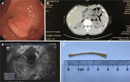
Upper endoscopy performed on March 6,2018 revealed an irregular submucosal tumor on the front wall of the gastric antrum with an area of 1.5 cm × 1.7 cm and a depressed appearance of the mucosal surface.Endoscopic ultrasonography showed abnormal and irregular thickening of the stomach wall characterized by a hypoechoic area,and an approximately 3.5-cm linear and hyperechoic lesion protruding through the thickened stomach wall and the neck of the pancreas.Computed tomography (CT)revealed that the thickened front wall of the stomach abutted against the pancreatic body,with a laterally oriented radiopaque foreign body inside(Figure 1).Considering the suspicion of foreign body ingestion,the patient was questioned,and he remembered that he had eaten fish 1 wk previously.
FINAL DIAGNOSIS
Abdominal pain resulting from a fish bone penetration of the stomach and migrating into the pancreas.

Figure1 lmages of the patient.
TREATMENT
We wanted to cut the mucosa and remove the embedded fish bone by endoscopic submucosal dissection on March 8,2018,but endoscopic ultrasonography showed that the fish bone was too deep to be easily removed.Finally,the patient underwent laparoscopic surgery seven days after hospital admission (March 12,2018),which revealed a thickened peritoneum and adhesion between the gastric wall and the pancreatic neck.We carefully separated the stomach wall and the pancreas,and after identifying a penetration of the serosa of the stomach wall,we found a fish bone of approximately 3.5 cm in length pinned in the neck of the pancreas.The fish bone was successfully removed,and a proton-pump inhibitor was routinely used for five days after surgery.
OUTCOME AND FOLLOW-UP
The patient was discharged without any complications and was in good clinical condition one week after the operation,and endoscopic reexamination had not found obvious abnormality one month after the surgery.
DISCUSSION
Unintentional,unconscious ingestion of foreign bodies in adults is usually dietary.Nearly two-thirds of foreign bodies are fish bones,and 75% of ingested foreign bodies become impacted in the oral cavity and laryngopharynx.If impaction does not occur in the upper gastrointestinal tract,the majority of foreign bodies pass asymptomatically within a week[6].Perforation of the digestive tract by an ingested fish bone is extremely rare (< 1%);when it does occur,the terminal ileum is the most common site of perforation,followed by the duodenal C-loop[4].Because patients usually cannot recall any recent history of foreign body ingestion and because clinical symptoms are nonspecific,gastrointestinal perforation may present as only odynophagia or abdominal pain.If the injury is not observed,a definite preoperative diagnosis is uncertain,and clinical intervention may be delayed.
Numerous reports of ingested fish bones penetrating the digestive tract and migrating to various parts of the chest,liver,or abdominal cavity can be found in the literature.These bones may be responsible for various complications,such as abscess,abdominal cavity infection,mediastinitis,and empyema[7].However,to date,only rare cases of fish bone migration to the pancreas have been described in the literature,and this injury may present as a pancreatic mass or suppurative infection of the pancreas[8,9].Laboratory analyses are nonspecific,and leukocytosis or increased blood amylase levels are observed.In our case,because of the inflammatory response and the adhesion of the surrounding tissue,perforation occurred in the stomach,where the thicker gut wall and the proximity of the omentum may have sealed the perforation;consequently,the patient did not present the classic symptoms of digestive tract perforation,and immediate correct preoperative diagnosis was very difficult.CT and endoscopic ultrasonography may be helpful for revealing the nature of foreign bodies,the location of migrated foreign bodies,and the relationship with surrounding tissues[10,11];therefore,imaging examinations can provide key information for delayed diagnosis of unexplained abdominal pain caused by foreign bodies.Laparoscopy was successfully used to identify the fish bone and extract it.In this case,the patient recovered without any postoperative complications.
CONCLUSION
Ingestion of foreign bodies is a common clinical problem,of which the fish bone is one of the most common;however,involvement of the pancreas is very rare.Perforation occurs in the stomach where a thicker gut wall and proximity of the omentum may seal the perforation,and clinical symptoms are nonspecific.Thus,a definite preoperative diagnosis and clinical intervention may be delayed.CT and endoscopic ultrasonography may be helpful for revealing the nature of foreign bodies,the location of migrated foreign bodies,and the relationship with surrounding tissues.Acute perforations usually require emergency surgery when the diagnosis becomes apparent.Laparoscopic surgery is the most effective option,and disease outcome is generally good after treatment.
杂志排行
World Journal of Clinical Cases的其它文章
- Photodynamic therapy as salvage therapy for residual microscopic cancer after ultra-low anterior resection:A case report
- Effective chemotherapy for submandibular gland carcinoma ex pleomorphic adenoma with lung metastasis after radiotherapy:A case report
- Primary hepatic follicular dendritic cell sarcoma:A case report
- Multiple gastric angiolipomas:A case report
- Lump type crossed fused renal ectopia with bilateral vesicoureteral reflux:A case report
- lnduction chemotherapy with docetaxel,cisplatin and fluorouracil followed by concurrent chemoradiotherapy for unresectable sinonasal undifferentiated carcinoma:Two cases of report
