Characteristics of multiple nodules in a patient with pulmonary Langerhans cell histiocytosis:A case report
2019-03-14NobuhiroKanajiYoshimasaTokunagaRyouIshikawaNaokiWatanabeNorimitsuKadowaki
Nobuhiro Kanaji,Yoshimasa Tokunaga,Ryou Ishikawa,Naoki Watanabe,Norimitsu Kadowaki
Abstract
Key words:Langerhans cell histiocytosis;Multiple;Non-cystic;Nodule;Distribution;Size;Case report
INTRODUCTION
Langerhans cell histiocytosis (LCH) is characterized by accumulations of large mononuclear cells named Langerhans cells that form granulomas in various organs such as bone,skin and lung[1-3].LCH can be clinically classified into three groups:Single-system,low-risk multisystem,and multisystem with risk-organ involvement[4,5].Pulmonary Langerhans cell histiocytosis (PLCH) is a diffuse lung disease whose clinical manifestations may involve a single organ or multisystem[4].The common findings of PLCH on chest computed tomography (CT) scan are multiple cysts predominantly in upper lung zones and a micronodular pattern of the middle-upper lobes[4].Non-cystic nodules and large nodules are rare in PLCH.Such characteristics of multiple nodules are common in metastatic cancers.
We here present a case of PLCH with multiple nodules including the non-cystic nodules > 20 mm in dia.We assessed the characteristics of multiple nodules including the number,distribution and sizes of cystic and non-cystic nodules.
CASE PRESENTATION
Chief complaints
A medical examination revealed an abnormal shadow on the chest radiograph of a 48-year-old Japanese man with no symptoms.
History of present illness
He visited to our hospital immediately after an abnormal shadow was pointed out.
History of past illness
He had been diagnosed with primary aldosterone disease and hypertension and had continued with treatment with tablets of potassium chloride,spironolactone,telmisartan and amlodipine besilate.He had no other notable medical history.
Personal and family history
He was a care assistant in a hospital and a current smoker (20 cigarettes/d for the past 28 years).He had no serious family history.
3.0 T MR磁敏感加权成像对脑干微量出血的诊断价值 … ………………………… 马清,许云,吴婷,等 207
Physical examination
The physical examination revealed no remarkable abnormalities.Normal breath sounds and no adventitious sounds were auscultated.
Laboratory examinations
Laboratory examinations revealed a low concentration of serum potassium (3.0 mmol/L).The C-reactive protein level was 0.58 mg/dL.There were no other abnormal findings,including circulating blood cell counts,blood biochemistry and routine urine tests.
Imaging examinations
The chest radiograph and CT showed multiple nodules in both lung fields (Figure 1).Although the nodules seemed to be distributed randomly,fewer nodules were adjacent to the pleura,and some were adjacent to bronchus (Figure 1B and C).Nodules existed from upper to lower lobes,dominantly in the right lower lobes(Figure 1B and C).Most of the nodules were non-cystic,and the small nodules were cystic (Figure 1C).The largest nodule was 22 mm in dia.,in the right middle lobe(Figure 1D).Mediastinal lymph nodes were approx.10 mm in size (not shown).The differential diagnosis included metastatic lung tumors,lymph proliferative disorder,granulomatous polyangiitis,and PLCH.
FINAL DIAGNOSIS
Transbronchial biopsy specimens showed non-specific features;only inflammatory cells (not shown).Several nodules in the right lower lobe were resected under thoracoscopic surgery,and tissue specimens showed abnormal cells with notched nuclei with the infiltration of many eosinophils (Figure 2A and B).Abnormal cells were positive for CD1a and S-100 (Figure 2C and D) and negative for CD68 and CD21(not shown).Based on these findings,we made the diagnosis of PLCH.
TREATMENT
The patient began to quit smoking after we encountered him.He received no additional medical treatment.
OUTCOME AND FOLLOW-UP
He continued the smoking cessation,and 6 months later,all nodules had almost disappeared (Figure 3).
INVESTIGATION OF CHARACTERISTICS OF MULTIPLE NODULES
To investigate the patient's CT findings at the diagnosis in detail,we counted the number of nodules in all lung lobes and classified the nodules into cystic and noncystic nodules (Figure 4).Of the total of 349 nodules,116 were in upper lobes and 199 were in lower lobes (right plus left lobes).Ninety-six (27.5%) were cystic,and the remaining 253 (72.5%) were non-cystic.The prevalence of cystic nodules was higher in upper lobes than in lower lobes (Figure 4A;right upper 37.5%vslower 18.2%,P=0.0068;left upper 48.1%vslower 24.4%,P= 0.0078).The average size (dia.) of cystic nodules was smaller than that of non-cystic nodules (5.03 mmvs7.40 mm,respectively;P< 0.0001,Figure 4B).
DISCUSSION
Our patient was diagnosed with PLCH and exhibited the following rare patterns on CT scans:(1) Multiple nodules dominantly in lower lobes;(2) Nodules > 20 mm in dia.Our investigation of the CT findings in detail revealed that (3) The prevalence of cystic nodules was higher in upper lobes than in lower lobes;and (4) The cystic nodules were smaller than the non-cystic nodules.

Figure1 Radiological findings at the initial diagnosis.
The two common CT findings of PLCH are multiple cysts and micronodules predominantly in middle-to-upper lung zones[4].Either one or both of the findings were recognized in all 40 of the reported cases[4].Other abnormal findings include patchy or cord shadows,pleural thickening,pleural effusion,and mediastinal lymph node enlargement[5].PLCH cases with multiple nodules without cystic formation have also been reported[6-8].However,the sizes of the nodules in those cases were usually <10 mm[6-8].Thus,the non-cystic large nodules observed our patient are rare in PLCH.In addition,a greater number of nodules were found in lower lobes than in upper lobes.Such characteristics are common in metastatic lung tumors (cancers) because they usually developviaa hematogenous route and the bloodstream is more abundant in lower lobes,although transbronchial dissemination in lung cancer was also occasionally reported[9].
Many studies have shown that a subset of LCH has genetic mutations including VRaf murine sarcoma vial homolog B1 (BRAF)[3,10].BRAF V600E was detected in approximately one-half of patients with LCH[10].MAPKK1 mutations were detected in 6 of 13 (46%) BRAF-negative cases[3].These findings suggest that LCH has some genetic monoclonality,which was considered to be a type of tumor.
Langerhans cells can develop in organs such as bone,skin,brain,liver and lung[1-3],suggesting that they can spread to the whole body hematogenously.It is possible that some of the nodules observed in the present case developed as a result of hematogenous metastases.Consistent with this,the CT findings in several prior cases with multiple nodules showed a distribution pattern mimicking hematogenous pulmonary metastases[7,8].We speculate that the distribution pattern as hematogenous lung metastases is consistent with PLCH as well as metastatic cancers.
There are several notable points for the differential diagnosis of PLCH from metastatic cancers in the present case.First,the distribution pattern of multiple nodules was not a completely hematogenous pattern,and some nodules were distributed around or adjacent to bronchi.It was reported that many small nodules were distributed in the centers of secondary lobules around small airways in PLCH[11].It is quite unusual for a plurality of primary tumors to occur simultaneously.However,the existence/non-existence of a transbronchial dissemination of Langerhans'cells remains unclear.
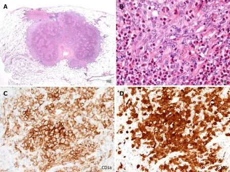
Figure2 Histopathological findings obtained from a nodule in right lower field.
Second,there were small cystic nodules.In cases of metastatic cancers,cystic changes tend to appear in larger nodules rather than in smaller nodules.Based on the follow-ups of PLCH cases,it was reported that nodular lesions transform into cysts[6].In progressed cases,cystic changes fuse and develop emphysema-like lesions.However,in our patient's case,the cystic nodules were smaller than the non-cystic nodules,and there were no fused cystic nodules.On the other hand,some non-cystic nodules fused and formed a snowman-like shape.In the present case,cystic changes inside nodules might appear at an early phase and disappear at a late phase at which Langerhans' cells proliferate and form a large,filled nodule.
Third,another important finding concerning the differential diagnosis between PLCH and metastatic malignant tumors is the patient's symptoms.Patients with advanced cancer often present subjective symptoms such as anorexia,body weight loss,fever,and fatigue.Our patient had no symptoms.PLCH as well as pulmonary alveolar proteinosis[12]and sarcoidosis[13]is a disease by which a symptom may relatively lack even though the evidence of the extensive radiological findings.
We counted the number of nodules and evaluated the ratio of cystic to non-cystic nodules visually.However,the computer-aided automatic evaluation of radiological findings may bring the diagnosis that is more exact than a human evaluation.Recently,trials of deep learning-based diagnosis of pulmonary nodules have been reported[14,15].The introduction of such artificial intelligence in the medical settings will be realized in the near future.
CONCLUSION
Multiple non-cystic nodules including large nodules (> 20 mm) can occur in PLCH.Knowledge of atypical findings in PLCH,taken together with other findings (e.g.,cystic formation in small nodules dominantly in upper lobes and no symptom) may help pulmonologists consider PLCH in differential diagnoses.
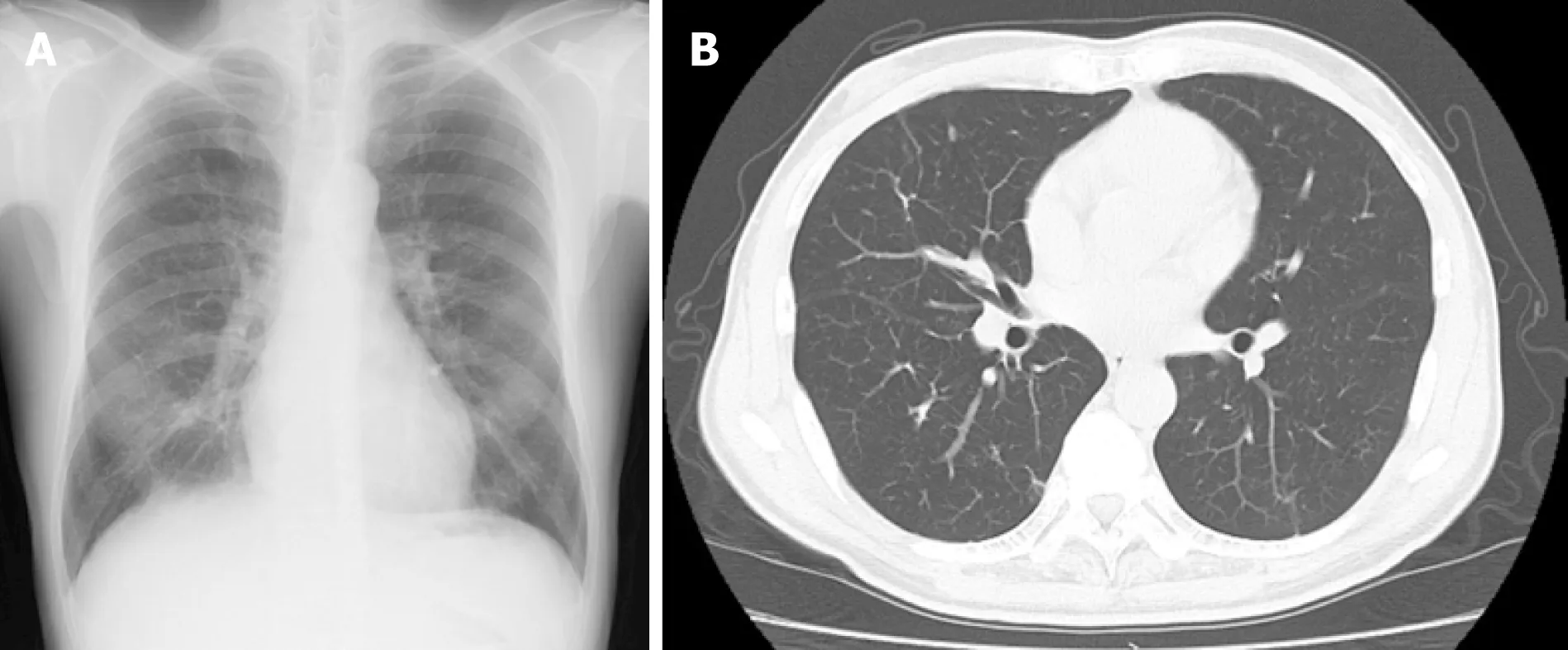
Figure3 Radiological findings at 6 mo after the patient quit smoking.
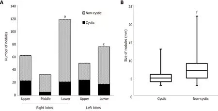
Figure4 Numbers and sizes of cystic and non-cystic nodules.
猜你喜欢
杂志排行

World Journal of Clinical Cases的其它文章
- Multifocal G1-G2 gastric neuroendocrine tumors:Differentiating between Type I,II and III,a clinicopathologic review
- Attention deficit hyperactivity disorder and comorbidity:A review of literature
- Dietary manipulation and testosterone replacement therapy may explain changes in body composition after spinal cord injury:A retrospective case report
- Risk factors,clinical features,and short-term prognosis of spontaneous fungal peritonitis in cirrhosis:A matched case-control study
- Incidence of portal vein thrombosis after splenectomy and its influence on transjugular intrahepatic portosystemic shunt stent patency
- Multiplex gene expression profile in inflamed mucosa of patients with Crohn’s disease ileal localization:A pilot study