Effect of moxibustion at Shenque (CV 8) on free radical metabolism in rat’s hippocampus after different degrees of exhaustive exercise
2018-09-12ZhouXiaohong周小红DuXiaoyi杜潇怡ZhuJie祝婕GaoFei高飞ZhangZhifang张治方XuXiaokang许晓康LiangYulei梁玉磊SunLihong孙立虹SunHao孙浩
Zhou Xiao-hong (周小红), Du Xiao-yi (杜潇怡), Zhu Jie (祝婕), Gao Fei (高飞), Zhang Zhi-fang (张治方),Xu Xiao-kang (许晓康), Liang Yu-lei (梁玉磊), Sun Li-hong (孙立虹), Sun Hao (孙浩)
Abstract Objective: To investigate the effect of moxibustion at Shenque (CV 8) on free radical metabolism in rat’s hippocampus after different degrees of exhaustive exercise.Methods: A total of 72 male Sprague-Dawley (SD) rats were randomly divided into a normal group (n=8), a model group(n=32) and a moxibustion group (n=32). According to the times of modeling or treatment, the model group was further randomly divided into different subgroups of a 1-time model subgroup, a 4-time model subgroup, a 7-time model subgroup and a 10-time model subgroup (n=8); the moxibustion group was also further randomly divided into different subgroups of a 1-time moxibustion subgroup, a 4-time moxibustion subgroup, a 7-time moxibustion subgroup and a 10-time moxibustion subgroup (n=8). Rats in both model and moxibustion subgroups were subjected to establishing the swimming exhaustive model. Rats in each moxibustion subgroup received mild moxibustion at Shenque (CV 8) for 15 min immediately after modeling, once every other day. The concentration of malic dialdehyde (MDA), as well as the activities of superoxide dismutase (SOD), glutathione peroxidase (GSH-Px) and total antioxidant capacity (T-AOC) in rat’s hippocampus in each group were detected 24 h after the exhaustive exercise.Results: Compared with the 1-time model subgroup, the exhaustive swimming time of rats was significantly prolonged in the 4-time model subgroup (P<0.01), while it was significantly shortened in the 7-time and 10-time model subgroups (both P<0.01). Compared with the matched model subgroup, the exhaustive swimming time of rats in the 7-time and 10-time moxibustion subgroups was significantly prolonged (both P<0.01). Compared with the normal group, the MDA concentration was increased significantly (P<0.01), and the activities stress response of SOD and T-AOC were increased in the 1-time model subgroup (both P<0.05); the MDA concentration was increased (all P<0.01), and the activities of SOD,GSH-Px and T-AOC were decreased differently (P<0.05 or P<0.01) in the 4-time, 7-time and 10-time model subgroups.Compared with the matched model subgroup, the concentration of MDA was significantly reduced (P<0.05 or P<0.01), and the activities of SOD, GSH-Px and T-AOC were significantly increased in the 4-time, 7-time and 10-time moxibustion subgroups (all P<0.01).Conclusion: Moxibustion at Shenque (CV 8) can improve the fatigue status of the body after long-term exhaustive exercise by regulating free radical metabolism in rat’s hippocampus. To some extent, this provides an experimental basis for moxibustion at Shenque (CV 8) against exercise-induced fatigue.
Keywords: Moxibustion Therapy; Mild Moxibustion; Point, Shenque (CV 8); Exhaustive Exercise; Fatigue; Free Radicals; Rats
Exercise-induced fatigue refers to a temporary decline in exercise capacity caused by physiological changes in the body after exercise. Both high-intensity exercise training and overloaded work can lead to the occurrence of exercise fatigue. The incidence of fatigue was as high as 81.86% according to a survey on the fatigue status in 2 823 healthy population with medical examination conducted by a medical examination center in Beijing in 2013[1]. To prevent exercise-induced fatigue has become a topic of current medical concern.
Recent studies have found that excessive exercise increases free radicals and lipid peroxidation in brain tissues, resulting in imbalance of oxidation and antioxidation, and oxidative damage to brain tissues followed by the exercise fatigue[2-3]. Our previous study found that moxibustion at Shenque (CV 8) effectively prolonged the swimming time; improved the concentrations of serum malondialdehyde (MDA) and blood urea nitrogen (BUN), and the activities of aspartate transaminase (AST), alanine transaminase(ALT) and lactate dehydrogenase (LDH); and relieved the fatigue status of rats after exhaustive exercise[4-5]. To further explore the mechanism of anti-fatigue effect of moxibustion at Shenque (CV 8), rats with different degrees of exhaustion were used to detect the concentration of MDA, activities of SOD, GSH-Px and T-AOC in the hippocampus in our current study.
1 Materials and Methods
1.1 Experimental animals and grouping
A total of 72 clean-grade male Sprague-Dawley (SD)rats, weighing (200±10) g, were purchased from Beijing Weitong Lihua Experimental Animal Technology Co.,Ltd., China [license number: SCXK (Beijing) 2012-0001].Rats were housed in separate cages with 4 in each cage.The temperature of the rearing room was maintained at 20-24 ℃ with the humidity of about 50%, and 12 h light and 12 h darkness (light time was 07:00-19:00). All the rats had free access to water. The rats were fed adaptively for 7 d and then received adaptive swimming on the 3rd and 6th day, respectively, 3 min/time. The whole experiment procedure followed the relevant regulations for laboratory animal management of Hebei University of Chinese Medicine.
The rats were randomly divided into a normal group(n=8), a model group (n=32) and a moxibustion group(n=32) according to the random number table.According to the times of treatment, the model group was randomly divided into a 1-time model subgroup, a 4-time model subgroup, a 7-time model subgroup, and a 10-time model subgroup, with 8 rats in each subgroup;and the moxibustion group was randomly divided into a 1-time moxibustion subgroup, a 4-time moxibustion subgroup, a 7-time moxibustion subgroup, and a 10-time moxibustion subgroup, with 8 rats in each subgroup.
1.2 Main reagents and instruments
Kits of MDA, SOD, GSH-Px and T-AOC (Nanjing Jiancheng Institute of Bioengineering, China); self-made thermostatic water tank (200 cm × 80 cm); moxa stick(specification: 7 mm × 117 mm, Henan Nanyang Chinese Medicine Moxa Co., Ltd., China); homemade moxibustion box for rats (national patent of utility model, patent number: ZL201120193244.8)[6]; TDL-5-A centrifuge (Shanghai Anting Scientific Instrument Factory, China); PT1300D homogenizer (Shanghai Scholar Biotechnology Co., Ltd., China); 722 spectrophotometer (Shanghai Optical Instrument Factory, China).
1.3 Model preparation
After 7 d of adaptive feeding, an exhaustive exercise model was created referring to the literature[7]. Rats in the model group and the moxibustion group were placed in a thermostatic water tank [50 cm deep and temperature at (30±2) ℃], with a piece of lead(weighing 5% of the body weight) wrapping around the tail (at 1-5 cm from the base of the tail] for the exhaustive exercise (the exhaustion criteria: the swimming movement of the rats was obviously out of balance and could no longer be maintained; rat’s nose was under water for 5 s and unable to return to the surface of water).
Rats in the model and moxibustion subgroups were subjected to exhaustive exercise according to the methods mentioned above for the designated times.The exhaustive exercise was performed once every other day. After each exhaustive exercise, rats in the moxibustion group received moxibustion once.
1.4 Intervention method
Normal group: Rats were kept in the moxibustion box without any other intervention, once every other day for 10 times in total, 15 min each time.
The 1-time moxibustion subgroup: After 1-time exhausting swim, the rats were placed in a moxibustion box immediately for mild moxibustion at Shenque(CV 8), 15 min only once[8].
The 4-time moxibustion subgroup: After each exhausting swim, the rats were placed in a moxibustion box immediately for mild moxibustion at Shenque (CV 8)for 15 min every other day, for a total of 4 treatments[8].
The 7-time moxibustion subgroup: After each exhausting swim, the rats were placed in a moxibustion box immediately for mild moxibustion at Shenque (CV 8)for 15 min every other day, for a total of 7 treatments[8].
The 10-time moxibustion subgroup: After each exhausting swim, the rats were placed in a moxibustion box immediately for mild moxibustion at Shenque (CV 8)for 15 min every other day, for a total of 10 treatments[8].
The 1-time model subgroup: After the first exhausting swim, the rats were placed in a moxibustion box immediately for 15 min without moxibustion, only once.
The 4-time model subgroup: After each exhausting swim, the rats were placed in a moxibustion box immediately for 15 min without moxibustion, once every other day, for a total of 4 times.
The 7-time model subgroup: After each exhausting swim, the rats were placed in a moxibustion box immediately for 15 min without moxibustion, once every other day, for a total of 7 times.
The 10-time model subgroup: After each exhausting swim, the rats were placed in a moxibustion box immediately for 15 min without moxibustion, once every other day, for a total of 10 times.
1.5 Indicator detection
1.5.1 Exhaustion time
The last exhaustive swim duration of rats in the model and moxibustion subgroups was recorded by a designated person with the unit of second (being accurate to 0.1 s).
1.5.2 Free radical concentrations in hippocampus
The rats were sacrificed by dislocation (rats in the normal group and in the 10-time subgroups were sacrificed at the same time) 24 h after the exhaustive exercise in each subgroup; the whole brain was quickly removed on ice, washed with ice-cold saline and dried with filter paper. The hippocampus was immediately isolated and stored in liquid nitrogen for later use. The MDA concentration was determined by thiobarbituric acid (TBA) colorimetric analysis, the SOD activity was measured by hydroxylamine method, the activity of GSH-Px was measured by an enzymatic reaction method, and the T-AOC activity was measured by a chemical colorimetric method. All biochemical indicators were examined by the Scientific Research Center of Hebei University of Chinese Medicine.
1.6 Statistical processing
The SPSS version 13.0 software was used for statistical analysis. Measurement data were expressed as mean ± standard deviation (±s). Multiple groups were compared using variance analysis and multiple comparison q-test. The pairwise comparison was performed using the least significant difference (LSD)test. The t-test was used for the heterogeneity of variance. P<0.05 showed a statistically significant difference.
2 Results
2.1 Comparison of the exhaustive swimming time of rat
Compared with rats in the 1-time model subgroup,the exhaustive swimming time of rats was significantly prolonged in the 4-time model subgroup (P<0.01), while it was significantly shortened in the 7-time model subgroup and the 10-time model subgroup (both P<0.01); compared with rats in the matched model subgroup, the swimming time of rats in the 1-time moxibustion subgroup and 4-time moxibustion subgroup was insignificantly changed (all P>0.05), while the swimming time of rats in the 7-time moxibustion subgroup and 10-time moxibustion subgroup was significantly prolonged (Table 1).
Table 1. Comparison of the exhaustive swimming time (±s,s)
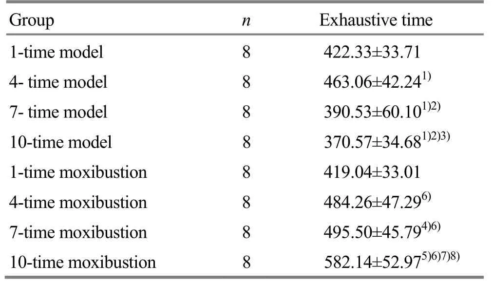
Table 1. Comparison of the exhaustive swimming time (±s,s)
Note: Compared with the 1-time model subgroup, 1) P<0.01;compared with the 4-time model subgroup, 2) P<0.01; compared with the 7-time model subgroup, 3) P<0.05, 4) P<0.01; compared with the 10-time model subgroup, 5) P<0.01; compared with the 1-time moxibustion subgroup, 6) P<0.01; compared with the 4-time moxibustion subgroup, 7) P<0.01; compared with the 7-time moxibustion subgroup, 8) P<0.01
?
2.2 Comparison of the changes in MDA, SOD, GSH-Px and T-AOC in rat’s hippocampus
Compared with the normal group, the MDA concentrations in all model subgroups were increased significantly (all P<0.01); the activities of SOD and T-AOC in the 1-time model subgroup were increased (both P<0.05); the activities of SOD, GSH-Px and T-AOC in the 4-time, 7-time and 10-time model subgroups were all decreased differently (P<0.05 or P<0.01). Compared with the normal group, the MDA concentrations in the 4-time and 7-time moxibustion subgroups were increased significantly (all P<0.01); the activities of SOD and T-AOC in each moxibustion subgroup was all significantly increased (P<0.05 or P<0.01); the activity of GSH-Px in the 7-time and 10-time moxibustion subgroups were significantly increased (P<0.05 or P<0.01), (Table 2).
Compared with the 1-time model subgroup, the MDA concentrations in the 4-time, 7-time, and 10-time model subgroups were increased significantly (all P<0.01); the activities of SOD, GSH-Px and T-AOC in the 4-time, 7-time and 10-time model subgroups were decreased to different degrees (P<0.05 or P<0.01).Compared with the 4-time model subgroup, the MAD concentrations in the 7-time and 10-time model subgroups were increased significantly (all P<0.01); the SOD activities in the 7-time and 10-time model subgroups were significantly reduced (all P<0.01); the activities of GSH-Px and T-AOC in the 10-time model subgroup was significantly reduced (all P<0.01).Compared with the 7-time model subgroup, the MDA concentration in the 10-time model subgroup was increased significantly (P<0.01), and the activities of SOD, GSH-Px and T-AOC were decreased significantly(P<0.05 or P<0.01), (Table 2).
Compared with the 1-time moxibustion subgroup,the MDA concentrations in the 4-time and 7-time moxibustion subgroups were significantly increased (all P<0.01); the activities of SOD in the 4-time, 7-time and 10-time moxibustion subgroups were significantly increased (all P<0.01); the activities of GSH-Px and T-AOC in the 7-time and 10-time moxibustion subgroups were significantly increased (P<0.05 or P<0.01). Compared with the 4-time moxibustion subgroup, the MDA concentration in the 10-time moxibustion subgroup was significantly lower (P<0.01),and the activities of SOD and T-AOC were significantly increased (P<0.01). The activities of GSH-Px in the 7-time and 10-time moxibustion subgroups were significantly increased (all P<0.01). Compared with the 7-time moxibustion subgroup, the MDA concentration in the 10-time moxibustion subgroup was significantly lower (P<0.01), and the activities of SOD, GSH-Px and T-AOC were enhanced (P<0.05 or P<0.01), (Table 2).
Compared with the model subgroup receiving the same number of interventions, there was no significant change in the 1-time moxibustion subgroup (P>0.05);the concentrations of MDA in the 4-time, 7-time and 10-time moxibustion subgroups were significantly decreased (P<0.05 or P<0.01), and the activities of SOD,GSH-Px and T-AOC were significantly increased (P<0.01),(Table 2).
Table 2. Comparison of the changes in MDA, SOD, GSH-Px and T-AOC in rat’s hippocampus (±s)
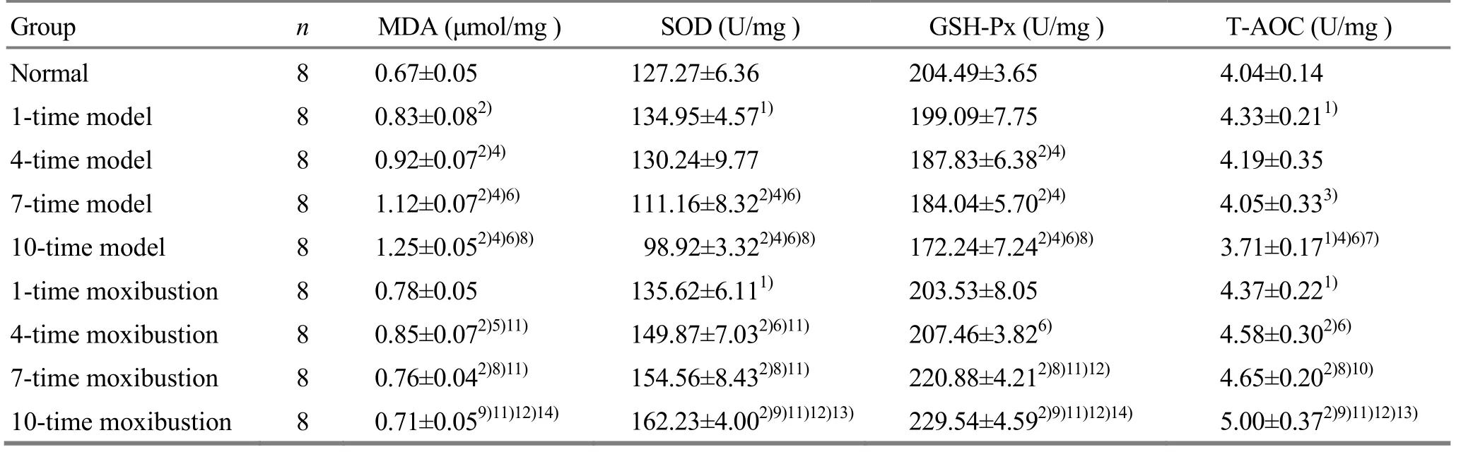
Table 2. Comparison of the changes in MDA, SOD, GSH-Px and T-AOC in rat’s hippocampus (±s)
Note: Compared with the normal group, 1) P<0.05, 2) P<0.01; compared with the 1-time model subgroup, 3) P<0.05, 4) P<0.01; compared with the 4-time model subgroup, 5) P<0.05, 6) P<0.01; compared with the 7-time model subgroup, 7) P<0.05, 8) P<0.01; compared with the 10-time model subgroup, 9) P<0.01; compared with the 1-time moxibustion subgroup, 10) P<0.05, 11) P<0.01; compared with the 4-time moxibustion subgroup, 12) P<0.01; compared with the 7-time moxibustion subgroup, 13) P<0.05, 14) P<0.01
?
3 Discussion
Brain tissue is one of the most seriously attacked body parts by the free radical[9]. The hippocampus tissue is vulnerable but has strong plasticity, and its structure can be affected by the stress. Studies have shown that hippocampus is prone to appear oxidative stress injury under the cerebral ischemic and hypoxic conditions[10]. Under normal conditions, the generation and elimination of free radicals in the body are in a dynamic equilibrium to maintain the normal physiological functions[11]. Similar to ischemiareperfusion injury[12], exhaustive sport injury also causes the brain tissue to undergo an ‘anoxic-reoxygenation’process, resulting in the production of a large number of free radicals to damage the hippocampal tissues. The utilization of mitochondrial oxygen in the brain tissues is increased dramatically and mediates a large number of free radicals upon strenuous exercise, when the synthesis and utilization of adenosine triphosphate (ATP)in the brain is accelerated. At the same time, the relatively- hypoxic brain tissues cause decreased activity of mitochondrial respiratory chain components and blocked respiratory electron-transport chains, which induce the ubiquinone to generate free radicals by single electron reduction during electron transfer,resulting in MDA production by peroxidation of unsaturated lipid[13]. During the free radical metabolism,MDA represents the level of free radicals in the body.Increased MDA level indicates increased lipid peroxidation in the body, as well as increased damage to mitochondria and cell membranes. An enzyme antioxidant system in the body prevents the tissues in the body from damage, in which SOD and GSH-Px represent the function of body's free radical scavenging system. Increased activities of SOD and GSH-Px promote body's ability to clear the free radicals. T-AOC represents the total antioxidant capacity of the local tissues, and a higher T-AOC level indicates a stronger antioxidant capacity in the body[2-3].
Exercise fatigue belongs to ‘exhaustive consumption’in Chinese medicine. Shenque (CV 8) has the effect of tonifying the spleen and kidney, cultivating Yuan-Primordial qi to consolidate the root, restoring yang to save life and reinforcing qi to rescue the collapse. Moxibustion has the effect of restoring yang and prostration. Studies have shown that moxibustion removes free radicals from muscle tissues inexercise-induced fatigue[14-15], increases energy supply to muscle tissues[16-17], corrects disordered endocrine function[18-19], and reduces accumulation of metabolites in vivo[20-21], thus to play a role in preventing exercise fatigue.
The results of our current experiment showed changes in free radical metabolism of rat’s hippocampus in the 1-time model subgroup, including an increase in MDA concentration, rapid activation of the antioxidant enzyme system, secretion of a large amount of stress-induced SOD and GSH-Px to remove tissue free radicals, and an increase in T-AOC level. With the continuation of exercise, the concentration of free radical MDA was increased, the SOD and GSH-Px were continuously depleted with continuously decreased enzyme activity and anti-oxidant ability, and T-AOC level was decreased in the hippocampus in the 4-time,7-time and 10-time model subgroups. At the same time,the exhaustive swimming time of the 7-time and 10-time model subgroups was significantly shorter than that of the 4-time model subgroup. It was speculated that after 4 times of exhaustive exercises, the generation and clearance of free radicals in rat’s hippocampus were inconsistent. A large amount of free radicals were generated and antioxidant enzymes were consumed, and the body entered a fatigue state.However, the free radical concentration of the hippocampus in the 4-time moxibustion subgroup was decreased; the levels of SOD and GSH-Px were increased. The concentration of free radical MDA was significantly decreased, and the levels of SOD, GSH-Px and T-AOC were significantly increased in the 7-time and 10-time moxibustion subgroups. Exhaustive swimming time of the 7-time and 10-time moxibustion subgroups was significantly longer than that of the 4-time moxibustion subgroup. Our results provided an experimental basis for moxibustion at Shenque (CV 8)to resist exercise fatigue by regulating free radical metabolism in rat’s hippocampus and improving the fatigue status of the body after long-term exhaustive exercise.
Conflict of Interest
The authors declared that there was no potential conflict of interest in this article.
Acknowledgments
This work was supported by Youth Scientists Fund of Natural Science Foundation of Education Department of Hebei Province (河北省教育厅青年自然科学基金项目,No. QN2016021, No. Z2017074); Science and Technology Support Project of Hebei Provincial Administration of Traditional Chinese Medicine (河北省中医药管理局科技支撑项目, No. 2017011); Undergraduate Ⅰnnovation and Entrepreneurship Training Program of Hebei University of Chinese Medicine (河北中医学院大学生创新创业训练计划项目, No. 2017044).
Statement of Human and Animal Rights
The treatment of animals conformed to the ethical criteria.
Received: 28 December 2017/Accepted: 25 January 2018
猜你喜欢
杂志排行
Journal of Acupuncture and Tuina Science的其它文章
- Modern quantum theories and experimental achievements motivate new exploration of acupuncture in traditional Chinese medicine
- Application and exploration of suspended magnetic moxibustion cup for obesity
- Therapeutic efficacy observation on acupuncture for persistent allergic rhinitis
- Correlation analysis on clinical effects of acupuncture for elderly patients with sensorineural deafness and ear distending sensation
- Effect of Governor Vessel-unblocking and mindrefreshing acupuncture plus functional training on neural development in infants with brain damage
- Effects of acupuncture on ovarian blood supply and pregnancy outcomes in patients receiving assisted reproduction