Injury of the superior longitudinal fasciculus by ventriculoperitoneal shunt: a diffusion tensor tractography study
2018-07-21SungHoJang,HanDoLee
In this study, we report on a patient who suffered from injury of the superior longitudinal fasciculus following ventriculoperitoneal (VP) shunt operation, which was demonstrated with diffusion tensor tractography (DTT).
An 82-year-old female patient was diagnosed as suffering from normal pressure hydrocephalus (NPH) and underwent VP shunt operation approached through the right posterior parietal area in the brain 3 years ago(Figure 1A). Brain MRI at 3 years after the shunt operation showed that the VP shunt passed through the right inferior parietal lobule and adjacent subcortical white matter (Figure 1B). Her cognition was so poor that the Mini-Mental State Exam score was not even checked. Therefore, her neglect could not be evaluated using a formal evaluation tool such as the line bisection test, however, she did not show symptoms of neglect clinically. The study was approved by the Institutional Review Board of Yeungnam University Hospital (IRB No. YUMC-2015-07-064).
Diffusion tensor imaging (DTI) data were acquired 2 weeks after VP shunt operation using a 6-channel head coil on a 1.5 T Philips Gyroscan Intera (Philips,Ltd, Best, The Netherlands) with single-shot echo-planar imaging. For each of the 32 non-collinear diffusion sensitizing gradients, 70 contiguous slices were acquired parallel to the anterior commissure-posterior commissure line. Sixty contiguous slices (acquisition matrix = 96 × 96; reconstruction matrix = 192 × 192 matrix; fi eld of view = 240 × 240 mm2; repetition time= 10,398 ms; echo time = 72 ms,b= 1000 s/mm2, number of excitations = 1, slice gap = 0 mm and thickness =2.5 mm) were acquired for each of the 32 noncollinear diffusion-sensitizing gradients. Fiber tracking was performed using the fi ber assignment continuous tracking algorithm implemented within the DTI task card soft-ware (Philips Extended MR Work Space 2.6.3). Each DTI replication was intra-registered to the baseline “b0”images to correct for residual eddy-current image distortions and head motion effect, using a diffusion registration package (Philips Medical Systems). For reconstruction of the superior longitudinal fasciculus (SLF),the seed region of interest was placed on the triangular shape just lateral to the corticospinal tract near the anterior horn of the lateral ventricle and the target region of interest was placed on the triangular shape near the posterior horn of the lateral ventricle (Bernal and Altman,2010). Termination criteria were fractional anisotropy(FA) < 0.15 and an angle change > 2°. The lower portion of the right SLF was not reconstructed compared with the left SLF (Figure 1C). In addition, the value of fi ber number (FN) of the SLF showed difference between the right and left hemispheres except for the values of fractional anisotropy and mean diffusivity (MD) the right SLF— FA: 0.37, MD: 0.91, FN: 4015 and left — FA: 0.38,MD: 0.88, FN: 4950).
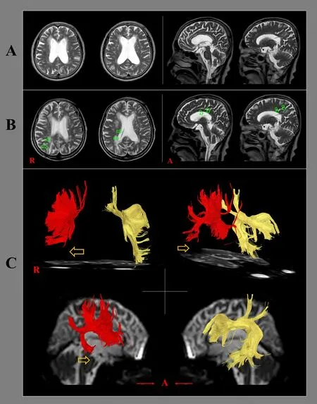
Figure 1 Brain MR imaging and diffusion tensor tractography for a 82-year-old female patient with normal pressure hydrocephalus following ventrioculoperitoneal (VP) shunt.
VP shunt operation is a common neurosurgical procedure for management of NPH. In this study, using DTT, injury of the right SLF following VP shunt operation was demonstrated in a patient with NPH. Because the VP shunt passed through the lower portion of the remaining SLF, it appeared that the injury of the lower portion of the right SLF resulted from the VP shunt.However, the symptom of neglect, which is the most representative symptom of the right SLF injury, was not observed in this patient, although the patient’s neglect could not be precisely estimated due to poor cognition(Shinoura et al., 2009; Lunven et al., 2015). Since introduction of DTT, several studies have reported that passage of a VP shunt caused injury of adjacent neural tracts (Gold et al., 2008; Kwon and Jang, 2012; Jang and Yeo, 2013; Jang and Seo, 2015, 2016). These neural tracts comprised the corticospinal tract, limbic system,cingulum, corticoreticulospinal tract, and thalamocingulate tract among the Papez circuit (Gold et al., 2008;Kwon and Jang, 2012; Jang and Yeo, 2013; Jang and Seo,2015, 2016). Thus, to the best of our knowledge, this is the fi rst case report to demonstrate an injury of the SLF following VP shunt operation. However, limitations of DTT should be considered: it can produce both false negative and false positive results due to crossing fi ber,partial volume effect, or instrument-induced artifact(Parker and Alexander, 2005).
In conclusion, injury of the SLF was demonstrated following VP shunting in a patient with NPH, using DTT. It appears that DTT could be a useful imaging tool in detection of underlying injury of neural tracts after VP shunting. In-depth studies including a large number of subjects and development of safe neurosurgical procedures to save the neural tracts should be encouraged (Jang and Kwon, 2014; Jang et al., 2015; Kwon and Jang, 2015).
Sung Ho Jang, Han Do Lee*
Department of Physical Medicine and Rehabilitation, College of Medicine, Yeungnam University, Namku, Daegu, Republic of Korea
*Correspondence to:Han Do Lee, M.S., lhd890221@hanmail.net.
orcid:0000-0002-1668-2187 (Han Do Lee)
Accepted:2017-05-10
doi:10.4103/1673-5374.235093
Author contributions:SHJ was responsible for conception and design of this study and development and writing of the paper. HDL participated in conception and design of this study, was responsible for acquisition and analysis of data, and authorized the paper. Both of these two authors approved the fi nal version of this paper.
Con fl icts of interest:None declared.
Financial support:This work was supported by the Medical Research Center Program (2015R1A5A2009124) through the National Research Foundation of Korea (NRF) funded by the Ministry of Science, ICT and Future Planning. The funder had no involvement in the study design;data collection, analysis, and interpretation; paper writing; or decision to submit the paper for publication.
Institutional review board statement::The study was approved by the Institutional Review Board of Yeungnam University Hospital (approval No. YUMC-2015-07-064). The study followed the principles of the Declaration of Helsinki.
Data sharing statement:Datasets analyzed during the current study are available from the corresponding author on reasonable request.
Copyright transfer agreement:The Copyright License Agreement has been signed by all authors before publication.
Plagiarism check:Checked twice by iThenticate.
Peer review:Externally peer reviewed.
Open access statement:This is an open access journal, and articles are distributed under the terms of the Creative Commons Attribution-Non-Commercial-ShareAlike 4.0 License, which allows others to remix, tweak,and build upon the work non-commercially, as long as appropriate credit is given and the new creations are licensed under the identical terms.
杂志排行
中国神经再生研究(英文版)的其它文章
- Stem cells: a promising candidate to treat neurological disorders
- Modulation of microglial functions by methyl jasmonate
- Degree of dopaminergic degeneration measured by 99mTc-TRODAT-1 SPECT/CT imaging
- Differences in brain pathological changes between rotenone and 6-hydroxydopamine Parkinson’s disease models
- Zishenpingchan granules for the treatment of Parkinson’s disease: a randomized, double-blind,placebo-controlled clinical trial
- Puerarin ameliorates allodynia and hyperalgesia in rats with peripheral nerve injury