二氢杨梅素通过STAT3/Bcl-2信号通路促进HNSCC细胞自噬和凋亡
2017-09-22王满英丁小军余优成
王满英, 丁小军, 孙 扬, 余优成
复旦大学附属中山医院口腔科, 上海 200032
·短篇论著·
二氢杨梅素通过STAT3/Bcl-2信号通路促进HNSCC细胞自噬和凋亡
王满英, 丁小军*, 孙 扬, 余优成
复旦大学附属中山医院口腔科, 上海 200032
目的: 探讨二氢杨梅素(DHM)对人头颈鳞癌(HNSCC)细胞增殖及凋亡的影响。方法: 分别用不同浓度(12.5、25、50 μmol/L)DHM处理人HNSCC SCC-25细胞6、12、24 h,用流式细胞仪检测SCC-25细胞凋亡情况;用细胞免疫荧光法观察SCC-25细胞中自噬小体产生情况;Western 印迹法检测SCC-25凋亡和自噬标志物的表达。结果: 12.5、25、50 μmol/L的DHM处理24 h后,SCC-25细胞凋亡逐渐增加;细胞中凋亡标志物CL-PARP、Bax、CL-casp3逐渐增加,Bcl-2逐渐减少(P<0.05)。12.5、25、50 μmol/L的DHM处理24 h后,SCC-25细胞自噬逐渐增强;自噬标志指标p-STAT3表达上调,LC3Ⅱ/LC3Ⅰ的比值逐渐加大,Beclin 1增加,p62逐渐减少(P<0.05)。抑制自噬能促进DHM诱导的SCC-25细胞凋亡,细胞凋亡标志物CL-PARP、Bax增加,Bcl-2减少(P<0.05)。结论: DHM能诱导HNSCC细胞凋亡及自噬,其机制与STAT3相关信号通路有关;抑制自噬能促进DHM诱导的HNSCC细胞凋亡。
头颈鳞状细胞癌;二氢杨梅素;凋亡;自噬
头颈部鳞状细胞癌(head and neck squamous cell carcinoma,HNSCC)是世界范围内第六大常见的癌症,每年新增病例约600 000例[1]。尽管随着分子生物学和细胞生物学技术的不断发展,HNSCC的诊断和治疗方法也在不断进步,然而近30年来并未明显提高HNSCC患者的生存率[1]。其原因在于HNSCC易远处转移、局部区域复发,且对现阶段化疗方案的耐药性较高[2]。
二氢杨梅素(dihydromyricetin,DHM),又名蛇葡萄素,其化学名为3,5,7,3’,4’,5’-六羟基2,3双氢黄酮醇,是一种天然的黄酮类化合物,广泛存在于葡萄科蛇葡萄属植物中[3]。近年来,大量研究表明DHM对人肝癌、人乳腺癌、卵巢癌、肺癌细胞等具有较强的体外抑制作用[4-9],DHM的抗肿瘤活性作用不断得到重视。本研究通过分析DHM对HNSCC细胞凋亡及自噬的影响,初步探讨DHM抗HNSCC的机制,以期为HNSCC的临床治疗提供新的靶点,提高疗效。
1 材料与方法
1.1 材 料 SCC-25细胞株购买于中国科学院上海生命科学研究院细胞资源中心。DMEM培养基购买于美国Gibco公司,胎牛血清购买于杭州四季青生物工程材料有限公司;DHM购买于美国Selleck公司 ;CL-PARP、Bcl-2、Bax、LC3、Beclin 1、p62、兔抗人STAT3、p-STAT3抗体购买于美国CST公司,鼠抗人GAPDH抗体购买于英国Abcam公司;3-MA、ATG5-siRNA购买于美国Santa公司;Caspase-3试剂盒购自碧云天生物有限公司。
1.2 细胞培养 SCC-25细胞接种在胎牛血清(体积浓度为10%)并含100 U/L青霉素、100 mg/L链霉素的新鲜DMEM培养液中培养,培养条件为37℃、饱和湿度、5% CO2[10]。
1.3 流式细胞学分析 选取对数生长期的SCC-25细胞制成单细胞悬液,接种于12孔板,密度为每孔2×10⁵/mL。培养24 h 后,分别加入含有12.5、25、50 μmol/L DHM的培养液,于离心管251.55×g离心10 min,弃上清,加入固定液(预冷的75%乙醇)1 mL,4℃过夜。第2天转移至5 mL EP管中,111.8×g离心10 min,弃上清。用标记凋亡细胞的异硫氰酸荧光素(fluorescein isothiocyanate,FITC)标记膜联蛋白Ⅴ或标记坏死细胞的荧光染料碘化丙啶处理后,4℃避光条件下静置2 h,置于流式细胞仪进行检测。每组重复3个样本,重复实验3次。
1.4 Western 印迹法检测凋亡指标 取对数生长期的SCC-25细胞,消化后以每孔2 mL的量接种于6孔板中,细胞密度为(2.0~3.0)×106/mL。培养条件为37℃、5%CO2。培养24 h后,分别加入12.5、25、50 μmol/L的DHM,培养 6、12、24 h后,提取细胞总蛋白,Western 印迹法检测凋亡指标(CL-PARP、Bcl-2、Bax)及自噬指标(LC3、Beclin 1、p62)蛋白的表达。用Image J 软件扫描各条带进行半定量分析,以上述目的蛋白电泳条带灰度值与内参照GAPDH条带灰度值的比值作为各蛋白的相对表达水平。实验重复3次。
1.5 酶法检测caspase-3活性 细胞加入自噬的抑制剂3-MA和敲除自噬成膜特异性基因ATG5处理后,加入50 μmol/L的DHM处理24 h后,胰酶消化,并用细胞裂解液裂解,离心取上清,按照碧云天说明书进行caspase-3活性的检测[11]。

2 结 果
2.1 DHM诱导SCC-25细胞凋亡 12.5、25、50 μmol/L 的DHM处理SCC-25细胞24 h后,细胞凋亡率分别为6.00%、24.93%及38.17%(图1A)。Western 印迹检测发现,CL-PARP、Bax、CL-casp3随着DHM浓度的增加而增加,Bcl-2则随着DHM浓度的增加而减少(P<0.05,图1B)。用50 μmol/L的DHM分别处理SCC-25细胞6、12、24 h后,CL-PARP、Bax及CL-casp3逐渐增加,而Bcl-2逐渐减少(P<0.05,图1C)。
2.2 DHM诱导SCC-25细胞自噬 免疫荧光检测结果显示,培养 24 h后,LC3阳性细胞数随DHM浓度的增加而增加(P<0.05,图2A)。Western 印迹检测发现,随着DHM浓度的增加或时间的延长,LC3Ⅱ/LC3Ⅰ的比值逐渐加大,Beclin 1增加,而p62逐渐减少(P<0.05,图2B、2C)。
2.3 DHM通过靶向影响STAT3促进SCC-25细胞自噬 Western 印迹结果表明,培养24 h后,随着DHM浓度的增加,磷酸化STAT3蛋白逐渐增加(P<0.05, 图3A)。DHM 50 μmol/L处理SCC-25细胞6、12、24 h后,p-STAT3表达逐渐增加(P<0.05, 图3B)。用STAT3的特异性抑制剂NSC74859抑制SCC-25细胞中p-STAT3的表达24 h后,再用50 μmol/L的DHM处理SCC-25细胞24 h,结果发现,细胞自噬水平下降(LC3Ⅱ/LC3Ⅰ比值下降),细胞凋亡水平升高(CL-PARP、Bax的表达增加,Bcl-2表达降低;P<0.05, 图3C)。
2.4 抑制自噬显著增强DHM引起的SCC-25细胞凋亡 通过使用自噬的抑制剂3-MA和敲除自噬成膜特异性基因ATG5观察自噬抑制后对凋亡的影响。50 μmol/L的 DHM处理SCC-25细胞后,细胞中的caspase-3活性增强;3-MA和ATG5 si-RNA处理后,caspase-3活性进一步增强(P<0.05,图4)。

图1 DHM诱导SCC-25细胞凋亡

3 讨 论
部分癌症患者对化疗药物不敏感或耐受,因此,亟需发现一些潜在的治疗癌症的药物。DHM有多种生物学效应[12-16],如抗毒性、抗肿瘤及抗乙醇中毒活性。但是,DHM抗肿瘤的具体机制目前尚不清楚。研究[17]显示,DHM可以通过p53相关信号通路促进人类胃癌细胞凋亡。研究[18]发现,在人类肝癌细胞中,DHM通过p53/Bcl-2信号通路协同萘达铂的杀肿瘤效应。本研究中,DHM可以诱导SCC-25细胞自噬及凋亡,凋亡相关蛋白CL-PARP、Bcl-2、Bax、Cl-casp3及自噬相关蛋白LC3、Beclin 1、p62表达改变,且p-STAT3表达增加。
细胞自噬普遍存在于真核细胞中[19]。自噬在生物体存活中起着双刃剑的作用:一方面细胞可以通过自噬吸取能量来维持细胞正常的新陈代谢;另一方面,自噬可以导致细胞死亡。本研究中,首先用3-MA抑制细胞的自噬过程,再用DHM处理肿瘤细胞,发现细胞中caspase-3活性增强;然后,在基因水平上沉默自噬过程中起决定作用的ATG5基因,再用DHM处理肿瘤细胞,发现细胞中caspase-3的活性也增强。结果说明,在HNSCC细胞中,DHM引起的细胞自噬对肿瘤细胞起保护作用。
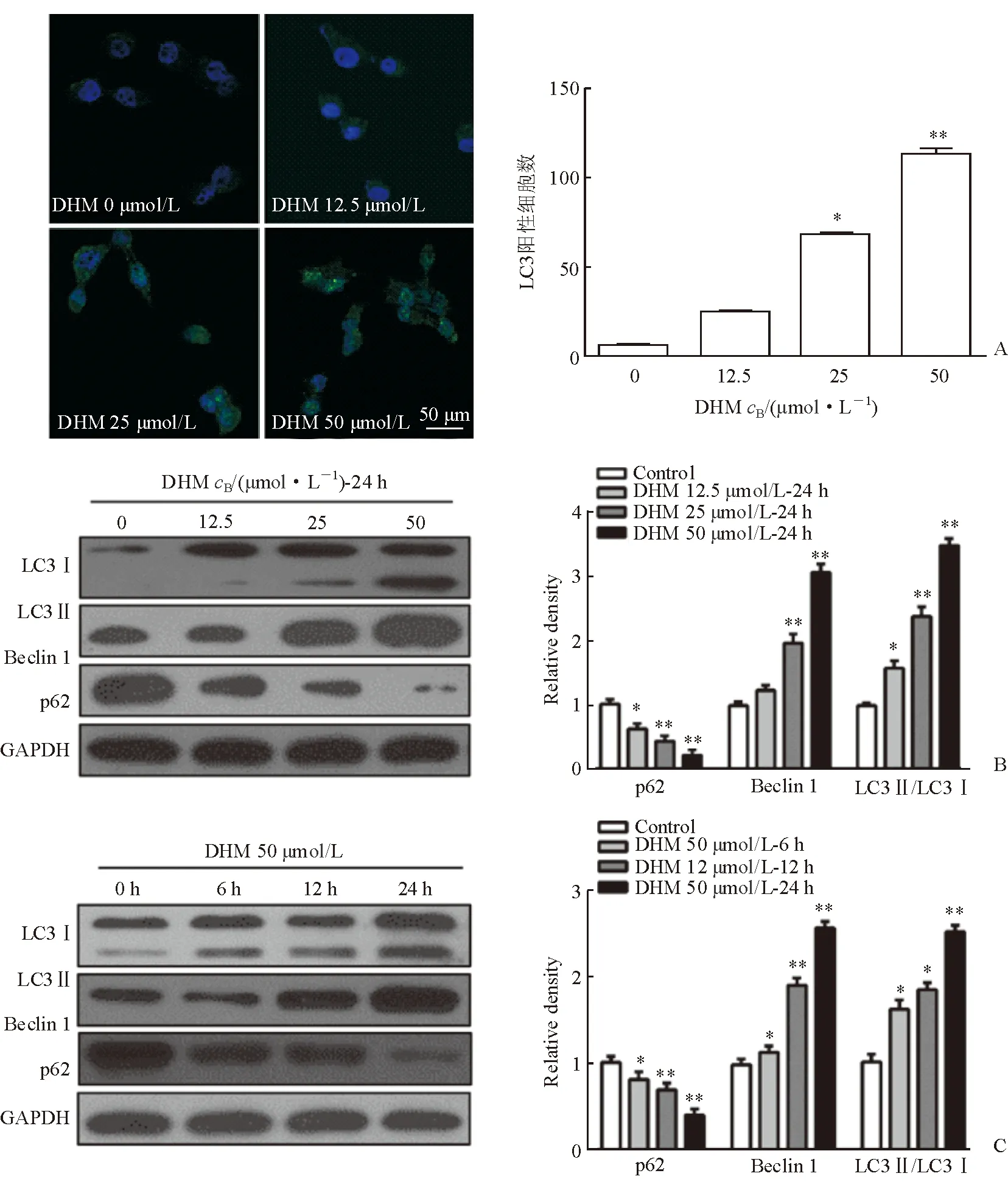
图2 DHM诱导SCC-25细胞自噬
STAT3是信号转导和转录激活因子家族的重要成员之一。持续激活STAT3信号转导通路可导致肿瘤发生、发展。体内外使某些肿瘤细胞中的STAT3信号通路被阻断,可使细胞的增殖和存活被抑制,并诱导细胞自噬[20]。本研究发现,p-STAT3的表达随DHM浓度的增加而增加;用p-STAT3特异性抑制剂NSC74859抑制p-STAT3的活性后,DHM所导致的肿瘤细胞自噬减少,而DHM所引起的肿瘤细胞凋亡增多。
综上所述,本研究结果表明,人HNSCC细胞凋亡及自噬水平可在DHM所诱导下升高,其机制可能与STAT3信号通路相关,提示抑制自噬可使DHM的抗癌能力显著增强,DHM联合应用抗自噬药物会加强抗肿瘤效应。但是,目前国内外基于DHM抗肿瘤效应的研究中,各种肿瘤细胞对DHM的敏感性机制并不一致[4-9]。本研究证明,DHM可诱导自噬和凋亡,而STAT3信号的研究说明引起的自噬起保护作用,为了增加DHM的杀肿瘤效应可以联合使用自噬抑制剂。
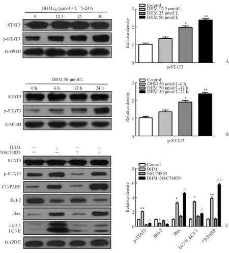
图3 DHM通过促进p-STAT3的表达影响SCC-25细胞自噬及凋亡蛋白的表达
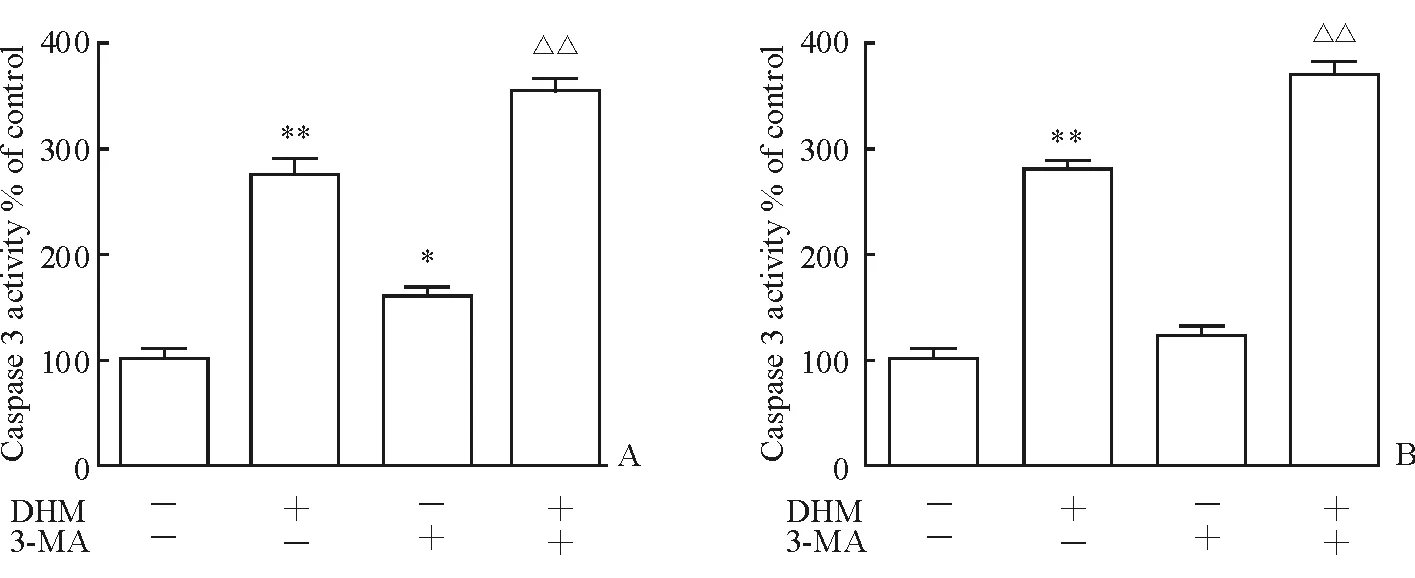
图4 3-MA(A)和ATG5 si-RNA(B)抑制自噬增强DHM对SCC-25细胞中caspase-3活性的增强效应
[ 1 ] AMINUDDIN A, NG P Y. Promising druggable target in head and neck squamous cell carcinoma: wnt signaling[J]. Fronti in Pharmacol, 2016,7:244.
[ 2 ] MARTIN A, SALVADOR F, MORENO-BUENO G, et al. Lysyl oxidase-like 2 represses Notch1 expression in the skin to promote squamous cell carcinoma progression[J]. EMBO J, 2015,34(8):1090-1109.
[ 3 ] WONG I L, WANG B C, YUAN J, et al. Potent and nontoxic chemosensitizer of P-glycoprotein-mediated multidrug resistance in cancer: synthesis and evaluation of methylated epigallocatechin, gallocatechin, and dihydromyricetin derivatives[J]. J Med Chem, 2015,58(11):4529-4549.
[ 4 ] ROY K, KANWAR R K, KRISHNAKUMAR S, et al. Competitive inhibition of survivin using a cell-permeable recombinant protein induces cancer-specific apoptosis in colon cancer model[J]. Int J Nanomedicine, 2015,10:1019-1043.
[ 5 ] ZHANG K, LI Y, LIU W, et al. Silencing survivin expression inhibits the tumor growth of non-small-cell lung cancer cells in vitro and in vivo[J]. Mol Med Rep, 2015,11(1):639-644.
[ 6 ] LIU B, ZHOU W, CHEN X, et al. Dihydromyricetin induces mouse hepatoma Hepal-6 cell apoptosis via the transforming growth factor-beta pathway[J]. Mol Med Rep, 2015,11(3):1609-1614.
[ 7 ] ZHOU Y, SHU F, LIANG X, et al. Ampelopsin induces cell growth inhibition and apoptosis in breast cancer cells through ROS generation and endoplasmic reticulum stress pathway[J]. PLoS One, 2014,9(2):e89021.
[ 8 ] ZHANG Q Y, LI R, ZENG G F, et al. Dihydromyricetin inhibits migration and invasion of hepatoma cells through regulation of MMP-9 expression[J]. World J Gastroenterol, 2014,20(29):10082-10093.
[ 9 ] XU Y, WANG S, CHAN H F, et al. Dihydromyricetin Induces Apoptosis and Reverses Drug Resistance in Ovarian Cancer Cells by p53-mediated Downregulation of Survivin[J]. Sci Rep, 2017,7:46060.
[10] WANG Y F, ZHANG W, HE K F, et al. Induction of autophagy-dependent cell death by the survivin suppressant YM155 in salivary adenoid cystic carcinoma[J]. Apoptosis, 2014,19(4):748-758.
[11] LI Z L, HU J, LI Y L, et al. The effect of hyperoside on the functional recovery of the ischemic/reperfused isolated rat heart: potential involvement of the extracellular signal-regulated kinase 1/2 signaling pathway[J]. Free Radic Biol Med, 2013,57:132-140.
[12] LIU B, TAN X, LIANG J, et al. A reduction in reactive oxygen species contributes to dihydromyricetin-induced apoptosis in human hepatocellular carcinoma cells[J]. Sci Rep, 2014,4:7041.
[13] MENG G, YANG S, CHEN Y, et al. Attenuating effects of dihydromyricetin on angiotensin Ⅱ-induced rat cardiomyocyte hypertrophy related to antioxidative activity in a NO-dependent manner[J]. Pharm Biol, 2015,53(6):904-912.
[14] SHEN Y, LINDEMEYER A K, GONZALEZ C, et al. Dihydromyricetin as a novel anti-alcohol intoxication medication[J]. J Neurosci, 2012,32(1):390-401.
[15] YE L, WANG H, DUNCAN S E, et al. Antioxidant activities of Vine Tea (Ampelopsis grossedentata) extract and its major component dihydromyricetin in soybean oil and cooked ground beef[J]. Food Chem, 2015,172:416-422.
[16] LIU B, ZHOU W, CHEN X, et al. Dihydromyricetin induces mouse hepatoma Hepal-6 cell apoptosis via the transforming growth factor-beta pathway[J]. Mol Med Rep, 2015,11(3):1609-1614.
[17] JI F J, TIAN X F, LIU X W, et al. Dihydromyricetin induces cell apoptosis via a p53-related pathway in AGS human gastric cancer cells[J]. Genet Mol Res, 2015,14(4):15564-15571.
[18] XIE J, LIU J, CHEN T M, et al. Dihydromyricetin alleviates carbon tetrachloride-induced acute liver injuryviaJNK-dependent mechanism in mice[J]. World J Gastroenterol, 2015,21(18):5473-5481.
[19] KLIONSKY D J, ABDALLA F C, ABELIOVICH H, et al. Guidelines for the use and interpretation of assays for monitoring autophagy[J]. Autophagy, 2012,8(4):445-544.
[20] MALI S B. Review of STAT3 (signal transducers and activators of transcription) in head and neck cancer[J]. Oral Oncol, 2015,51(6):565-569.
[本文编辑] 姬静芳
Dihydromyricetin promotes autophagy and apoptosis in head and neck squamous cell carcinoma cells through STAT3/Bcl-2 signaling pathway
WANG Man-ying, DING Xiao-jun*, SUN Yang, YU You-cheng
Department of Stomatology, Zhongshan Hospital, Fudan University, Shanghai 200032, China
Objective: To study the effects of dihydromyricetin (DHM) on cell proliferation and apoptosis of head and neck squamous cell carcinoma (HNSCC).Methods: SCC-25 cancer cells were treated with 12.5, 25, 50 μmol/L DHM for 6, 12, and 24 h. SCC-25 cell apoptosis was detected by flow cytometry. Immunofluorescence was used to observe the emergence of autophagosomes in HNSCC cells. Western blotting was used to detect the expression of apoptosis and autophagy markers in SCC-25.Results: After treatment of 12.5, 25, and 50 μmol/L DHM for 24 h, the apoptosis of SCC-25 cells gradually increased; the apoptotic markers CL-PARP, Bax, and CL-casp3 gradually increased and Bcl-2 gradually decreased (P<0.05). After treatment of 12.5, 25, and 50 μmol/L DHM for 24 h, in line with the apoptosis, the autophagy markers of SCC-25 cells gradually changed, the p-STAT3 expression was up-regulated; the ratio of LC3 Ⅱ /LC3 Ⅰ gradually increased, Beclin 1 increased, p62 gradually decreased (P<0.05). Inhibition of autophagy could promote apoptosis of SCC-25 cells which were induced by DHM, increase apoptosis markers CL-PARP and Bax, and decrease Bcl-2 (P<0.05).Conclusions: DHM can induce apoptosis and increase autophagy of HNSCC cells. The mechanism is related to STAT3 signaling pathway. Inhibition of autophagy can promote apoptosis of HNSCC cells induced by DHM.
head and neck squamous cell carcinoma;dihydromyricetin; apoptosis; autophagy
R 739.91
A
2016-10-17 [接受日期] 2017-01-14
王满英, 硕士生. E-mail:14211210070@fudan.edu.cn
*通信作者(Corresponding author). Tel: 021-64041990-2585, E-mail:dingxiaojun@zs-hospital.sh.cn
10.12025/j.issn.1008-6358.2017.20160961