先天或原(特)发性(真性)肢体肌肥大
2017-08-25田光磊田文赵俊会
田光磊,田文,赵俊会
(北京积水潭医院 手外科,北京 100035)
(本文接上期)
9 掌长肌肥大
掌长肌,于灵长类动物来说,是块不断退化的肌肉,极具变异性,如二腹肌、双肌腹、副肌、肌腹-肌腱位置互换、肌腹缺失、缺如等,其中多数为返祖现象。掌长肌肥大,目前仅报道5例,且都是单侧发生。
⑴1948年Goulding[57]报道1例:患者 男,22岁,发育正常,体健,右前臂掌面偏尺侧有卵圆形隆起5年,发现突然,无诱因,手术探查见掌长肌肥大,未予处理。之后,其大小无变化,长轴与前臂一致,主动屈腕时体积略增且质地变硬。近1年出现钝痛,但无碍手功能及工作,查体未见其他异常,无家族病史。再次手术探查,见掌长肌甚巨大,肌腹抵近肌腱止点,予以大部切除。术后恢复正常。
⑵1964年Ashby[58]报道1例:患者 女,13岁,(非优势)右前臂远1/3掌侧中央条状膨隆,拇示中环指麻痛3年,膨隆略呈蓝色,无压痛,感觉无丧失,肌力无减退,只是双手掌呈“猿手”状,双手、躯干、双下肢皮肤粗糙、皮纹深,其母、其弟也如此。手术探查见正中神经正常,而掌长肌却肥大,肌腹附着在屈肌支持带上,予以切除。组织学检查:正常肌肉。术后恢复正常。Ashby认为,肥大似无关返祖。
⑶1992年杨国庆[59]报道1例:患者 男,24岁,工人,因右腕管综合征2个月行松解手术,见掌长肌肥大,肌腹远端远侧延展,接近肌腱止点,并压迫正中神经。术前X线平片未显示任何异常。
⑷1998年Polesuk和Helms[60]报道1例:患者男,26岁,手工业者,(优势)右前臂掌侧无痛膨隆2年,MRI见掌长肌极度肥大,肌腹向远侧延展,直至止点。未处理。随访2年无变化。
⑸2015年Barkats[61]在一项关于掌长肌发生与缺如率的调查中发现1例:患者 女,22岁,左前臂掌侧有条状膨隆,位置略偏尺侧,触诊为质软组织。根据体检,他们认为是掌长肌肥大所致。患者主诉示、中指及腕部有刺痛感,屈指握拳时有疼痛感;从事某些活动时,环、小指也常有疼痛不适。Barkats认为这些都是正中、尺神经受压的表现,需做进一步的诊疗,但患者以宗教因素为由拒绝了Barkats的建议。肥大何时出现,作者未述,但认为属肌肉变异。未作影像学检查,即诊断肥大,是否合适有待商榷。
10 尺侧腕屈肌肥大
1975年Harrelson和Newman[62]报道1例:患者男,22岁,右腕尺管综合征2个月,腕掌尺侧横纹近侧膨隆,且有压痛,认为是神经肿瘤,手术才知是尺侧腕屈肌肥大,肌腹延长,直至豌豆骨,并压迫尺神经,予以部分切除。术后随访1年,一切正常。
11 上肢肌肥大[4]
上肢肌肥大,文献中无此称谓,是我们自撰,暂用于此,仅为表述方便。此肌肥大出生即有,多累及一侧上肢,即一侧上肢多块肌肉非进行性肥大,且与性别无关;现共报道40余例,可谓是肢肌肥大中的大户。其中,大部分病例来自日本和中国。此肌肥大病例较多,综而述之,不再堆砌病例数据了。
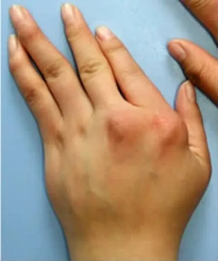
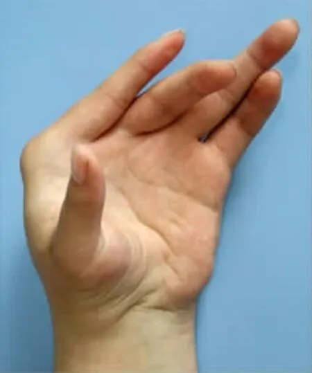
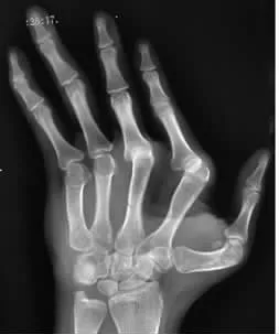
上肢肌肥大,既有知名肌肉也包括迷走(副)肌肉,尤以手、前臂为甚,其结果:⑴手通常肥于前臂,前臂又硕于上臂及肩部,即肢体远端肥大重、近端轻;⑵肩及前臂活动多正常,肘与腕偶有屈曲、背伸挛缩,手则是畸形严重,功能缺失,如拇指过度外展、背伸时无法与其他手指捏合,示指旋前、屈曲时会与中指相叠罗,手指掌指关节掌屈、尺偏时形同吹风手,伸直受限,不经手术很难有所恢复(图6-8)。
上肢肌肥大,还有许多别称,如:⑴鱼际和小鱼际重复肌 (duplication of the thenar and hypothenar muscles);⑵迷走肌肉综合征(aberrant muscle syndrome);⑶副肌肉综合征 (accessory muscle syndrome);⑷先天性单上肢肌肥大(congenital unilateral upper limb muscular hypertrophy/congenital monomelic muscular hypertrophy of the upper extremity);⑸先天性单侧上肢肌源性肥大综合征(unilateral congenital upper limb myohypertrophy)。

图6 左手背侧

图7 左手掌侧

图8 左手X线片
上肢肌肥大者,智力、身体发育均正常,肌力正常或增加,骨骼基本正常,且无家族遗传病史。2013-2014年Casiglioni等[63,64]报道 1 例 6 岁女童左上肢肌肥大,出生即存在,包括肩带肌,发育正常,无海神、CLOVES综合征等过度生长性疾患,更无家族遗传病史。X线平片见骨骼正常。超声、MRI检查见上肢诸肌肥大,手部还有肥大的迷走肌肉。第1背侧骨间肌活检见肌纤维增粗,纤维粗细差异增大,束膜、内膜纤维化增多;Ⅰ型纤维为优势纤维;肌原纤维间网(intermyofibrillar network)杂乱。超微检查见肌原纤维排列无序,肌质管扩张。AKT1(蛋白激酶B)、PIK3CA(磷脂酰肌醇-3-激酶催化亚基α)和PTEN(一种抑癌基因)Sanger测序,见肌肉PIK3CA基因p.H1047R激活突变,位点在c.3140A>G,而血细胞PIK3CA基因则无此突变。因此Castiglioni等认为,单侧上肢肌肥大,与PIK3CA基因突变有关。其结论可靠与否,还需验证。因为一系列过度发育综合征,甚至结肠癌、乳腺癌,均可检测到PIC3CA基因的突变,即此基因突变未必是致肌肥大的根本原因。
2016年Kalay等[65]报道1例双侧上肢肌肥大。看来,上肢肌肥大还是可以双侧发生的。
治疗上肢肌肥大,首选依然是肥大肌肉切除,之后是旋转切骨内固定矫正手指旋前,松解植皮矫正皮肤挛缩,肌腱移位矫正关节尺偏等。
12 肩带肌肥大
现有3例,由Almansoor等[66]在1998年报道。
患者1:男,4岁,不好运动,右肩胛骨区膨隆,无痛,超声、CT检查见肩胛骨周围肌肉局限性肥大,尤其是背阔肌、小圆肌和冈下肌,前者显著,后二者稍轻。9个月后复查CT及MRI,所见同前;活检结果是正常的骨骼肌组织。随访2年,膨隆无变化。
患者2:女,10岁,体操运动员,右腋窝无痛膨隆数月,无压痛,超声、MRI检查见背阔肌局限性肥大。2个月后复查超声,所见同前;活检结果是正常的骨骼肌组织。随访9个月,无变化。
患者3:男,10岁,右腋窝膨隆数周,观察18个月无变化,超声检查见腋窝肌肉弥漫性肥大,信号强度正常。皮下组织活检,未见异常;肩胛下肌肉外观正常,未做组织学检查。
作者认为,肌肉肥大,原因不明;病例2活动虽多,但似与肥大无关。
13 上下肢肌肥大
2004年Schuelke等[67,68]报道1例正常妊娠分娩男婴,体重正常,但腱反射亢进,肌肉强壮,尤以大腿、上臂为著。刺激诱发性肌阵挛(stimulus-induced myoclonus),生后数小时即能引出,2个月后逐渐消失。体检未见其他异常。血糖、睾丸素、胰岛素样生长因子检测结果均正常。超声检查见肌肉肥大。超声心动、心电图检查均正常。4.5岁时复检,运动、智力发育正常,肌力及体积持续增加,双上肢平举,每手可各持3.0 kg重的哑铃。与同龄、同性别正常儿相比,其肌肉超声回波强度正常,无纤维化、脂肪浸润迹象;股四头肌肥大,皮下脂肪垫减薄,股骨粗细无差别;基因检测未检测到抑肌素(myostatin),一种骨骼肌生长发育负向调控基因,位于染色体2 q 32.2上,1997年由美国科学家McPherron所发现。抑肌素基因缺失或失活突变,其表达量或表达产物活性也会随之消失或下降,致使骨骼肌纤维肥大性增生,间或数量也增多,即肌肉肥大。Schuelke认为,此肥大源于抑肌素的缺失。
14 偏身肌肥大
目前仅有1例,1989年由胡兆昆[69]报道。患者女,20岁,右侧面部、躯干及上下肢肌肉渐进性肥大1年;发育正常,营养良好,体健,无头部外伤史,月经正常;头颅大小正常,舌肌正常,甲状腺不肿大;感觉无异常,右侧面肌、躯干肌、上下肢肌均肥于左侧,右侧肢体肌力、肌张力及腱反射均正常,无病理反射及肌强直现象;右侧肢体比过去有力,出汗也较左侧明显。血液化验正常,X线平片、脑电图、肌电图检查均无异常。未做组织学检查。何为病因,作者未述。
上面所列肌肥大,有些可能源于先天,有些则可能属“工作性肌肥大”,只是没有确凿证据,无法给出诊断,唯能冠名“原发”罢了。源于先天的单块肌肉肥大,似不能排除变异的可能,毕竟肢体肌肉变异很常见,受累肌肉数量也有限;而多块肌肉肥大,则有可能就是病症了,极有可能为基因突变所致。不管怎样,这些“例外”肥大,日后还会出现,肢体畸形严重、运动功能障碍者恐怕还得寻求手术治疗。知其本质,予之定义并分类,建立合理的治疗方案,则是临床医生目前所期盼的。总览上述资料,细究病史、生活史,在常规检查的基础上增扩组织、基因学检查,以排除法,即以排除假性肥大为前提做诊断,似乎是目前当选之径。此举是否合适,医疗费用是否能被接受,一切都还属未知。
[1]沈定国,吴士文.神经病学肌肉疾病[M].北京:人民军医出版社,2007.33-34,28-598.
[2]丁锡琴.工作性肌肥大的研究进展[J].中国运动医学杂志,1991,10(4):225-228.
[3]Iconomou T,Tsoutsos D,Spyropoulou G,et al.Congenital hypertrophy of the abductor digiti minimi muscle of the foot[J].Plast Reconstr Surg,2005,115(4):1223-1225.
[4]田文,赵俊会,田光磊,等.先天性单侧上肢肌源性肥大综合症—形态学特点及治疗[J].中华手外科杂志,2014,30(3):161-165.
[5]Shiraishi T,Park S,Niu A,et al.Congenital hypertrophy of multiple intrinsic muscles of the foot[J].J Plast Surg Hand Surg,2014,48(6):437-440.
[6]Boelmans K,Fischbach F,Mirastschijski U,et al.Bilateral idiopathic hypertrophy of the first dorsal interosseous muscles in a 43-year-old man[J].J Neurol Neurosurg Psychiatry,2008,79(9):996.
[7]Clay NR,Austin S.Idiopathic thenar muscle hypertrophy[J].J Hand Surg Br,1988,13(1):100-101.
[8]Atroshi I,Persson PE.Idiopathic hypertrophy of the first dorsal interosseous and thenar muscles presenting as a tumor in a 12-year-old boy[J].Acta Orhtop,2005,76(6):939-940.
[9]Kroeldrup L,Kjaergaard S,Kerchhoff M,et al.Duplication of 7q36.3 encompassing the Sonic Hedgehog(SHH)gene is associated with congenital muscular hypertrophy[J].Eur J Med Genet,2012,55(10):557-560.
[10]Silver HK,Schroeder FA.Congenital muscular hypertrophy:An association with increased urinary mucopolysaccharides[J].Am J Dis Child,1964,108:406-412.
[11]Jahss MH.Pseudotumors of the foot[J].Orthop Clin North Am,1974,5(1):67-87.
[12]Ringelman PR, G oldberg NH.Hypertrophy of abductor hallucis muscle:An unusual congenital foot mass[J].Foot Ankle,1993,14(6):366-369.
[13]鳥越雄史,寺本司,中村智,等.踇 趾外転筋部の腫瘤を主訴として来院した2症例[J].形外科と災害外科,1995,44(4):1437-1441.
[14]Kim DH,Hrutkay JM,Grant MP.Radiologic case study.Diagnosis:hypertrophic abductor hallucis muscle(causing tarsal tunnel syndrome)[J].Orthopedics,1997,20(4):365-366,376.
[15]Reed SC,Wright CS.Compression of the deep branch of the peroneal nerve by the extensor hallucis brevis muscle:a variation of the anterior tarsal tunnel syndrome[J].Can J Srug,1995,38(6):545-546.
[16]Evans RD,Biever J.Hypertrophy of the extensor brevis[J].J Am Podiatr Med Assoc,1999,89(9):485-487.
[17]Tennant JN,Rungprai C,Phisitkul P.Bilateral anterior tarsal tunnel syndrome variant secondary to extensor hallucis brevis muscle hypertrophy in a ballet dancer:A case report[J].Foot Ankle Surg,2014,20(4):e56-e58.
[18]Estersohn HS,Agins SW,Ridenour J.Congenital hypertrophy of an intrinsic muscle of the foot[J].J Foot Surg,1987,26(6):501-503.
[19]Raab P,Ettl V,Kozuch A,et al.Hypertrophy of the abductor digiti minimi muscle simulating a localized soft tissue mass[J].J Foot Ankle Surg,2008,14(1):43-46.
[20]Koussouris P.The extensor digitorum brevis manus and hypertrophy of the synonym muscle of the feet[J].Handchirurgie,1973,5(4):237-239.
[21]Montgomery F,Miller R.Hypertrophic extensor digitorum brevis muscles simulating pseudotumors:A case report[J].Foot Ankle Int,1998,19(8):566-567.
[22]Sevin AB,Deren O,Gencaga S,et al.Isolated unilateral hypertrophy of the plantar muscle:A case report[J].Foot Ankle Int,2005,26(9):767-770.
[23]Schmauss D,Harder Y,Machens HG,et al.Recurrence of hypertrophic abductor digiti minimi muscle of the foot after subtotal resection[J].J Foot Ankle Surg,2016,55(2):368-372.
[24]Rodriguez D,Devos Bevernage B,Maldague P,et al.Tarsal tunnel syndrome and flexor hallucis longus tendon hypertrophy[J].Orthop Traumatol Surg Res,2010,96(7):829-831.
[25]Patryn A,Dziak A.Genuine hypertrophy of the triceps surae muscle[J].Pol Orthop Traumatol,1967,32(1):85-88.
[26]Herlin C,Chaput B,Rivier F,et al.Bilateral idiophathic calf muscle hypertrophy:An exceptional cause of unsightly leg curvature[J].Ann Chir Plast Esthet,2015,60(2):160-163.
[27]Kim HT,Lee SH,Yoo CI,et al.The management of brachymetatarsia[J].J Bone Joint Surg Br,2003,85(5):683-690.
[28]Maestro M,Besse JL,Ragusa M,et al.Forefoot morphotype study and planning method for forefoot osteotomy[J].Foot Ankle Clin N Am,2003,8(4):695-710.
[29]Symeonides PP,Paschaloglou C.Localized hypertrophy of the semimembranous muscle simulating popliteal cyst[J].J Bone Joint Surg Br,1970,52(2):337-339.
[30]王全美.双侧半膜肌限局性肥大[J].江苏医药,1982,8(7):41.
[31]王宝顺.腘窝部半膜肌肥大症误诊及治疗探讨(附1例报告)[J].临床误诊误治,1989,4(2):17-18.
[32]Carrozza M,Giombini A,Dragoni S,et al.Localized hypertrophy of semimembranous muscle:A report of two cases in athletes[J].J Sports Med Phys Fitness,2001,41(3):415-418.
[33]Michez D,Spinoit A,Quintart C.Localized hypertrophy of the semimembranosus muscle in a young athlete:A case report[J].Orthop Traumatol Surg Res,2013,99(7):871-873.
[34]Mittal VA,Mittal BV.Idiopathic hypertrophy of the ten-sor fascia lata[J].Indian J Orhtop,1990,24(2):241-242.
[35]Ilaslan H,Wenger DE, Shives TC,et al.Unilateral hypertrophy of tensor fascia lata:a soft tissue tumor simulator[J].Skeletal Radiol,2003,32(11):628-632.
[36]Levine RB,Forrester D,Halpern M.Ureteral deviation due to iliopsoas hypertrophy[J].Am J Radium Ther Nucl Med,1969,107(4):756-759.
[37]Haines JO,Kyaw MM.Anterolateral deviation of ureters by psoas muscle hypertrophy[J].J Urol,l97l,106(6):83l-832.
[38]Ziter FM.Unilateral ureteral deviation due to unilateral iliopsoas muscle hypertrophy[J].J Can Assoc Radiol,1974,25(4):327-328.
[39]McLoughlin MJ.Pitfalls to avoid:Psoas hypertrophy mimicking retroperiloneal fibrosis[J].J Can Assoc Radiol,1981,32(1):56-57.
[40]Bree RL,Green B,Keiller DL,et al.Medial deviation of the ureters secondary to psoas muscle hypertrophy[J].Radiology,1976,118(3):691-695.
[41]Duprat G,Levesque HP,Seguin R,et al.Bowel displacement due to psoas muscle hypertrophy[J].J Can Assoc Radiol,1983,34(1):64-65.
[42]Chang SF.Pear shaped bladder caused by large iliopsoas muscles[J].Radiology,1978,128:349-350.
[43]Kuchta SG,Manco LG,Evans JA.Prominent iliopsoas muscles producing a gourd-shaped deformity of the bladder[J].J Urol,1982,127(6):1188-1189.
[44]Weschsler RJ,Brennan RE.Teardrop bladder:additional considerations[J].Radiology,1982,144(2):281-284.
[45]Cohen JM,Weinreb JC.Teardrop bladder:demonstration by magnetic resonance imaging[J].Urol,1987,30(2):168-170.
[46]Cover KL,Slasky BS,Bonadio PM.Ascending colon compression by psoas muscle hypertrophy[J].Am J Gasstrointest,1983,78(2):119-123.
[47]Zeiss J,Smith RR,Taha AM.Iliopsoas hypertrophy mimicking acute abdomen in a bodybuilder[J].Gastrointest Radiol,1987,12(4):340-342.
[48]Dawson DJ,Khan AN,Shreeve DR.Psoas muscle hypertrophy:mechanical cause for“jogger’s trots?”[J].Br Med J,1985,291(6498):787-788.
[49]Chuang CC,Tsai MC,Chen WC,et al.Psoas hypertrophy mimicking retroperitoneal tumor in a child with abdominal pain[J].Am J Emerg Med,2004,22(3):229-231.
[50]Erden I,Gogus O,Safak M,et al.Ureteral displacement due to congenital psoas muscle hypertrophy[J].Urol Int,1990,45(6):376-377.
[51]Lipscomb PR.Duplication of hypothenar muscle simulating soft tissue tumor of the hand:Report of a case[J].J Bone Joint Surg Am,1960,42(6):1058-1061.
[52]Stark HH,Otter TA,Boyes JH,et al.“Atavistic contrahentes digitorum”and associated muscle abnormalities of the hand:A cause of symptoms[J].J Bone Joint Surg Am,1979,61(2):286-289.
[53]Peh WC,Ip WY,Wong LL.Diagnosis of dorsal interosseous pseudotumors by magnetic resonance imaging[J].Australas Radiol,1999,43(3):394-396.
[54]Mirastschijski U,Damert HG,Mawrin C,et al.Myophathic changes in bilateral hypertrophy of the first dorsal interosseous muscle of the hand[J].J Neurol,2009,256(9):1551-1554.
[55]Yang WJ,Lee KE,Jin W,et al.Sonographic appearance of idiopathic hypertrophy of the first dorsal interosseous muscle of the hand[J].J Korean Soc Ultrasound Med,2013,32(3):193-197.
[56]Legan CJ,Shepler TR,Wind G.Congenital hypertrophy of the thenar eminence with accessory head of the abductor pollicis brevis in the forearm[J].J Hand Surg Am,1992,17(5):884-886.
[57]Goulding R.Gross hypertrophy of the palmaris longus muscle simulating a tumor of the forearm[J].Br J Surg,1948,36(142):213-214.
[58]Ashby BS.Hypertrophy of the palmaris longus muscle[J].J Bone Joint Surg Br,1964,46(2):230-232.
[59]杨国庆.先天性掌长肌肥大致腕管综合征一例[J].冶金医药情报,1992,9(6):376.
[60]Polesuk BS,Helms CA.Hypertrophied palmaris longus muscle,a pseudomass of the forearm:MR appearance-Case report and review of the literature[J].Radiology,1998,207(2):361-362.
[61]Barkats N.Hypertrophy of palmaris longus muscle,a rare anatomic aberration[J].Folia Morphol,2015,74(2):262-264.
[62]Harrelson JM,Newman M.Hypertrophy of the flexor carpi ulnaris as a cause of ulnar-nerve compression in the distal part of the forearm[J].J Bone Joint Surg Am,1975,57(4):554-555.
[63]Castiglioni C,Orellana P,Las Heras F,et al.PIK3CA somatic mutation in congenital monomelic muscular hypertrophy of the upper extremity:Case resort[J].Neuromuscular Dis,2013,23(9):837.
[64]Castiglioni C,Bertini E,Orellana P,et al.Activating PIK3CA somatic mutation in congenital unilateral isolated muscle overgrowth of the upper extremity[J].Am J Med Genet Part A,2014,164(9):2365-2369.
[65]Kalay T,Gilhuis HJ,Kraan G,et al.Congenital bimelic hypertrophy of the hands[J].Case Rep Neurol,2016,8(1):34-38.
[66]Almansoor J,Boothroyd AE,Carty H.Asymmetrical muscle hypertrophy simulating a localized soft tissue mass[J].J Pediatr Orhtop Part B,1998,7(1):86-88.
[67]Schuelke M,Wagner KR,Stolz LE,et al.Myostatin mutation associated with gross muscle hypertrophy in a child[J].New Engl J Med,2004,350(26):2682-2688.
[68]Uhlenberg B,Lucke B,Schuelke M.Myostatin mutation associated with gross muscle hypertrophy in a child[J].New Engl J Med,2004,351(10):1030-1031.
[69]胡兆昆.偏身肌肉肥大症1例[J].实用内科杂志,1989,9(4):221.
