3.0T MR DTI技术对外周带前列腺癌的定量分析
2017-08-16姬广海郑义叶平李鹏蔡磊陈志强
姬广海郑 义叶 平李 鹏蔡 磊陈志强
3.0T MR DTI技术对外周带前列腺癌的定量分析
姬广海1郑 义1叶 平1李 鹏2蔡 磊2陈志强2
目的:探讨扩散张量成像( DTI)对外周带前列腺癌(PCa)的诊断价值。方法:回顾性分析行常规MRI、DWI及DTI扫描,后经穿刺病理证实的25例外周带PCa患者(恶性组),40例良性前列腺增生(BPH)和/或慢性前列腺炎(CP)患者(良性组)影像学资料,采用ROC的曲线下面积(AUC)值比较ADC、FA值及两者联合诊断外周带PCa的诊断效能,并初步确定ADC、FA的诊断阈值。结果:前列腺外周带癌区与非癌区ADC值和FA值分别为(0.89±0.19)×10-3mm2/s,0.25±0.05和(1.46±0.23)×10-3mm2/s,0.17±0.04,两者差异均有统计学意义(t值分别为 10.414 和 -7.789,P值均<0.05)。ADC阈值为1.135×10⁻³mm2/s,敏感度、特异度分别为96.0%和95.0%;FA阈值为0.196,敏感度、特异度分别为96.0%和85.0%。ROC曲线上,ADC值、FA值及两者联合的AUC分别为0.974,0.907 和 0.990,95% 置信区间分别为 0.900 ~ 0.998,0.809 ~ 0.965 和 0.926 ~ 1.000。AUC 的两两比较,ADC值与FA值、ADC值与两者联合,FA值与两者联合,z值分别为1.498、1.312、2.151,P值分别为0.134、0.190、0.032。结论:DTI评价参数ADC、FA值均可为外周带PCa的诊断及鉴别诊断提供有价值的信息,ADC值较FA值对外周带PCa的诊断效能高,两者联合的诊断效能较ADC值无统计学差异,较FA值差异有统计学意义。
磁共振成像;扩散张量成像;前列腺肿瘤
前列腺癌(prostate cancer, PCa)是欧美国家老年男性最常见的恶性肿瘤,其预期新发及死亡病例分别居首位及次位[1]。在我国,PCa的发病率呈逐年上升趋势,已成为危害我国老年男性生命健康的重要疾病之一[2]。MRI具有良好的软组织分辨率,可较好显示前列腺生理及病理学改变,是目前公认诊断PCa最好的影像学检查方法[3-4]。DTI是以DWI技术为基础,多方向施加扩散敏感梯度来测量水分子的扩散程度的方向性,其主要评价参数包括表观扩散系数(apparent diffusion coefficient,ADC),可量化不同组织内水分子扩散运动程度,在前列腺疾病诊断和鉴别诊断中有较高的应用价值[5];各向异性分数( fractional anisotropy,FA),可反映活体组织水分子交换功能状况,从细胞及分子水平研究疾病的病理生理状况[6]。本研究探讨DTI主要参数指标,从影像学角度对外周带癌区及非癌区组织结构特点进行对照分析,旨在提高对外周带PCa的诊断效能。
方 法
1.一般资料
收集2013年7月—2014年10月于我院行常规MRI、DWI及DTI检查,后经病理学证实的外周带PCa患者25例,平均年龄69.44±8.54岁,中位年龄71岁,40例良性前列腺增生(BPH)和/或慢性前列腺炎(CP)患者,平均年龄68.68±8.32岁,中位年龄67岁,MR检查前所有患者均未行穿刺活检及接受任何治疗。本研究经我院伦理委员会批准,所有入组病例检查前均签署知情同意书。
2.检查方法
采 用 GE Signa Excite HD 3.0T MR 仪,心脏相控线圈,仰卧位。常规扫描序列包括前列腺和精囊腺范围的轴位、冠状位、矢状位压脂快速自旋回波(FSE)T2WI扫描以及盆腔大范围扫描(TR 4500ms,TE 115 ms;TR 3300ms,TE 115ms;TR 3300ms,TE 115ms),FOV 36cm,矩阵288×224,层厚3mm,层间距0。DWI采用轴位单次激发EPI序列,TR 3500ms,TE 90ms,FOV 36cm,矩阵128×128,层厚3mm,层间距0,b值为0、50、800s/mm2,取层面选择、频率及相位编码3个方向,单次扫描时间64s;DTI扫描采用单次激发自旋回波-回波平面序列(spin echoecho plane image,SE-EPI)横断面成像,参数:TR=8000ms;TE=75.7ms;FOV=36cm,层厚3.0mm,层距0mm;扩散敏感梯度方向数为25个方向;1次采集;b值取0、1000s/mm2,成像时间为460s。
3.图像分析和数据处理
将数据传至ADW4.3工作站,经Functool 2软件自动后处理获得ADC图,将轴位T2WI脂肪抑制序列作定位图,用ROI法测量ADC值及FA值。由2名MR医师在不知晓病理结果的情况下分析常规T2WI、ADC图及FA图上病灶的位置、信号特点,意见不同时经协商达成一致。ROI放置原则:恶性组ROI以T2WI及ADC图上低信号且与穿刺阳性对应的区域为准;良性组ROI根据病理结果结合T2WI表现,在T2WI及ADC图上以等高信号为主判定为非癌区,避开钙化、出血、尿道及坏死等信号不均区域。测量中ROI面积尽量保持一致,大小约25±3 mm2,重复测量3次,取平均值。
4.统计学分析
采用 MedCalc11.4.2.0统计软件进行分析。对前列腺癌外周带癌区与非癌区ADC值和FA值先行方差齐性检验,后行独立样本t检验,以P<0.05为差异有统计学意义。采用ROC的AUC分析比较ADC值、FA值及两者联合对外周带PCa 的诊断效能,并初步确定ADC值及FA值的诊断阈值。
结 果
1.病理结果
25例外周带PCa病例中,20例为单侧癌,5例为双侧癌,其中Gleason评分2~6分9例,7分9例,8~10分7例;40例良性病例中,T2WI上外周带信号减低,9例表现为单侧炎症,31例为双侧炎症,13例伴有BPH。
2.前列腺外周带癌区与非癌区ADC值与FA值的比较
前列腺外周带癌区与非癌区ADC值分别为(0.89±0.19)×10-3mm2/s 和(1.46±0.23)×10-3mm2/s;FA 值 分 别 为 0.25±0.05 和0.17±0.04(图 1,2),癌区与非癌区 ADC 值与FA值的比较差异均有统计学意义(t分别为10.414和 -7.789,P 均 <0.05)。
3.ROC曲线比较分析及确定ADC、FA阈值
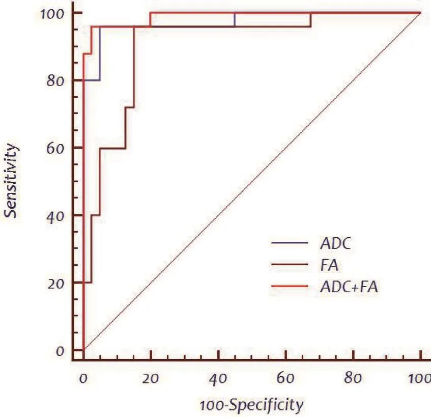
ADC值、FA值及两者联合ROC曲线上(图3):诊断外周带PCa的ADC阈值为1.135×10⁻³mm2/s,敏感度、特异度分别为96.0%和95.0%;FA阈值为0.196,敏感度、特异度分别为96.0%和85.0%;ADC值、FA值及两者联合的AUC分别为0.974,0.907和0.990,两者联合诊断效能较高;两两比较中,ADC值与FA值、ADC值与两者联合,差异均无统计学意义(z值分别为 1.498、1.312,P>0.05),FA值与两者联合比较,差异有统计学意义(z=2.151,P<0.05)。

图1 男,77岁,尿血2月余,右侧外周带PCa。A.压脂T2WI图,右侧外周带低信号结节(箭),形态不规则;B.DWI图呈明显高信号(箭);C.ADC图,病灶ADC值明显减低(箭),对应ROI1为0.789×10⁻³mm2/s; D.FA图,呈明显高信号(箭),对应FA值0.224。

图2 男,69岁,会阴部疼痛数月,左侧外周带慢性前列腺炎(CP)。A.压脂T2WI图,左侧外周带低信号弥漫性减低(箭);B.DWI图呈稍高信号(箭);C.ADC图,病灶ADC值稍减低(箭),对应ROI1为1.237×10⁻³mm2/s; D.FA图,呈稍高信号(箭),对应FA值0.168。
讨 论
1.DTI的理论基础及临床应用
DTI是DWI技术的延伸与发展,不但能测量组织水分子扩散运动速度,而且可显示其运动的方向性,对水分子的扩散运动描述更加精确,更好地推测组织内部微观结构状态的细微变化,其主要评价参数[7]:ADC和FA值,ADC值用于描述水分子扩散运动的速度和范围,其值越大,表明水分子的扩散能力越强,反之,则扩散受限;FA值指水分子各向异性成分占整个扩散张量的比例,取值范围0~1,FA值趋近于1,各向异性越大,接近于0,表示各向同性。

图3 ADC值、FA值及ADC+FA诊断外周带PCa的ROC曲线。
前列腺DTI对PCa成像的ADC参数变化基本与DWI相同,近年来的研究已达成初步共识。通过测定ADC值,可定性、定量评估PCa[8-9],Jie等[10]通过Meta分析表明测定ADC值可以为PCa的诊断提供丰富的信息,并表现出较高的准确性;Nagel等[11]通过比较正常前列腺、前列腺炎及低级别、高级别PCa的ADC值差异,提示ADC 值有助于PCa的鉴别诊断;Rud等[12]通过对42例体外放疗后复发PCa患者的研究表明,ADC值对PCa放疗后复发的诊断具有高度敏感性(95%);Tamada等[13]采用1.5T MR研究发现前列腺外周带癌区ADC值为(1.02 ±0.25)×10-3mm2/s,正常外周带 ADC 值为(1.80±0.27)×10-3mm2/s (P < 0.01)与本研究结果相仿。而对FA参数变化尚存在分歧,多数学者认为可能与样本差异、成像设备、癌灶的病理特点等多因素有关。Gürses 等[6]认为,PCa的FA值高于前列腺炎和正常前列腺组织,与笔者研究相符,而Manenti 等[14]认为,PCa 的FA值低于正常前列腺组织,Xu等[15]则报道前列腺外周带癌区和非癌区的FA值之间无显著差异。
2.DTI对外周带PCa诊断效能的ROC曲线分析
夏国金等[16]研究显示ADC值阈值为0.725×10-3mm2/s,敏感度为100.0%,特异度为96.0%,AUC为0.996;FA值阈值为0.311,敏感度为100.0%,特异度为68.7%,AUC为0.904。Yoon等[17]行ROC曲线分析显示,ADC值阈值为1.65×10-3mm2/s,AUC为0.914,敏感度为87.2%,特异度为71.9%;FA值阈值为0.55,AUC为0.983,敏感度和特异度分别为99.1%和88.5%,本研究与夏国金等研究结果接近,ADC值诊断效能(0.974)较之稍低,FA值诊断效能(0.907)较之稍高,而与Yoon等研究结果有所差异。这种结果的差异,笔者认为一方面源自抽样误差,另一方面可能是由于纳入的受检者年龄、患者数、设备和参数不同有关。Kozlowski等[18]对DTI用于PCa诊断的效能进行评估,研究显示DTI用于PCa诊断的敏感度和特异度分别为81%、85%,而单独应用ADC参数诊断PCa的ROC曲线下面积明显高于FA参数,提示ADC值较FA值更有助于PCa的诊断,与本研究结果符合,ADC值和FA值对外周带PCa的诊断都具有高效能,AUC分别为0.974、0.907,两者之间差异无统计学意义。FA值联合ADC值诊断效能更高,与单独使用FA值的AUC比较,差异有统计学意义,而与ADC值的AUC比较无统计学差异,与Gibbs等[19]研究的结果一致。
3.本研究的不足
最主要的是研究中作为诊断金标准的病理结果主要来源于前列腺的穿刺活检,取得的病理组织有限,容易漏掉微小癌灶,从而引起良恶性分组的混淆,与根治术的病理尚存在偏差。穿刺点与MR图像的匹配程度也有待商榷。 样本量有限,可能会存在抽样误差,导致研究结果存在一定的偏差。心脏相控阵线圈相对于直肠内线圈,信噪比稍低,DTI各参数的测值存在少许误差。
4.展望
DTI作为MRI功能成像可提供前列腺癌早期的功能改变,其评价参数的量化指标对组织微观结构的变化更能精准地反映,随着对前列腺疾病病理改变的深入研究、成像设备的升级及序列的优化,更多临床研究资料的积累,解决研究中的存在问题及争议,DTI在前列腺疾病诊治中的应用前景将更加广阔。
[1]Siegel RL, Miller KD, Jemal A.Cancer statistics,2016.CA Cancer J Clin,2016,66:7-30.
[2]Chen W, Zheng R, Baade PD, et al. Cancer statistics in China, 2015.CA Cancer J Clin,2016,66:115-132.
[3]Delongchamps NB, Rouanne M, Flam T, et al. Multiparametric magnetic resonance imaging for the detection and localization of prostate cancer: combination of T2-weighted, dynamic contrastenhanced and diffusion-weighted imaging. BJU Int,2011,107:1411-1418.
[4]Turkbey B,Choyke PL.Multiparametric MRI and prostate cancer diagnosis and risk stratification.Curr Opin Urol,2012,22:310-315.
[5]Esen M,Onur MR,Akpolat N,et al.Utility of ADC measurement on diffusion-weighted MRI in differentiation of prostate cancer,normal prostate and prostatitis.Quant Imaging Med Surg,2013,3:210-216.
[6]Gürses B,Tasdelen N,Yencilek F,et al.Diagnostic utility of DTI in prostate cancer.European Journal of Radiology,2011,79: 172-176.
[7]Li C,Chen M,Li S,et al. Diffusion tension imaging of prostate at 3.0 Tesla. Acta Radiol, 2011, 52: 813-817.
[8]李 鹏,刘家赵,陈志强,等.3.0 T MR扩散加权成像定量诊断前列腺疾病.临床放射学杂志,2014,33:536-539.
[9]Durmus T,Baur A,Hamm B.Multiparametric magnetic resonance imaging in the detection of prostate cancer.Aktuelle Urol,2014,45:119-126.
[10]Jie C, Rongbo L, Ping T. The value of diffusion-weighted imaging in the detection of prostate cancer: a meta-analysis . Eur Radiol, 2014,24: 1929-1941.
[11]Nagel KN,Schouten MG,Hambrock T,et al. Differentiation of prostatitis and prostate cancer by using diffusion weighted MR imaging and MR-guided biopsy at 3 T .Radiology,2013,267:164-172.
[12]Rud E, Baco E, Lien D, et al. Detection of radiorecurrent prostate cancer using diffusion -weighted imaging and targeted biopsies. AJR,2014, 202:W241-246.
[13]Tamada T,Sone T,Jo Y,et al.Apparent diffusion coef ficient values in peripheral and transition zones of the prostate comparison between normal and malignant prostatic tissues and correlation with histologic grade.J Magn Reson Imaging,2008,28: 720-726.
[14]Manenti G, Carlani M, Mancino S, et al. Diffusion tensor magnetic resonance imaging of prostate cancer . Invest Radiol,2007, 42: 412-419.
[15]Xu J, Humphrey PA, Kibel AS, et al. Magnetic resonance diffusion characteristics of histologically defined prostate cancer in humans.Magn Reson Med, 2009, 61: 842-850.
[16]夏国金,龚洪翰,曾献军,等.MR扩散张量成像在前列腺癌诊断中的价值.中华放射学杂志,2012,46:526-528.
[17]Yoon SK,Kim DW,Ha DH,et al.Value of diffusion tensor imaging of prostate cancer:comparison with systemic prostate biopsy.J Korean Soc Radiol,2011,64:179-184.
[18]Kozlowski P,Chang SD,Meng R,et a1.Combined prostate diffusion tensor imaging and dynamic contrast enhanced MRI at 3 T-quantitative correlation with biopsy.Magn Reson Imaging,2010,28:621-628.
[19]Gibbs P,Pickles M,Turnbull LW. Diffusion imaging of the prostate at 3.0 tesla.Invest Radiol,2006,41:185-188.
更 正
本刊2017年第2期《多排螺旋CT对儿童颅骨膜血窦的诊断价值》作者唐文伟的单位应为“南京医科大学附属妇产医院。特此更正并向作者致歉。
(本刊编辑部)
Quantitative Analysis of Prostate Cancer in Peripheral Zone with 3.0T MR Diffusion Tensor Imaging
JI Guang-hai1,ZHENG Yi1,YE Ping1, LI Peng2,CAI Lei2,CHEN Zhi-qiang2
Purpose:To explore the diagnostic values of diffusion tensor imaging (DTI) in prostate cancer in peripheral zone.Methods:Twenty-five patients with prostate cancer in peripheral zone (malignant group) and 40 with benign prostate hyperplasia (BPH) and/or chronic prostatitis (CP) (benign group), who were all confirmed by biopsy results, were undergone conventional MRI, diffusion-weighted Imaging (DWI) and diffusion tensor imaging(DTI). Receiver operating characteristic (ROC) curves were used to compare the diagnostic efficiency of ADC value,FA value and the combination of them. The cutoff threshold of ADC value and FA value were determined.Results:The mean ADC value and FA value of the cancerous and noncancerous regions were (0.89±0.19)×10-3mm2/s, 0.25±0.05 and (1.46±0.23)×10-3mm2/s, 0.17±0.04, respectively. There were statistical significant differences of ADC value and FA value between the cancerous and noncancerous regions (t=10.414, -7.789, P<0.05). Using the ADC value of 1.135×10⁻³mm2/s and the FA value of 0.196 as the cutoff threshold, the sensitivity and specificity were 96.0%, 95.0%and 96.0%, 85.0%, respectively. When ADC value, FA value and combination of them were used as criteria, the areaunder curve (AUC) of ROC were 0.974,0.907 and 0.990,95% CI were 0.900-0.998,0.809-0.965 and 0.926-1.000,respectively. For the comparison of the differences of AUC for the ADC and FA value, the ADC value and the combination, the FA value and the combination, their z values and P values were 1.498, 1.312, 2.151, and 0.134, 0.190,0.032, respectively.Conclusion:Both the ADC value and FA value provide valuable information for the diagnosis and differential diagnosis of PCa. The ADC value has higher diagnostic efficiency than the FA value. The difference of diagnostic efficacy between the combination criteria and the ADC value was with no statistical significant, and the difference between the combination criteria and the FA value was with statistical significant.
Magnetic resonance imaging; Diffusion tensor imaging; Prostatic neoplasms
Natural Science Foundation of Ningxia Province(NZ13280、NZ1234)
R445.2
A
1006-5741(2017)-03-0242-05
2016.07.08;修回时间:2016.09.30)
中国医学计算机成像杂志,2017,23:242-246
1 宁夏医科大学临床医学院
2 宁夏医科大学总医院放射科
通信地址:宁夏回族自治区银川市兴庆区胜利南街804号 ,银川750004
陈志强(电子邮箱:zhiqiang_chen99@163.com)
宁夏自然科学基金(NZ13280、NZ1234)
Chin Comput Med Imag,2017,23:242-246
1 Clinical Medicine School of Ningxia Medical University
2 Department of Radiology,General Hospital of Ningxia Medical University
Address: 804 Shengli Road, Yinchuan 750004,P.R.C
Address Correspondence to CHEN Zhi-qiang (E-mail: zhiqiang_chen99@163.com)
