晚期非小细胞肺癌原发灶和转移灶ROS1融合基因表达差异分析
2017-07-01刘晓辉
刘晓辉
晚期非小细胞肺癌原发灶和转移灶ROS1融合基因表达差异分析
刘晓辉
目的 分析ROS1(c-ros oncogene 1,receptor tyrosine kinase)融合基因在晚期非小细胞肺癌原发灶和转移灶中的差异表达。方法 选取非小细胞肺癌原发灶162例,其中配对转移灶139例。采用实时荧光定量PCR检测ROS1融合基因的表达,并分析其在晚期非小细胞肺癌原发灶和转移灶中的表达差异。结果 晚期非小细胞肺癌原发灶ROS1融合基因表达阳性率4.3%(7/162),配对原发灶ROS1融合基因表达阳性率2.6%(5/139),配对转移灶融合基因表达阳性率2.2%(3/139);原发灶较转移灶ROS1融合基因检出阳性率显著增高,差异具统计学意义(χ2=13.517,P=0.000);ROS1融合基因阳性表达在原发灶和转移灶一致性好(κ=0.479,P=0.000)。非小细胞肺癌原发灶中ROS1融合基因表达与病理类型存在密切关系(χ2=5.195,P=0.031),与患者性别、年龄等不存在明显相关性(P>0.05)。非小细胞肺癌配对原发灶以及转移灶中ROS1融合基因表达与患者性别、年龄、吸烟以及病理类型不存在明显相关性(P>0.05)。结论 晚期非小细胞肺癌ROS1融合基因的阳性表达在原发灶中与病理类型有关,其在配对原发灶和转移灶中阳性表达一致性较好,可作为检测ROS1融合基因的备选手段。
ROS1融合基因;非小细胞肺癌;原发灶;转移灶
(ThePracticalJournalofCancer,2017,32:891~893)
肺癌是恶性肿瘤死亡的主要原因,其中非小细胞肺癌占所有肺癌的80%以上,大约七成的非小细胞肺癌确诊时多为晚期,临床上采用综合治疗非小细胞肺癌疗效并不理想,尤其晚期非小细胞肺癌5年生存率仅为15%[1]。目前临床上关于ROS1的研究成为热点。ROS1融合基因是1个肿瘤驱动基因,属于酪氨酸激酶胰岛素受体基因家族[2],其在肺癌分子治疗靶点中的应用较广[3],因此检测晚期非小细胞肺癌患者ROS1的表达显得愈加重要。
1 材料与方法
1.1 一般资料
选取我院2013年至2015年非小细胞肺癌患者162例,所有病例经病理证实为Ⅲb~Ⅳ期非小细胞肺癌。所有肺癌患者术前未接受化疗、放疗或生物免疫治疗。取原发灶标本162例,其中与转移灶配对原发灶标本139例,转移灶主要包括淋巴结、脑、骨、肺内等,多以淋巴结为主。
1.2 方法
采用实时荧光定量PCR(Quantitative Real-time PCR)检测非小细胞肺癌患者原发灶和转移灶中ROS1融合基因。
1.3 统计学分析
采用统计学软件SPSS17.0处理数据,计数资料行卡方检验,以P<0.05为差异具统计学意义。采用Fisher确切概率法计算一致性,κ>0.75为一致性极好,κ<0.4为一致性差,κ居于两者之间为一致性良好。
2 结果
2.1 ROS1在晚期非小细胞肺癌原发灶和转移灶中的表达
晚期非小细胞肺癌原发灶ROS1融合基因表达阳性率4.3%(7/162),配对原发灶ROS1融合基因表达阳性率2.6%(5/139),配对转移灶融合基因表达阳性率2.2%(3/139)。原发灶较转移灶ROS1融合基因检出阳性率显著增高,差异具统计学意义(χ2=13.517,P=0.000)。ROS1融合基因阳性表达在原发灶和转移灶一致性好(κ=0.479,P=0.000)。见表1。

表1 ROS1在晚期非小细胞肺癌配对原发灶和转移灶中的表达/例
2.2 162例原发灶患者临床资料与ROS1融合基因的关系
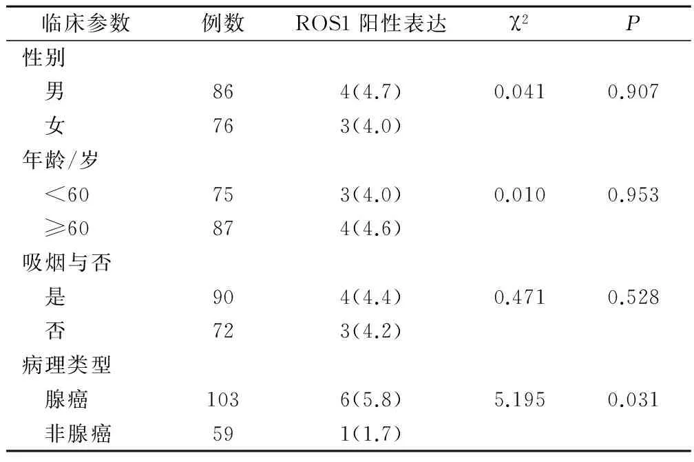
非小细胞肺癌原发灶中ROS1融合基因表达与病理类型存在密切关系(χ2=5.195,P=0.031),与患者性别、年龄等不存在明显相关性(P>0.05)。见表2。

表2 162例原发灶患者临床资料与ROS1融合基因的关系(例,%)
2.3 139例配对原发灶及转移灶患者临床资料与ROS1融合基因的关系
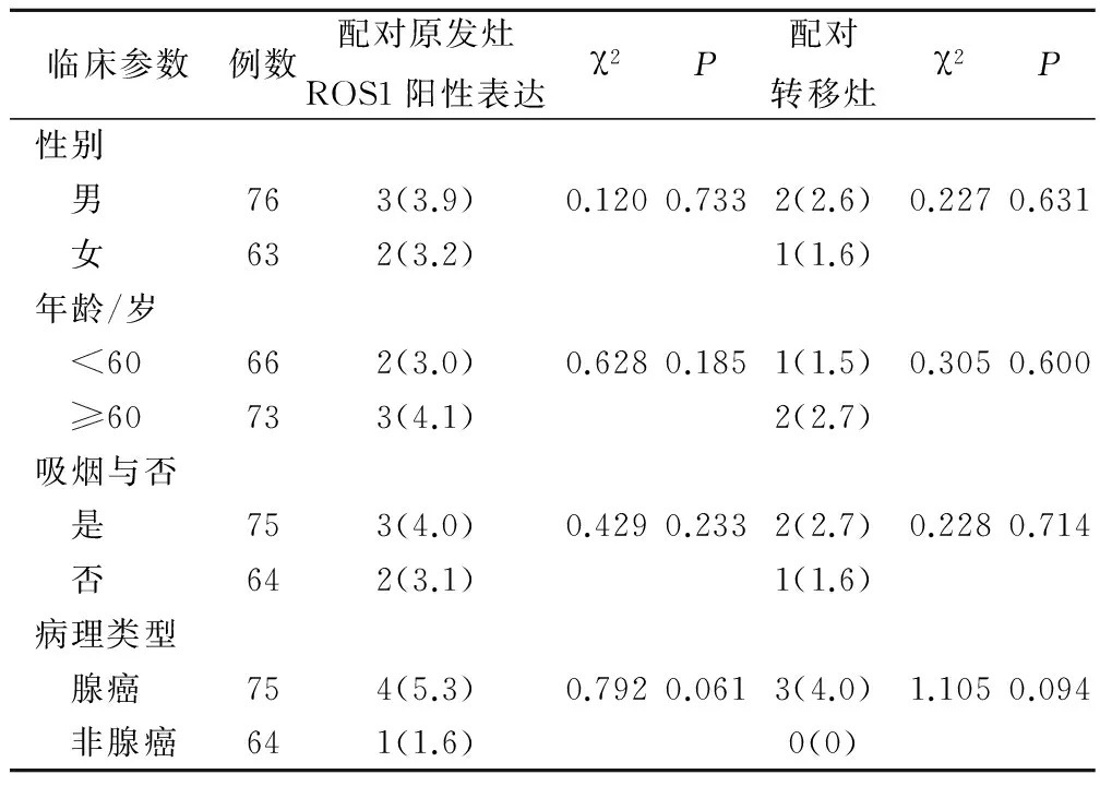
非小细胞肺癌配对原发灶及转移灶中ROS1融合基因表达与患者性别、年龄、吸烟以及病理类型均不存在明显相关性(P>0.05)。见表3。

表3 139例配对原发灶患者临床资料与ROS1融合基因的关系(例,%)
3 讨论
随着精准医学时代的到来,越来越多的肿瘤驱动基因被发现,人们对肺癌的发生发展也有极大了解,靶向治疗药物给临床癌症治疗带来了希望[4]。ROS1融合基因的产生是由于ROS1基因重排导致,ROS1基因重排发生在非小细胞肺癌、卵巢癌等多种恶性肿瘤中[5-6]。
ROS1融合基因表达阳性的肺癌患者例数较少,其在非小细胞肺癌中的发生率为1%~2%,所以目前临床上关于ROS1融合基因表达特征的大样本资料较少[7],本研究中晚期非小细胞肺癌原发灶ROS1融合基因表达阳性率4.3%(7/162),配对原发灶ROS1融合基因表达阳性率2.6%(5/139),配对转移灶融合基因表达阳性率2.2%(3/139),相较于文献报道本研究中ROS1融合基因表达阳性率较高[8],可能原因为本研究纳入研究的晚期非小细胞肺癌例数较少的缘故。ROS1融合基因在肺癌原发灶和转移灶中的表达存在差异,原发灶较转移灶ROS1融合基因检出阳性率显著增高,差异具统计学意义(χ2=13.517,P=0.000);但ROS1融合基因阳性表达在原发灶和转移灶一致性好(κ=0.479,P=0.000)。原发灶和转移灶具较好的表达一致性,则转移灶组织病理基因检测对靶向治疗有较好的预测性,但因肿瘤病灶存在一定的异质性,使得ROS1融合基因在肺癌原发灶和转移灶的表达并非完全一致,临床上我们应该对肿瘤转移灶多地取材以提高检测结果的可靠性[9]。非小细胞肺癌中ROS1重排具独特的临床病理特征,哈佛大学Bergethon等[10]的研究结果显示ROS1表达阳性患者具有年轻化、未吸烟以及亚洲占多数的临床特征,本研究中5例非小细胞肺癌中ROS1表达阳性患者中4例为腺癌,1例为非腺癌,得出非小细胞肺癌原发灶中ROS1融合基因表达与病理类型存在密切关系(χ2=5.195,P=0.031)。本研究结果显示非小细胞肺癌原发灶中ROS1融合基因表达与患者性别、年龄等不存在明显相关性,且非小细胞肺癌配对原发灶以及转移灶中ROS1融合基因表达与患者性别、年龄、吸烟以及病理类型亦不存在明显相关性。本研究结果与Cai等[11]的小样本研究认为ROS1阳性表达临床特征与年龄、性别以及吸烟无关的研究结果相一致,但临床上关于ROS1融合基因表达临床特征的研究还较少,有待于进一步考证。
综上所述,ROS1融合基因表达在晚期非小细胞肺癌患者中阳性表达率较低,其在原发灶中的表达与病理类型有关,在转移灶中与病理类型无关,但ROS1融合基因表达在晚期非小细胞肺癌患者原发灶和转移灶中具较好的表达一致性,转移灶ROS1融合基因检测可作为备选手段。
[1] De Santis CE,Lin CC,Mariotto AB,et al.Cancer treatment and survivorship statistics,2014〔J〕.CA Cancer J Clin,2014,64(4):252-271.
[2] Wu YL,Zhou C,Liam CK,et al.First-line erlotinib versus gemcitabine/cisplatin in patients with advanced EGFR mutation-positive non-small-cell lung cancer: analyses from the phase Ⅲ,randomized,open-label,ENSURE study〔J〕.Ann Oncol,2015,26(9):1883-1889.
[3] 赵 睿,常小红.分子靶向治疗在非小细胞肺癌中的研究进展〔J〕.实用癌症杂志,2014,29(3):364-366.
[4] Mitsudomi T,Morita S,Yatabe Y,et al.Gefitinib versus cisplatin plusdocetaxel in patients with non-small-cell lungcancer harbouring mutations of the epidermal growth factor receptor (WJTOG3405):an open label,randomised phase 3 trial〔J〕.Lancet Oncol,2010,11(2):121-128.
[5] Rimkunas VM,Crosby KE,Li D,et al.Analysis of receptor tyrosine kinase ROS1-positive tumors in non-small-cell lung cancer:identification of a FIG- ROS1 fusion〔J〕.Clin Cancer Res,2012,18(16):4449-4457.
[6] Birch AH,Arcand SL,Oros KK,et al.Chromosome 3 anomalies investigated by genome wide SNP analysis of benign,low malignant potential and low grade ovarian serous tumours〔J〕.PLoS One,2011,6(12):e28250.
[7] Go H,Kim DW,Kim D,et al.Clinicopathologic analysis of ROS1-rearranged non-small-cell lung cancer and proposalof a diagnostic algorithm〔J〕.J Thorac Oncol,2013,8(11):1445-1450.
[8] Takeuchi K,Choi YL,Togashi Y,et al.KIF5B-ALK,a novel fusion oncokinase identified by an immunohistochemistry-based diagnostic system for ALK-positive lung cancer〔J〕.Clin Cancer Res,2009,15(9):3143-3149.
[9] 王 琳,亓岽东,许春伟,等.非小细胞肺癌患者组织原发灶和转移灶中ROS1融合基因的研究〔J〕.临床与病理杂志,2016,36(2):148-153.
[10] Bergethon K,Shaw AT,Ou SH,et al.ROS1 rearrangements define a unique molecular class of lung cancers〔J〕.J Clin Oncol,2012,30(8):863-870.
[11] Cai W,Li X,Su C,et al.ROS1 fusions in Chinese patients with non-small-cell lung cancer〔J〕.Ann Oncol,2013,24(7):1822-1827.
(编辑:甘 艳)
Differential Analysis of the ROS1 Fusion Gene Expression in Patients with Advanced Non Small Cell Lung Cancer in Primary and Metastatic Foci
LIUXiaohui.
InnerMongoliaChifengInstituteofMedicineAffiliatedHospital,Chifeng,024000
Objective To study the differential expression of ROS(c-ros oncogene 1,receptor tyrosine kinase)fusion gene in primary tumor and metastasis foci of advanced non small cell lung cancer.Methods 162 cases with non-small-cell lung cancer were collected,which paired metastases 139 cases,detected the expression of ROS1 fusion gene using real-time fluorescence quantitative PCR,and analysis the differential expression of the ROS1 in primary and metastatic foci in advanced non-small cell lung cancer.Results The positive rate of ROS1 fusion gene expression in primary tumor with advanced non-small cell lung cancer was 4.3% (7/162),the positive rate of ROS1 fusion gene expression in metastasis foci was 2.6% (5/139),the positive expression rate of paired metastasis fusion gene was 2.2% (3/139);The positive rate of ROS1 fusion gene was significantly higher than that of the primary lesion,and the difference was statistically significant (χ2=13.517,P=0.000);There was a close relationship between ROS1 fusion gene expression and pathological type in non small cell lung cancer (χ2=5.195,P=0.031),and there was no significant correlation with the gender,age,and so on (P>0.05).There was no significant correlation with the gender,age,smoking and pathological type of the patients in the expression of ROS1 fusion gene in paired primary tumor and metastasis foci of the advanced non-small-cell lung cancer(P>0.05).Conclusion The positive expression of ROS1 fusion gene in patients with advanced non-small cell lung cancer is related to the pathological type,the consistency of positive expression of ROS1 is good in the primary tumor and metastatic lesions,it can be used as an alternative means of detecting ROS1 fusion gene.
ROS1 fusion gene;Non-small cell lung cancer;Primary tumor;Metastasis
024000 内蒙古自治区赤峰学院医学院;024000 内蒙古赤峰市赤峰学院附属医院
10.3969/j.issn.1001-5930.2017.06.006
R734.2
A
1001-5930(2017)06-0891-03
2016-08-08
2017-03-09)
