心电图参数预测心脏再同步治疗患者发生超反应的意义
2017-04-20王玉占
王玉占,刘 娟
心电图参数预测心脏再同步治疗患者发生超反应的意义
王玉占,刘 娟
目的 探讨12导联心电图(ECG)参数预测心脏再同步治疗患者发生超反应的意义。 方法 采用ECG记录心脏再同步治疗(CRT)设备植入前基线水平及植入后即刻心电活动,分析基线ECG参数(QRS波时限、束支形态、电轴、PQ间期、QT间期)及植入后起搏QRS时限,并计算QRS时限的相对变化。超反应定义为12个月后左室舒张末期容积降低≥30%。结果 本组101例患者,32例(31.7%)出现超反应。超反应组与非超反应组之间ECG基线水平差异无统计学意义,但超反应组起搏QRS时限短于非超反应组[(148±22)msvs(162±28)ms;P=0.010]。CRT植入后超反应组出现QRS波显著降低。超反应组出现相对QRS波缩短的比例显著高于非超反应组[12.1%(6.8~22.2)vs1.7%(-11.9~11.8);P=0.005]。采用多变量分析得出,NYHAⅡ级、起搏QRS时限及相对QRS波缩短是预测CRT患者发生超反应的因素。 结论 起搏QRS时限及相对QRS波缩短是与CRT患者发生超反应相关的参数。
心脏再同步治疗(CRT);心电图;超反应
不管是心脏再同步治疗(CRT)⁃起搏(CRT⁃P)或CRT⁃除颤(CRT⁃D),CRT都能够改善顽固性心衰患者的症状、提高运动量、引起左心室重构逆转,从而降低心衰患者住院率和死亡率[1⁃3]。目前,一些多中心随机研究已证实,CRT治疗可以改善超声心动图征象,其CRT反应指左心室收缩末容积(LVESV)下降≥15%[4]。然而,由于CRT的反应个体间存在着显著差异,有些患者临床症状改善显著,左室完全逆转重构,左室功能几乎正常,这在临床上称为“CRT超反应”[5]。鉴于超声心动图参数不具同步性,其预测CRT反应性较差[6]。与此相反,研究表明QRS时限及形态学与CRT治疗预后紧密相关[7]。本研究通过检测CRT超反应者的心电图(ECG)参数,探讨其预测CRT超反应的临床价值。
1 资料与方法
1.1 一般资料 收集我院2013年1月至2015年6月接受CRT的慢性充血性心力衰竭(CHF)患者。入选标准:12导联心电图QRS波时限≥120 ms,纽约心功能分级(NYHA)Ⅱ~Ⅳ级,左心室射血分数(LVEF)<35%,最佳药物治疗基础,窦性心律。排除标准:瓣膜病,最近3个月曾接受冠状动脉支架植入术或搭桥术,肝肾功能不全。分组:随访期间完善详尽的临床评估、ECG以及心超,根据CRT术后1年LVEF增加情况分为,研究组(超反应组):LVEF≥30%以及对照组(无反应组及有反应组):增加<5%及LVEF增加≥5%但LVEF<30%为CRT(CRT⁃R)。
1.2 CRT植入 锁骨下静脉穿刺成功后,制作囊袋,采用冠状静脉引导系统,定位冠状窦开口,应用静脉球囊导管行冠状静脉造影,显示心脏静脉后,将左心室心外膜电极导线送至心脏静脉的侧支(心脏侧静脉或侧后静脉)。测试左室起搏夺获的最满意参数后,再分别植入右心房电极导线于右心耳及右心室电极导线于右心室心尖部,固定电极导线后连接双室脉冲发生器,将起搏器埋藏于皮下囊袋中,缝合皮肤。
1.3 超声心动图 所有患者均在入院及CRT植入术后12个月行超声检查。采用GE Vivid 7超声诊断仪,M4S探头,频率为1.5~4.3 MHz;分别采集胸骨旁左心室长轴切面、左心室二尖瓣、乳头肌和心尖短轴切面及心尖四腔心、二腔心和三腔心切面3个心动周期二维和彩色多普勒图像进行测量,测量指标:左心室舒张末容积(LVEDV)、左心室收缩末容积(LVESV)及LVEF。
1.4 12导联心电图 患者取仰卧位,在双心室起搏器植入前及植入后即刻进行12导联ECG测量。测量结果均有本研究之外的两名心电图专业医师收集及分析,其主要包括:心率(HR)、QRS时限及电轴、PR及QTc间期。如果两者出现分歧,则重复测量直至结果达成统一。①左束支传导阻滞(LBBB)传统定义为符合以下三条标准中任意一条:V1导联呈QS或rS型,QRS时限≥120 ms,I、V6呈单向R波,前无Q波。②相对QRS时限变化定义为:QRSCRT-QRS基线/QRS基线(%),其中,负值表示相对QRS时限缩短。
1.5 统计学分析 所有数据采用SPSS 21.0软件包进行分析。采用柯尔莫诺夫⁃斯米尔诺夫检验来分析变量是否符合某种分布。正态分布连续变量以均数±标准差表示,比较采用t检验。非正态分布连续变量以中位数(四分位间距)表示,比较采用Wilcoxon符号秩和检验或Mann⁃Whitney U检验。分类变量以频率及百分比表示,比较采用卡方检验或Fisher精确检验。超反应预测因素采用单变量及多变量逻辑回归分析。将单变量分析中P<0.2变量纳入前进逐步回归程序。通过多变量逻辑回归分析获得最佳模型,把影响超反应的最佳独立变量设为独立变量。计算超反应比值比(OR)以及95%可信区间。采用受试者工作特征曲线(ROC)确定QRS时限变量预测CRT超反应最佳界限值。以双侧P<0.05为差异具有统计学意义。
2 结 果
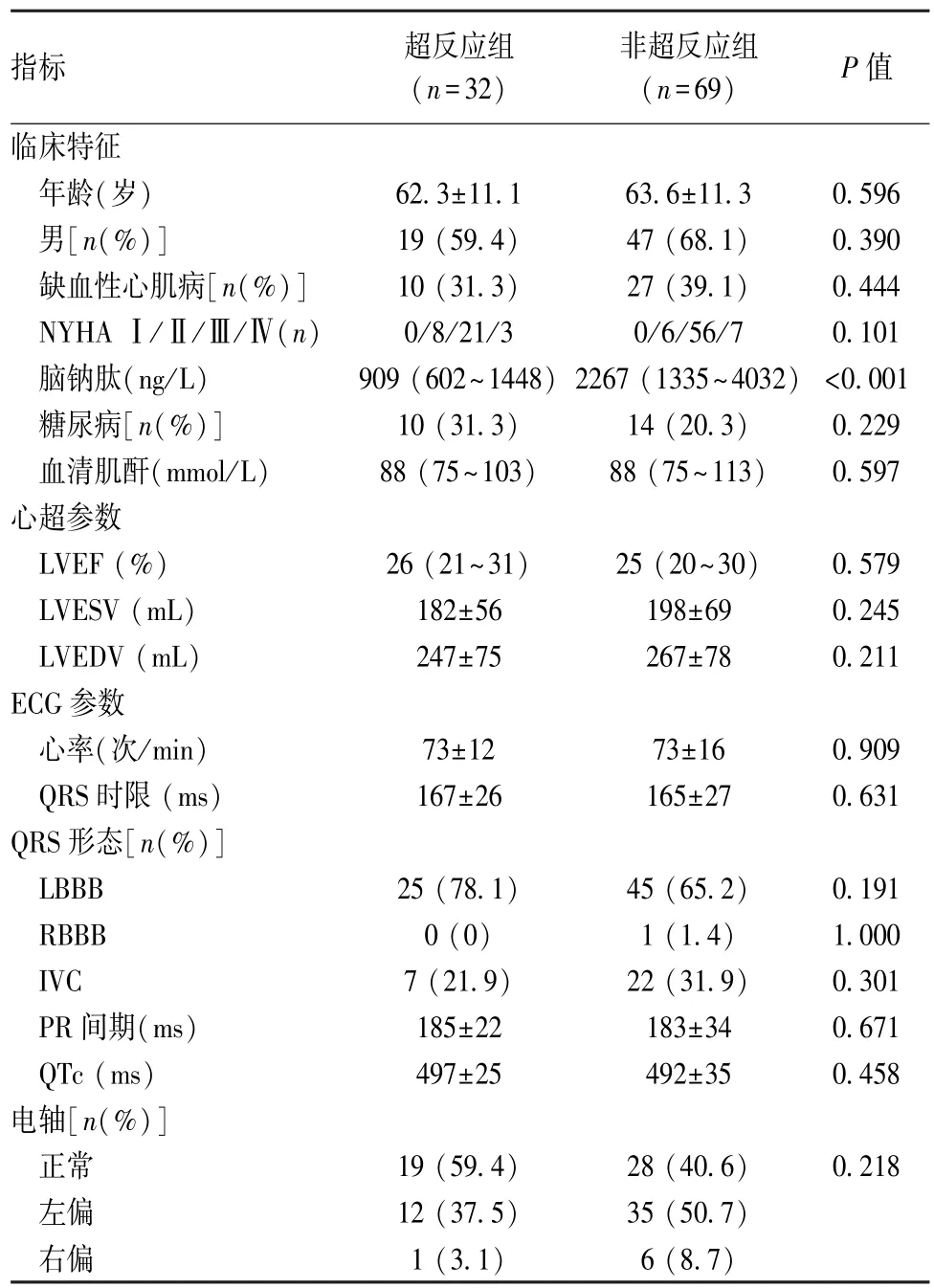
2.1 研究对象特征 本研究纳入101例合格患者,平均年龄(63.2±10.9)岁,其中男性占65.2%。缺血性心肌病37例(36.6%)。多数患者NYHA评分为Ⅲ(77例;76.2%)。LVEF中位值25%(20.0~30.0)。所有患者均为窦性心律。基线ECG特征及其他临床表现见表1。口服药物:99例服用ACEI或血管紧张素Ⅱ受体阻滞剂,100例服用β受体阻滞剂,90例服用螺内酯,70例使用利尿剂。

表1 超反应组及非超反应组基线特征及比较
2.2 超反应组及非超反应组临床表现、ECG及心超特征 双心室起搏植入后12个月,32例患者符合心超诊断超反应的标准。15例患者LVESV降低15%~29%,54例患者LVESV没有显著降低(0%~14%),部分甚至升高。超反应与非超反应组临床表现及基线心超参数比较见表1。超反应组植入前脑钠肽(BNP)显著低于非超反应组,两组在其余心超参数、临床表现及心衰药物治疗上差异无统计学意义。两组植入前LVESV见表1,植入后,超反应组LVESV(94.2±34.7)mL,而非超反应组为(194.1±70.1)mL。两组左心室电极位置(多位于侧后静脉)及无起搏方式显著差异(P=0.127)。超反应与非超反应组基线心率、PR间期、QRS时限、QRS轴及QTc间期差异均无统计学意义。两组患者QRS形态学无显著差异,且LBBB出现QRS切迹比例亦无显著差异[25(78.1%)vs 42(60.9%);P=0.088]。超反应组在植入后QRS时限短于非超反应组[(148± 22)ms vs(162±28)ms;P=0.010],与植入前相比,QRS时限显著降低[(167±26)ms降至(148±22)ms;P=0.010],而非超反应组则无显著降低[(165±27)ms到(162±28)ms;P=0.536]。超反应组QRS波形相对缩短比例显著高于非超反应组[12.1%(6.8~22.2)vs 1.7%(-11.9~11.8);P=0.005]。
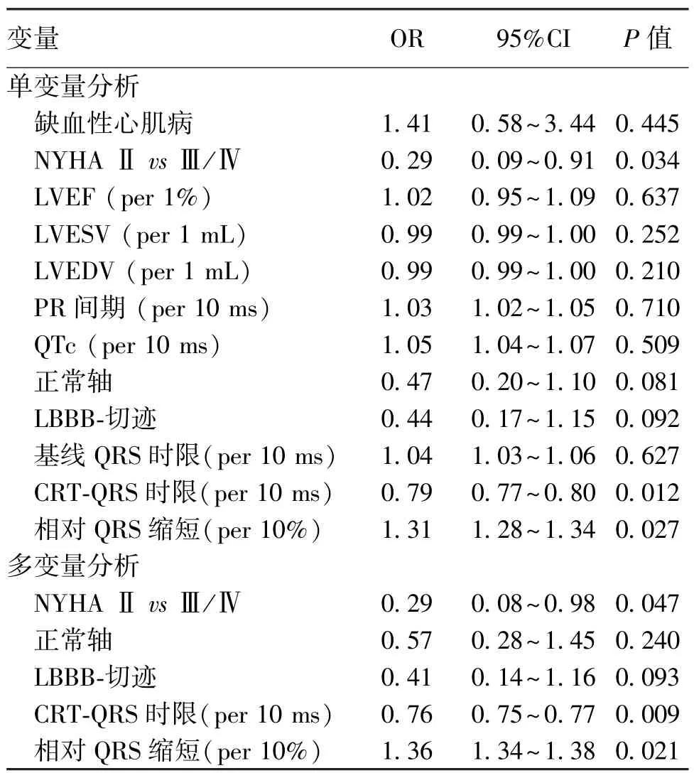
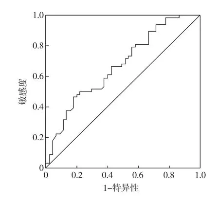
2.3 预测CRT超反应 单变量分析得出,与超反应相关的变量包括NYHA分级、QRS时限及相对缩短的QRS波。将单因素分析中P<0.2变量(NYHA分级、正常轴、LBBB出现切迹、植入后QRS时限、相对QRS缩短)采用多变量逻辑回归模式进行校正,得出QRS时限与QRS缩短与CRT超反应独立相关。见表2。为进一步明确QRS时间参数预测CRT超反应的最佳界值,采用ROC曲线分析。相对QRS缩短预测超反应的曲线(AUC=0.68,95%CI 0.57~0.78),相对QRS缩短预测CRT超反应界值>4.5%,其敏感性及特异性分别为81%、58%。见图1。

表2 单因素及多因素回归模型预测CRT超反应

图1 相对QRS时限缩短预测CRT超反应ROC曲线
3 讨 论
已有研究证实,CRT治疗可使心衰患者心超表现得以改善[8]。但临床中也发现,不同患者个体对CRT反应的程度差异较大,部分患者出现CRT超反应[9]。目前关于 CRT超反应的定义无同一标准。由于对超反应的定义不同,相应研究报道的超反应患者比例亦不尽相同(13%~38%)[10⁃11]。CRT治疗后LVESV减小反应LV结构逆重构,其是预测CRT长期预后的重要因素[12⁃13],结合文献报道,我们定义LVESV降低≥30%为CRT超反应。本研究发生CRT超反应患者比例(31.7%)与采用相同定义的研究基本一致。此外,各项研究间CRT后随访时间范围广(2个月到12个月)[14⁃15],CRT治疗后,LV逆重构至少需要9个月的时间[16],过早随访可能会导致出现超反应数目较实际减少,从而造成结果差异。同样,超反应定义的不同也会导致超反应预测因素的多样化。先前有研究发现,CRT植入前一些临床特征及心超参数对于预测CRT超反应有一定的意义,如女性、非缺血性心肌病、低BNP水平、基线LV直径短及心室机械不同步[17]。虽然本研究结果也发现,超反应组具有较低的BNP水平以及NYHA分级,但其他基线临床特征及心超特征与非超反应组比较均无显著差异。
CRT通过改变心电活动的顺序从而改善左心室机械功能[18],因此,CRT可以说是一种“电活动”治疗。心超通过检测二尖瓣反流程度和左心室舒张模式,可为临床选择CRT适应证提供的信息[19],但其不能同步反应心电活动制约了预测CRT反应性,而ECG参数可以表现心电活动紊乱及改善的状态,从这点来看,这应该是预测CRT反应的良好预测工具。虽然诸多研究证实,CRT后QRS时限及QRS时限缩短与心脏重构的总体改善存在显著相关,但其研究没有涉及超反应。本研究中,我们得出植入后QRS时限与相对QRS时限缩短是预测超反应的独立因素。相对QRS缩短预测CRT超反应界值>4.5%,其敏感性及特异性分别为81%、58%。
目前,基线QRS时限对CRT超反应的预测价值也存在差异,本研究得出,宽大的QRS波群对于CRT超反应无预测价值,此结论与相关研究[20⁃21]结果一致,但也有些研究则认为,宽大的QRS波群可降低临床事件及CRT超反应[22],其差异原因考虑与本研究组中QRS波群相对更宽(平均165 ms)。
根据心脏电⁃机械活动紊乱的病生学机制,CRT后引起的QRS时限的变化反应了再同步的质量,且可以间接的反应心脏电⁃机械紊乱的修复程度[23]。QRS明显缩短表明心肌基质与LV起搏电极的位置高度同步[24⁃25]。如果起搏位置不佳会导致传导缓慢,从而造成一个宽大的起搏QRS波群。Molhoek等[26]认为,植入后即刻及半年后QRS时限缩短可以预测CRT反应性。另外,Lecoq等[27]报道了类似结果,他们通过调整导丝位置以获得最短的QRS时限,并最终发现QRS缩短是预测CRT反应性的唯一因素,且对于早期预测CRT反应,QRS缩短是较易获得的 ECG参数。Rickard等[21]采用多因素分析得出,QRS时限差值与心脏重构显著相关,但是,ROC曲线未能进一步证实其具有良好的特异性和敏感性。由于QRS时限缩短绝对值因个体差异,并受基线QRS时限的影响,因此其预测意义受到影响。我们认为,就预测CRT反应性而言,相对QRS时限缩短优于绝对值。因此,通过检测细微的QRS时限缩短也可能辨别出那些存在潜在预后不良的患者,相应的对这些患者进行密切监护及增加治疗干预(正性肌力药物及抗心律失常药物、CRT优化等)可能会提高预后。
另外,本研究采用多因素模型分析后得出,除了ECG参数之外,NYHA分级也是预测超反应的独立因素,此结果与van Bommel等[28]研究一致,他们发现轻度HF患者(NYHA分级低)易发生CRT超反应。2012年最新指南关于CRT适应证较2008版宽泛,早期的研究由于当时治疗指南的局限性,它们排除了 NYHAⅡ患者。另一方面,有研究表明[29],LBBB形态不仅与临床不良事件有关,还是预测CRT超反应的重要指标。但本研究没有得出相似结果,此差异考虑与本研究中LBBB人数多有关,本研究约70%患者有LBBB。
[1] Siciliano M,Migliore F,Badano L,et al.Cardiac resynchroniza⁃tion therapy by multipoint pacing improves response of left ven⁃tricular mechanics and fluid dynamics:a three⁃dimensional and particle image velocimetry echo study[J].Europace,2016,DOI:10.1093/europace/euw331.
[2] Zanon F,Baracca E,Pastore G,et al.Multipoint pacing by a left ventricular quadripolar lead improves the acute hemodynamic response to CRT compared with conventional biventricular pacing at any site[J].Heart Rhythm,2015,12(5):975⁃981.
[3] Osca J,Alonso P,Cano O,et al.The use of multisite left ven⁃tricular pacing via quadripolar lead improves acute haemodynamics and mechanical dyssynchrony assessed by radial strain speckle tracking:initial results[J].Europace,2016,18(4):560⁃567.
[4] Solomon SD,Foster E,Bourgoun M,et al.Effect of cardiac re⁃synchronization therapy on reverse remodeling and relation to out⁃come: multicenterautomaticdefibrillatorimplantation trial:cardiac resynchronization therapy[J].Circulation,2010,122(10):985⁃992.
[5] Foley PW,Chalil S,Khadjooi K,et al.Left ventricular reverse remodelling,long⁃term clinical outcome,and mode of death after cardiac resynchronization therapy[J].Eur J Heart Fail,2011,13(1):43⁃51.
[6] Amorim S,Rodrigues J,Campelo M,et al.Left ventricular re⁃verse remodeling in dilated cardiomyopathy⁃ maintained subclinical myocardial systolic and diastolic dysfunction[J].Int J Cardiovasc Imaging,2016,DOI:10.1007/s10554⁃016⁃1042⁃6.
[7] Waring AA,Litwin SE.Redefining reverse remodeling:can ech⁃ocardiography refine our ability to assess response to heart failure treatments?[J]J Am Coll Cardiol,2016,68(12):1277⁃1280.
[8] van der Heijden AC,Höke U,Thijssen J,et al.Long⁃term echo⁃cardiographic outcome in super⁃responders to cardiac resynchroni⁃zation therapy and the association with mortality and defibrillator therapy[J].Am J Cardiol,2016,118(8):1217⁃1224.
[9] Zareba W,Klein H,Cygankiewicz I,et al.Effectiveness of car⁃diac resynchronization therapy by QRS morphology in the Multi⁃center Automatic Defibrillator Implantation Trial—Cardiac Resyn⁃chronization Therapy(MADIT⁃CRT)[J].Circulation,2011,123(10):1061⁃1072.
[10] Birnie DH,Ha A,Higginson L,et al.Impact of QRS morphology and duration on outcomes after cardiac resynchronization therapy:results from the Resynchronization⁃Defibrillation for Ambulatory Heart Failure Trial(RAFT)[J].Circ Heart Fail,2013,6(6):1190⁃1198.
[11] Dickstein K,Vardas PE,Auricchio A,et al.2010 Focused up⁃date of ESC guidelines on device therapy in heart failure:an up⁃date of the 2008 ESC guidelines for the diagnosis and treatment of acute and chronic heart failure and the 2007 ESC guidelines for cardiac and resynchronization therapy.Developed with the special contribution of the Heart Failure Association and the European Heart Rhythm Association[J].Europace,2010,12(11):1526⁃1536.
[12] Surawicz B,Childers R,Deal BJ,et al.AHA/ACCF/HRS rec⁃ommendations for the standardization and interpretation of the e⁃lectrocardiogram:part III:intraventricular conduction disturb⁃ances:a scientific statement from the American Heart Association Electrocardiography and Arrhythmias Committee,Council on Clin⁃ical Cardiology;the American College of Cardiology Foundation;and the Heart Rhythm Society.Endorsed by the International So⁃ciety for Computerized Electrocardiology[J].J Am Coll Cardiol,2009,53(11):976⁃981.
[13] Del⁃Carpio Munoz F,Powell BD,Cha YM,et al.Delayed intrin⁃sicoid deflection onset in surface ECG lateral leads predicts left ventricular reverse remodeling after cardiac resynchronization therapy[J].Heart Rhythm,2013,10(7):979⁃987.
[14] Serdoz LV,Daleffe E,Merlo M,et al.Predictors for restoration of normal left ventricular function in response to cardiac resyn⁃chronization therapy measured at time of implantation[J].Am J Cardiol,2011,108(1):75⁃80.
[15] Sweeney MO,van Bommel RJ,Schalij MJ,et al.Analysis of ventricular activation using surface electrocardiography to predict left ventricular reverse volumetric remodeling during cardiac re⁃synchronization therapy[J].Circulation,2010,121(5):626⁃634.
[16] António N,Teixeira R,Coelho L,et al.Identification of‘super⁃responders’to cardiac resynchronization therapy:the importance of symptom duration and left ventricular geometry[J].Europace,2009,11(3):343⁃349.
[17] Zusterzeel R,Selzman KA,Sanders WE,et al.Cardiac resyn⁃chronization therapy in women:US Food and Drug Administration meta⁃analysis of patient⁃level data[J].JAMA Intern Med,2014,174(8):1340⁃1348.
[18] Hsu JC,Solomon SD,Bourgoun M,et al.Predictors of super⁃re⁃sponse to cardiac resynchronization therapy and associated im⁃provement in clinical outcome:the MADIT⁃CRT(Multicenter Automatic Defibrillator Implantation Trial with Cardiac Resyn⁃chronization Therapy)study[J].J Am Coll Cardiol,2012,59(25):2366⁃2373.
[19] Gasparini M,Regoli F,Ceriotti C,et al.Remission of left ven⁃tricular systolic dysfunction and of heart failure symptoms after cardiac resynchronization therapy:temporal pattern and clinical predictors[J].Am Heart J,2008,155(3):507⁃514.
[20] Stefan L,Sedlácˇek K,Cˇerná D,et al.Small left atrium and mild mitral regurgitation predict super⁃response to cardiac resynchroni⁃zation therapy[J].Europace,2012,14(11):1608⁃1614.
[21] Rickard J,Kumbhani DJ,Popovic Z,et al.Characterization of super⁃response to cardiac resynchronization therapy[J].Heart Rhythm,2010,7(7):885⁃889.
[22] Sipahi I,Carrigan TP,Rowland DY,et al.Impact of QRS dura⁃tion on clinical event reduction with cardiac resynchronization therapy:meta⁃analysis of randomized controlled trials[J].Arch Intern Med,2011,171(16):1454⁃1462.
[23] Brunet⁃Bernard A,Maréchaux S,Fauchier L,et al.Combined score using clinical,electrocardiographic,and echocardiographic parameters to predict left ventricular remodeling in patients having had cardiac resynchronization therapy six months earlier[J].Am J Cardiol,2014,113(12):2045⁃2051.
[24] Hsing JM,Selzman KA,Leclercq C,et al.Paced left ventricular QRS width and ECG parameters predict outcomes after cardiac re⁃synchronization therapy:PROSPECT⁃ECG substudy[J].Circ Arrhythm Electrophysiol,2011,4(6):851⁃857.
[25] Rickard J,Popovic Z,Verhaert D,et al.The QRS narrowing in⁃dex predicts reverse left ventricular remodeling following cardiac resynchronization therapy[J].Pacing Clin Electrophysiol,2011,34(5):604⁃611.
[26] Molhoek SG,Van Erven L,Bootsma M,et al.QRS duration and shortening to predict clinical response to cardiac resynchronization therapy in patients with end⁃stage heart failure[J].Pacing Clin Electrophysiol,2004,27(3):308⁃313.
[27] Lecoq G,Leclercq C,Leray E,et al.Clinical and electrocardio⁃graphic predictors of a positive response to cardiac resynchroniza⁃tion therapy in advanced heart failure[J].Eur Heart J,2005,26(11):1094⁃1100.
[28] van Bommel RJ,Bax JJ,Abraham WT,et al.Characteristics of heart failure patients associated with good and poor response to cardiac resynchronization therapy:a PROSPECT(Predictors of Response to CRT)sub⁃analysis[J].Eur Heart J,2009,30(20):2470⁃2477.
[29] Sipahi I,Chou JC,Hyden M,et al.Effect of QRS morphology on clinical event reduction with cardiac resynchronization therapy:meta⁃analysis of randomized controlled trials[J].Am Heart J,2012,163(2):260⁃267.
Electrocardiographic parameters predict super⁃response in cardiac resynchronization therapy
WANG Yu⁃zhan,LIU Juan
(Department of Clinical Electrophysiology,Ha Li Xun International Peace Hospital,Hengshui053000,Hebei,China)
Objective The aim of the study was to assess the value of ECG parameters to predict super⁃response in CRT pa⁃tients. Methods A 12⁃lead surface ECG was recorded at baseline and immediately after CRT⁃device implantation.Baseline ECG pa⁃rameters(QRS duration,bundle branch morphology,axis,PR interval,QTc)and post⁃implant paced QRS duration were analyzed;relative change in QRS duration was calculated.Decrease of left ventricular end⁃systolic volume≥30%after 12 months was classified as super⁃response. Results In group of 101 patients,32(31.7%)were super⁃responders.There were no significant differences in baseline ECG parameters between super⁃responders and other patients.Post⁃implant QRS duration was shorter in super⁃responders[(148±22)msvs(162±28)ms;P=0.010).Only in super⁃responders was significant QRS reduction observed after implantation.Rela⁃tive QRS shortening was higher in super⁃responders[12.1% (6.8 to 22.2)vs1.7% (-11.9 to 11.8);P=0.005].In a multivariable a⁃nalysis NYHA,post⁃implant QRS duration and relative QRS shortening remained independent predictor of super⁃response. Conclusion Post⁃implant QRS duration and relative QRS shortening are the only ECG parameters associated with super⁃response in CRT.
Cardiac resynchronization therapy(CRT);Electrocardiographic;Super⁃response
R541.6
A
1672⁃271X(2017)01⁃0062⁃05
10.3969/j.issn.1672⁃271X.2017.01.017
2016⁃07⁃14;
2016⁃11⁃29)
(本文编辑:叶华珍; 英文编辑:王建东)
053000衡水,哈励逊国际和平医院临床电生理科
王玉占,刘 娟.心电图参数预测心脏再同步治疗患者发生超反应的意义[J].东南国防医药,2017,19(1):62⁃66.
