Measurement of thymus in normal fetuses using two- and three-dimensional ultrasound
2017-02-21,,,
, , ,
(Department of Ultrasound, Obstetrics and Gynecology Hospital Affiliated toNanjing Medical University, Nanjing 210004, China)
Measurement of thymus in normal fetuses using two- and three-dimensional ultrasound
WANGYongmei,WUYun*,CAOLi,ZHAWen
(DepartmentofUltrasound,ObstetricsandGynecologyHospitalAffiliatedtoNanjingMedicalUniversity,Nanjing210004,China)
Objective To evaluate the best indicator for detecting normal fetal thymus using prenatal two-dimensional (2D) and three-dimensional (3D) ultrasound. Methods During 22—39 weeks of gestational ages, the 2D and 3D ultrasound measurement of thymus were performed on a total of 360 normal singleton fetuses. The transverse diameter, anterior-posterior diameter, perimeter and area of fetal thymus were detected by 2D ultrasound. The volume of fetal thymus were obtained by 3D ultrasound. Regression analysis was used to explore the relationship between gestational age and different measurement indicators. And theSteiger'sttext was done to compare the correlations. Results The transverse diameter, anterior-posterior diameter, perimeter, area and volume of fetal thymus increased with gestational age in linear fashion. The best equations of regression were as follows: Transverse diameter (cm)=-1.98+0.16×gestational age (week), antero posterior diameter (cm)=-0.80+0.08×gestational age (week), perimeter (cm)=-5.00+0.42×gestational age (week), area (cm2)=-1.49+0.35×gestational age (week), volume (ml)=-2.12+0.45×gestational age (week) (allP<0.01). TheSteiger'sttest showed that the correlation between gestational age and thymus volume was higher than other indicators (P<0.01). Conclusion For normal fetuses of 22—39 gestational weeks, 3D ultrasound measurement of fetal thymus volume is the best indicator to evaluate the developmental state of thymus.
Ultrasonography, prenatal; Thymus gland; Fetus

表1 孕22~39周胎儿胸腺超声测量值
胸腺是人体重要的免疫器官。近年来,胎儿胸腺发育不良与发生其他器官畸形等相关疾病的相关性受到广泛关注。高分辨率超声可清晰显示中孕期以后的胎儿胸腺,为产前超声评估胎儿胸腺发育并诊断胎儿胸腺发育不良提供了一种便捷、有效的方法[1]。本研究拟应用二维、三维超声对孕22~39周正常胎儿胸腺进行测量,计算不同孕周胎儿胸腺各径线长度及胸腺体积,并从中筛选出评估胎儿胸腺发育情况的最佳指标,以期为临床诊断胎儿胸腺发育不良及相关疾病提供可靠的参考依据。
1 资料与方法
1.1一般资料 选取2013年4月—2016年2月于我院接受产检的孕妇360名,年龄20~40岁,平均(29.0±4.9)岁,孕22~39周,每一孕周20名。均为单胎妊娠,孕早期超声检查胎儿大小符合孕周,孕中期胎儿系统超声筛查未见明确胎儿畸形。孕妇无妊娠合并症,如糖尿病、高血压等;孕期无胎膜早破。
1.2仪器与方法 采用Philips iU22彩色多普勒超声仪,二维超声应用C5-1探头,频率2.0~5.0 MHz,三维超声应用容积探头,频率1.0~6.0 MHz。首先行产科常规二维超声扫查,于胎儿胸部横断面的三血管-气管切面测量胎儿胸腺。胸腺前后径为胸骨后缘至大血管前缘的距离。胸腺横径为胸腺两端与胸骨和椎体连线相垂直的最大距离。应用轨迹球于声像图中描记胸腺边缘后,系统可自动获得胸腺周长和面积值。而后进行胎儿胸腺的三维采集,应用仪器自带软件包QLAB的GI3DQ进行分析,以三血管-气管切面显示胸腺横径为基准,自胸腺的一侧到另一侧生成间距相等的7个切面,并用电子游标勾画7个切面胸腺的边界后,以虚拟器官计算机辅助分析(virtual organ computer-aided analysis, VOCAL)技术获得胎儿胸腺的三维体积。

2 结果
于胸部横断面三血管-气管切面均可清晰显示360胎胎儿胸腺。胸腺后方自左向右依次为主肺动脉、升主动脉、上腔静脉。二维超声显示胎儿胸腺内部回声较均匀,与肺组织分界清晰,随着孕周的增加回声较肺组织略低。三维超声成像显示胎儿胸腺形态不规则,形似椭圆或纺锤形,呈分叶状(图1)。
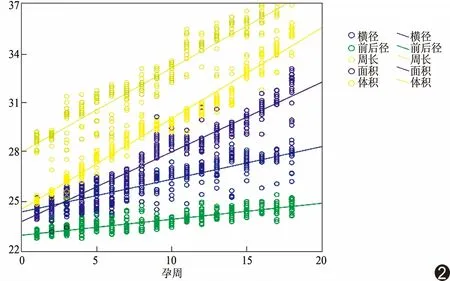
二维、三维超声测量胎儿胸腺的横径、前后径、周长、面积及体积结果见表1。胎儿胸腺的各径线、面积及体积的超声测值均随孕周的增加而增加,与孕周呈线性相关(图2)。最适回归方程为:横径(cm)=-1.98+0.16×孕周(r=0.85,P<0.01);前后径(cm)=-0.80+0.08×孕周(r=0.85,P<0.01);周长(cm)=-5.00+0.42×孕周(r=0.93,P<0.01);面积(cm2)=-1.49+0.35×孕周(r=0.95,P<0.01);体积(ml)=-2.12+0.45×孕周(r=0.98,P<0.01)。Steiger'st检验结果显示,三维超声检测胎儿胸腺体积与孕周的相关性明显优于二维超声所测胸腺各经线、周长及面积(P均<0.01)。
3 讨论
有研究[2]报道,胎儿胸部横断面三血管-气管切面是检测胎儿胸腺的最佳断面。本研究中,对所有胎儿胸腺的二维、三维超声检测均采用三血管-气管切面。超声检查中,当声束与胸腺两侧边界垂直时图像显示最为清晰,即胎儿侧位或仰卧位时最易进行超声探查,而俯卧位时由于胎儿胸腺远离探头,且被脊柱及肋骨声影所遮挡可能导致扫查困难。

图1 正常胎儿胸腺的二维(A、B)及三维超声图像(C、D)

图2 胎儿胸腺各指标与孕周相关性的散点图
目前国内外产前超声对胎儿胸腺的研究[3-5]多采用二维超声,均显示胎儿胸腺横径、前后径、周长及面积等正常参考值具有随孕周增长而增大的特点,但各研究对评估胎儿胸腺发育的最佳指标观点不一。李伯义等[3]认为用胸腺面积代表胸腺大小评估其发育情况较为合适。Zalel等[1]则认为以周长评估胎儿胸腺大小更为可靠。Chaoui等[6]测量胎儿胸腺-胸廓前后径的比值,发现比值减小时提示胸腺发育不良。Cho等[5]认为检测胸腺横径评估胎儿胸腺发育情况更方便可行。近年来,随着三维超声技术飞速发展,尤其是三维容积测量技术的进步,为产前研究胎儿胸腺提供了一种全新的方法。通过三维容积测量中的VOCAL技术可精确描记靶组织的边界,并根据需要任意调整增减切面数目,且具有更方便、省时的优点,更适合测量不规则形态的实质性器官的体积(如胸腺)[7-9]。二维超声只能反映单一平面而不能反映胸腺的全貌;而三维容积超声则可完整显示胎儿胸腺的分叶状结构,对器官体积的测量也是综合多个切面的信息后所获得,相对更为精准。本研究应用二维超声及三维超声的VOCAL技术,产前测量孕22~39周360胎正常胎儿的胸腺,包括二维超声所测胸腺横径、前后径、周长、面积及三维超声所测胸腺体积,经相关性分析发现胎儿胸腺的三维体积与孕周的相关性明显优于二维指标(P<0.01),提示三维超声测量胎儿胸腺体积为产前超声评估胸腺发育的最佳指标,与Li等[10]的研究结果基本一致。
胎儿胸腺发育不全与多种疾病有关,如染色体22q11.2微缺失综合征、感染、营养不良、先兆子痫等。染色体22q11.2微缺失综合征的心脏畸形主要表现为圆锥动脉干异常。Di Naro等[2]发现圆锥动脉干畸形先天性心脏病胎儿胸腺体积缩小的检出率明显增高。Li等[10]首先采用三维超声测量胎儿胸腺体积,并通过对比发现染色体22q11.2微缺失综合征胎儿胸腺体积明显缩小。Volpe等[11]认为胎儿胸腺发育异常与胎儿宫内生长迟缓和胎儿主动脉弓异常有关。Yinon等[12]研究报道,宫内感染胎儿的胸腺体积普遍缩小。因此,一旦发现胎儿胸腺体积短期内明显缩小,应高度警惕胎儿宫内感染的风险。另有学者[13-14]发现先兆子痫孕妇胎儿胸腺体积也明显缩小。因此,产前超声检测胎儿胸腺,尤其三维超声对胎儿胸腺体积的测量,可为诊断胎儿畸形或宫内异常提供线索,为临床正确、及时处理提供参考。
本研究结果显示,对孕22~39周胎儿采用三维超声测量胎儿胸腺体积为产前超声评估胸腺发育的最佳指标。目前国内胎儿胸腺的产前研究尚处于起步阶段,胸腺的超声观察、检测还未纳入常规产科超声检查中,有待进一步完善、规范。
[1] Zalel Y, Gamzu R, Mashiach S, et al. The development of the fetal thymus: An in utero sonographic evaluation. Prenat Diagn, 2002,22(2):114-117.
[2] Di Naro E, Cromi A, Ghezzi F, et al. Fetal thymic involution: A sonographic marker of the fetal inflamm atory response syndrome. Am J Obstet Gynecol, 2006,194(1):153-159.
[3] 李伯义,凌乐文,吕国荣,等,产前高分辨力超声检测正常胎儿胸腺的方法及临床意义.中国医学影像技术,2011,27(2):361-363.
[4] Gamez F, De Leon-Luis J, Pintado P, et al. Fetal thymus size in uncomplicated twin and singleton pregnancies. Ultrasound Obstet Gynecol, 2010,36(3):302-307.
[5] Cho JY, Min JY, Lee YH, et al. Diameter of the normal fetal thymuson ultrasound. Ultrasound Obstet Gynecol, 2007,29(6):634-638.
[6] Chaoui R, Heling KS, Lopez AS, et al. The thymic-thoracic ratio in fetalheart defects: A simple way to identify fetuses at high risk formicrodeletion 22q11. Ultrasound Obstet Gynecol, 2011,37(4):397-403.
[7] 玄英华,吴青青,王莉.超声检测胎儿胸腺的意义及研究进展.中华医学超声杂志(电子版),2013,10(10):791-793.
[8] Araujo Júnior E, Nardozza LM, Rolo LC, et al. Assessment of yolk sac volume by 3D-sonography using the XI VOCAL method from 7 to 10+6weeks of pregnancy. Arch Gynecol Obstet, 2011,283(Suppl 1):1-4.
[9] Barreto EQ, Milani HF, Araujo Júnior E, et al. Reproducibility of fetal heart volume by 3D-sonography using the XI VOCAL method. Cardiovasc Ultrasound, 2010,8:17.
[10] Li L, Bahtiyar MO, Buhimschi CS, et al. Assessment of the fetalthymus by two-and three-dimensional ultrasound during normal humangestation and in fetuses with congenital heart defects. Ultrasound Obstet Gynecol, 2011,37(4):404-409.
[11] Volpe P, Marasini M, Caruso G, et al. 22q11 deletions in fetuses with malformations of the outflow tracts or interruption of theaorticarch: Impact of additional ultrasound signs. Prenat Diagn, 2003,23(9):752-757.
[12] Yinon Y, Zalel Y, Weisz B, et al. Fetal thymus size as a predictor of chorioamnionitis in women with preterm premature rupture of membranes. Ultrasound Obstet Gynecol, 2007,29(6):639-643.
[13] Mohamed N, Eviston DP, Quinton AE, et al. Smaller fetal thymuses in pre-eclampsia: A prospective cross-sectional study. Ultrasound Obstet Gynecol, 2011,37(4):410-415.
[14] Eviston DP, Quinton AE, Benzie RJ, et al. Impaired fetal thymic growth precedes clinical preeclampsia: A case-control study. J Reprod Immunol, 2012,94(2):183-189.
王咏梅(1976—),女,江苏南京人,硕士,副主任医师。研究方向:妇产科超声。E-mail: 13912923229@163.com
吴云,南京医科大学附属妇产医院超声科,210004。E-mail: 405877652@qq.com
2016-07-10
2016-10-10
产前二维及三维超声测量正常胎儿胸腺
王咏梅,吴 云*,曹 荔,查 文
(南京医科大学附属妇产医院超声科,江苏 南京 210004)
目的 探讨产前二维及三维超声评估胎儿胸腺的最佳指标。方法 对360胎孕22~39周正常单胎胎儿,应用二维超声检测胎儿胸腺的横径、前后径、周长、面积,三维超声检测胎儿胸腺体积。采用线性回归分析各测量指标与孕周的关系。并采用Steiger'st检验比较各指标与孕周的相关性。结果 胎儿胸腺的横径、前后径、周长、面积及体积均随孕周的增加而增大,与孕周均呈线性相关。回归方程为:横径(cm)=-1.98+0.16×孕周;前后径(cm)=-0.80+0.08×孕周;周长(cm)=-5.00+0.42×孕周;面积(cm2)=-1.49+0.35×孕周;体积(ml)=-2.12+0.45×孕周(P均<0.01)。Steiger'st检验显示,三维超声检测胎儿胸腺体积与孕周的相关性明显优于其他指标(P<0.01)。结论 对孕22~39周正常胎儿,采用三维超声测量胎儿胸腺体积是评估胸腺发育情况的最佳指标。
超声检查,产前;胸腺;胎儿
R714.53; R445.1
A
1672-8475(2017)01-0031-04
10.13929/j.1672-8475.201607010
猜你喜欢
杂志排行
中国介入影像与治疗学的其它文章
- Conservative treatment of graft infection after endovascular repair of aortoiliac aneurysm
- Sorafenib administration combined with TACE in treatment of unresectable hepatocellular carcinoma
- Short-term and long-term efficacy of HIFU ablation for diffuse and focal adenomyosis
- Efficacy of uterine artery chemoembolization in treatment of cesarean scar pregnancy
- Ultrasonography in diagnosis of right hand deficiency and right upper limb most deficiency in fetuses: Two cases report 超声诊断胎儿右手缺如及右上肢大部分缺如2例
- Ultrasound in diagnosis of hydatidiform mole coexisting with live fetuses
