Human lgG Fc promotes expression, secretion and immunogenicity of enterovirus 71 VP1 protein
2016-12-13JuanXuChunhuaZhang
Juan Xu, Chunhua Zhang
1Department of Immunology, Nanjing Medical University, Nanjing, Jiangsu 210029, China;
2Department of Infectious Diseases;
3China-US Vaccine Research Center, The First Affiliated Hospital, Nanjing Medical University, Nanjing, Jiangsu 210029, China.
Human lgG Fc promotes expression, secretion and immunogenicity of enterovirus 71 VP1 protein
Juan Xu1,✉, Chunhua Zhang2,3
1Department of Immunology, Nanjing Medical University, Nanjing, Jiangsu 210029, China;
2Department of Infectious Diseases;
3China-US Vaccine Research Center, The First Affiliated Hospital, Nanjing Medical University, Nanjing, Jiangsu 210029, China.
Enterovirus (EV71) can cause severe neurological diseases, but the underlying pathogenesis remains unclear. The capsid protein, viral protein 1 (VP1), plays a critical role in the pathogenicity of EV71. High level expression and secretion of VP1 protein are necessary for structure, function and immunogenicity in its natural conformation. In our previous studies, 5 codon-optimized VP1 DNA vaccines, including wt-VP1, tPA-VP1, VP1-d, VP1-hFc and VP1-mFc, were constructed and analyzed. They expressed VP1 protein, but the levels of secretion and immunogenicity of these VP1 constructs were significantly different (P<0.05). In this study, we further investigated the protein levels of these constructs and determined that all of these constructs expressed VP1 protein. The secretion level was increased by including a tPA leader sequence, which was further increased by fusing human IgG Fc (hFc) to VP1. VP1-hFc demonstrated the most potent immunogenicity in mice. Furthermore, hFc domain could be used to purify VP1-hFc protein for additional studies.
enterovirus 71, VP1, DNA vaccine, human IgG Fc, immunogenicity
Introduction
Hand foot and mouth disease (HFMD), which is caused by enterovirus 71 (EV71), is often associated with severe neurological diseases[1–2], including poliomyelitis-like paralysis, fatal encephalitis with cardiopulmonary complications, meningitis and brain stem encephalitis, which occur mainly in infants and young children[3]. In recent years, EV71 infections in the Asia-Pacific region have shown increasing incidence and mortality[4-5]. However, there is a lack of effective antiviral agents and protective measures[4,6]. Additionally, underlying mechanisms of virus-host interaction and viral pathogenesis remain unclear[7-8].
The family of enteroviruses shares similar morphological characteristics, containing 60 copies of each of the 4 structural proteins viral protein (VP) 1 to 4. Of these 4 proteins, VP1 is more exposed than the other 3 proteins and plays a critical role in the pathogenicity of EV71[9]. In the mature picornavirus virion, VP1 is arrayed around the 5-fold axis of symmetry of the icosahedral virion and is the major surface-accessible protein of EV71[10]. VP1 contains the major epitopes recognized by neutralizing antibodies[11–12]and is used to make subunit vaccines[13–14]. The VP1 gene is serotype-specific and considered the most suitable region for sequence analysis[15–16]. The canyon hypothesis[10,17]suggests that a hydrophobic pocket contained withinVP1 could stabilize the capsid and was important for viral un-coating and attachment[18]. One theory suggests that the canyon is a key receptor-binding site and could function as part of a general strategy facilitating viral escape from immune surveillance[19].
Although thorough research of the structure of VP1 protein is necessary to understand the mechanism of early virus-host interactions and the pathogenesis of enteroviruses[19], the structure of VP1 remains limited. Structure and function, as well as immunogenicity, generally rely on high-level expression and secretion of antigenic proteins. However, VP1 antigen is typically expressed at very low levels[20].
In our previous research, a pilot antigen engineering study was conducted to explore novel VP1 designs to improve expression and immunogenicity of VP1 antigen[21]. Five codon-optimized VP1 DNA vaccines, including wt-VP1, tPA-VP1, tPA-VP1-dimer (VP1-d), tPA-VP1-hFc (VP1-hFc) and tPA-VP1-mFc (VP1-mFc), were constructed and tested for in vitro expression and in vivo immunogenicity. Our data suggest that different VP1 antigen designs show different levels of secretion and contain structural differences that could affect immunogenicity of these VP1 proteins.
In this study, we investigated possible factors that could affect the expression, secretion, and immunogenicity of VP1 DNA vaccines. In our previous study, we showed that VP1-mFc expressed at significantly lower levels and failed to mount an effective immune response[21]. In subsequent experiments, an EV71 VP1 monoclonal antibody raised in mice was investigated to further identify the optimal configuration of VP1. However, VP1-mFc DNA vaccine could not be used in mice, as the mouse IgG Fc fragment reacts with the secondary antibody.
Materials and methods
In vitro expression of VP1 antigens in 293T cells
The expression of VP1 DNA vaccines was verified by transient transfection in 293T cells as previously described[21]. Cells were seeded and grown to 50%-70% confluence on 100 mm culture dishes, and then plasmids (20 μg per dish) were mixed and incubated with 100 μL of polyethylenimine for 15 minutes before adding to the 293T cells. The supernatants and cell lysates were harvested 72 hours after transfection.
Immunization of mice
The protocol was approved by the local institutional board at the authors’ affiliated institutions and animal studies were carried out in accordance with the established institutional guidelines regarding animal care and use. Animal welfare and the experimental procedures were carried out strictly in accordance with the Guide for Care and Use of Laboratory Animals (National Research Council of USA, 1996). Four codon-optimized VP1 DNA vaccines, including wt-VP1, tPA-VP1, tPA-VP1-dimer (VP1-d) and tPA-VP1-hFc (VP1-hFc)[21], were used to immunize mice to further investigate their immunogenicity. BALB/c mice, 6–8 weeks of age, (purchased from Shanghai Animal Center at the Chinese Academy of Sciences) were housed by the Department of Animal Medicine at Nanjing Medical University (5 mice per cage, specefic pathogen free) and were inoculated with DNA vaccines. Briefly, following intramuscular injection of VP1 DNA vaccines or vector control at a dose of 100 μg per immunization, the injection sites were electroporated using the following parameters:100 V, 60 ms and 60 Hz. DNA plasmids were delivered at 2 different sites in the quadriceps muscle for each immunization. The immunizations were administered at week 0, 2, 4 and 8. Serum samples were obtained before the first immunization and 2 weeks after each immunization to study VP1-specific antibody responses.
Enzyme-linked immunosorbent assay (ELISA)
A total of 96-well plates were coated in duplicate with 100 μL of lysates at a dilution of 1:10 and supernatants at a dilution of 1:5 of VP1 DNA vaccines or control-transfected cells. EV71 VP1 protein (100 μL, Abnova, USA; working concentration 50 μg/L) was used as coating antigen to detect the immunized sera. After blocking, 100 μL of anti-EV71 VP1 monoclonal mouse antibody (Abnova) was used to determine the production of VP1 protein from the vaccines, and a rabbit anti-human IgG Fc (Jackson ImmunoResearch, West Grove, PA, USA) was performed to determine the production of VP1-hFc protein. After washing, 100 μL of biotinylated secondary antibody (Multi Sciences Biotech Co., Ltd., Hangzhou, Zhejiang, China) and 100 μL of horseradish peroxidase (HRP)-conjugated streptavidin (Southern Biotech, Birmingham, AL, USA) were added to each well. The plates were developed using a 3,3ʹ,5,5ʹ-tetramethylbenzidine (Sigma, St Louis, MO, USA) solution. For one step ELISA assays, HRP-conjugated goat anti-rabbit or mouse secondary antibody (Multi Sciences Biotech Co., Ltd.) was added after incubation with the appropriate primary antibody followed immediately by development. The plates were read spectrophotometrically at 450 nm, and the wells were scored as positive when the absorbance value was greater than twice the value of the negative control.
Western blotting assays
Western blotting assays were performed as described previously[21]. After incubation with the primary antibody, membranes were washed with PBST and then were allowed to react with HRP-conjugated secondary antibody (at a dilution of 1:10,000). After the final wash, chemiluminescent substrate was applied to the membranes, and Kodak radiographic films were exposed to the membrane and then developed.
To further determine the levels of expression and secretion of the different VP1 constructs, equal amounts of each of the constructs were transfected into 293T cells. Then, equivalent amounts of lysates were detected by Western blotting assays. Beta-actin was used as an internal control at a concentration of 1:500 (Multi Sciences Biotech Co., Ltd.). Before cell lysis, 40 μL of supernatant was collected from the cells and concentrated (Centrifugal Filter Unit, Millipore, Bedford, MA, USA) to 1 mL, after which 10 μL of the concentrated supernatants were analyzed by Western blotting assay.
Purification and identification of VP1-hFc protein
Supernatant (200 mL) from VP1-hFc-transfected 293T cells (VP1-hFc S) was purified using a protein A column (GE Healthcare, Piscataway, NJ, USA). Before passage through the column, supernatants were filtered through a 0.45-μm filter to avoid blockage. The protein A column was equilibrated with binding buffer (phosphate buffer, pH 7.0) at a rate of 1 mL/minute. Then, VP1-hFc S was diluted with binding buffer at 1:1 and added into the column at a rate of 1 mL/minute. The effluent was harvested for future analysis. To wash the column, binding buffer was added again, and the effluent was also harvested. Thereafter, elution buffer (0.1 M citric acid, pH 4.0) was added to the column, and the purified proteins were harvested using 1.5-mL tubes containing 60–200 μL of 1M Tris-HCl (pH 9.0) per mL of fraction to be collected to maintain neutral pH. All of the purified proteins were mixed for subsequent detection.
Sodium dodecyl sulfate polyacrylamide gel electrophoresis (SDS-PAGE)
Purified proteins were analyzed by SDS-PAGE. First, the purified samples and bovine serum albumin (BSA, Sigma) were subjected to electrophoresis on SDS-PAGE gel. After staining with Coomassie brilliant blue G-250 (Beyotime, Shanghai, China) the gel was then analyzed using a Bio-Rad Agarose Image Pattern Analysis System. Based on preliminary experiments, secreted VP1-hFc proteins were not observed by SDS-PAGE due to this method is not sensitive enough. Therefore, all of the purified proteins were mixed and concentrated to 1 mL using a 30-kDa protein centrifugal filter unit (Millipore). Concentrated proteins were detected by SDS-PAGE, Western blotting assay and ELISA.
Results
All VP1 DNA vaccines secrete VP1, and fusion with human IgG Fc fragment further increases VP1 secretion
An anti-VP1 monoclonal antibody was performed to recognize VP1 proteins by ELISA and Western blotting assays (Fig. 1). When leader sequence from human tissue plasminogen activator (tPA) was included as a fusion with VP1, we observed an increase in the expression of VP1 protein. Fusion of human IgG Fc fragment (hFc) further increased VP1 protein expression levels. Although all the 4 VP1 DNA vaccines were expressed and secreted by 293T cells as measured by ELISA (Fig. 1A), only the supernatant of cells expressing VP1-d (VP1-d S) and VP1-hFc (VP1-hFc S) were detected by Western blotting assays (Fig. 1B).
To compare the protein expression levels of the 4 VP1 DNA vaccines and to determine if the other 2 VP1 constructs were secreted, the 4 VP1 constructs were transfected under identical conditions. Then, equal amounts of lysates and concentrated supernatants were analyzed. Transfection of wt-VP1 and tPA-VP1 constructs also resulted in secretion of the VP1 protein, and VP1-hFc achieved the highest level of secretion both in the lysates (Fig. 1C) and in the supernatants (Fig. 1D). In addition to a high molecular weight band in the VP1-hFc supernatants (Fig. 1D), several bands which were obviously specific VP1 proteins were recognized by the anti-VP1 antibody.
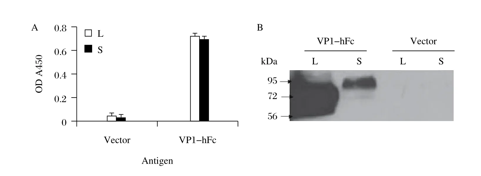
VP1-hFc were also detected using an antibody against human IgG Fc fragment (Fig. 2), but more protein was observed in the lysates than in the supernatants (Fig. 2B).
VP1-hFc DNA vaccine achieved the highest antibody response in mice
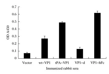
In our previous study[21], sera of immunized rabbit were tested using commercially purified VP1 protein to compare the immunogenicity of vaccine constructs. Antibodies in the sera recognized commercially purified VP1 protein as well as the lysates obtained from cells transfected with VP1 DNA vaccines (Fig. 3).
Then, the immunogenicity of the 4 VP1 DNA vaccines was verified in BALB/c mice. We observed VP1-specific antibodies in response to immunization(Fig. 4A), and the antibody response peaked after the 4thvaccination (Fig. 4B). tPA-VP1 construct showed increased immunogenicity when compared with wt-VP1 construct, and VP1-hFc construct resulted in the highest antibody levels. VP1-d construct approximately had no detectable antibody response. Immunized sera recognized the transfected VP1 proteins by Western blotting assays, but only VP1-hFc-immunized sera recognized VP1 protein in the supernatants of VP1-d and VP1-hFc-transfected cells (Fig. 4C).

Fig. 1 The expression and secretion of VP1 DNA vaccines. Mouse monoclonal VP1 antibody was used as the primary antibody (at a dilution of 1:1,000). HRP-conjugated goat anti-mouse IgG was diluted to 1:10,000. A: ELISA. Lysates (L, at a dilution of 1:10) and supernatants (S, at a dilution of 1:5) were used as antigens to coat the wells. The results represent the data from 3 biological replicates and are expressed as the mean ± SD (standard deviation). B: Western blotting assay: 20 μL original lysates or supernatants were loaded in each lane. The molecular weights of the proteins are as follows: wt-VP1 L ~35kD, tPA-VP1 L ~38kD, VP1-d L ~71kD and VP1-hFc L ~62kD. The molecular weights of the secreted proteins in the supernatant are greater than the corresponding intracellular forms due to protein glycosylation. To compare the protein expression levels of the different VP1 constructs and to determine if wt-VP1 and tPA-VP1 could be secreted, equal amounts of the 4 VP1 constructs were transfected into 293T cells. Lysates (L) and supernatants (S) of transfected cells were harvested and assayed using identical conditions. C: Comparison of the protein levels in the lysates. Twenty μL lysates were loaded in each lane. Anti-beta-actin monoclonal antibody was diluted 1:500. D: Detection of protein in the supernatants. Forty mL of supernatant from transfected cells was concentrated to 1 mL; 10 μL of the concentrated supernatants was analyzed by Western blotting.
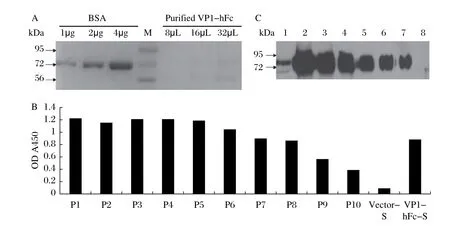
VP1-hFc protein could be purified by hFc fragment Supernatants of cells transfected with VP1-hFc construct were purified using protein A. After purification, we failed to detect VP1 protein by SDS-PAGE in thesupernatants and the non-concentrated purified protein samples (results not shown). Thus, all of the purified VP1-hFc proteins were mixed and concentrated to 1 mL for analysis by SDS-PAGE. However, few VP1proteins were observed in the concentrated protein preparations (Fig. 5A).

Fig. 2 Detection of the expression of VP1-hFc protein. The primary antibody used to detect VP1-hFc protein was rabbit anti-human IgG Fc (H+L) used at a dilution of 1:3,000. HRP-conjugated goat anti-rabbit IgG was diluted to 1:10,000. L: lysates, S: supernatants. A: ELISA. Lysates were diluted to 1:10, and supernatants were diluted to 1:5. The results represent the data from 3 biological replicates and are expressed as the mean ± SD. B: Western blotting assay. 20 μL lysates or supernatants was loaded in each lane.

Fig. 3 ELISA of the immunized rabbit sera. One hundred μL commercial EV71 VP1 protein (50 μg/L) was performed to coat ELISA plates. The immunized rabbit sera obtained from the previous experiments were diluted to 1:200. Biotinylated anti-rabbit IgG was diluted to 1:2000, HRP-conjugated streptavidin was diluted to 1:2000. The values represent the antibody response of 5 rabbits in the same group (average OD values with the associated SD).

Fig. 4 The immunogenicity of VP1 DNA vaccines in BALB/c mouse. One hundred μL EV71 VP1 protein (50 μg/L) was used to coat the wells to analyze the mouse sera for immunization (at a dilution of 1:200). HRP-conjugated goat anti-mouse IgG was diluted to 1:5,000. A: Temporal VP1-specific antibody responses in mouse immune sera. The arrows indicate the time points of DNA immunizations. Each curve represents the antibody response of 5 mice in the same group (average OD values with the associated SD). B: Peak level antibody titer in the mouse sera (average OD values with the associated SD). C: Western blotting assay of VP1 constructs. 20 μL of lysates (L) or supernatants (S) was added in each lane. Mouse sera immunized with different VP1 DNA vaccines were diluted to 1:200. HRP-conjugated goat anti-mouse IgG was diluted to 1:10,000.
To determine if the proteins were lost during their purification, we analyzed each effluent sample that passed through the column and ensured that there were few proteins that were washed off of the column (results not shown). Then, the purified products were serially diluted and analyzed using ELISA (Fig. 5B) and Western blotting assays (Fig. 5C). Our results showed that VP1 protein contents were proportional to the original supernatants (VP1-hFc S).
Discussion
DNA vaccines have become an attractive immunization strategy due to their potency in inducing cellular and humoral immune responses[22]. However, the most significant barrier for DNA vaccines is the lower levels of expression and secretion and the limited immunogenicity of the produced antigen. Optimized antigen expression and secretion is critical for maximal immunogenicity of DNA vaccines. A number of approaches, including optimization of codon usage[23]and fusion of secretory leader sequences[24], have been proposed toenhance antigen expression and/or immunogenicity of DNA vaccines. However, a limited fraction of injected DNA molecules are taken up by antigen presenting cells (APCs), which is a key step for effective initiation of the immune response[25]. Thus, enhancement of antigen presentation by APCs offers another attractive strategy to increase the potency of DNA vaccines[26]. APCs (e.g., dendritic cells and macrophages) express receptors for the Fc fragment of IgG (FcyR). IgG Fc fragment binds to Fc receptor with high affinity and triggers effector functions[27], including the internalization of antigen-antibody complexes and increased antigen presentation[28]. IgG Fc fragment is also considered a good fusion tag for facilitating the purification of target proteins due to its strong and specific binding for protein A or protein G of Staphylococcus. Therefore, antigen constructs that are fused to IgG Fc fragment and targeted to APCs can theoretically induce an effective immune response[29-30]and be used for the rapid purification of protein for intensive research.

Fig. 5 Detection of purified VP1-hFc protein. Primary antibody used to detect VP1-hFc protein was wt-VP1-immunized rabbit serum (at a dilution of 1:500). HRP-conjugated goat anti-rabbit IgG was diluted to 1:10,000. A: SDS-PAGE analysis of purified VP1-hFc protein. All of the purified VP1-hFc proteins were mixed and concentrated to 1 mL for SDS-PAGE analysis. Different amounts of the protein samples (8 μL, 16 μL and 32 μL; initial concentration of 0.16 μg/μL) were detected. Different amounts of BSA (1 μg, 2 μg and 4 μg) were added as a semi-quantitative gauge. B: ELISA. P1 represents 1 μg of purified and concentrated VP1-hFc protein (0.16 μg/μL) in 100 μL of PBS. Then, P1 was serially diluted 2-fold (P2-P10). Vector-S and VP1-hFc-S represent the original supernatants diluted to 1:5. C: Western blotting assay. Lane 1 shows 20 μL of the original VP1-hFc-S; Lanes 2-7 indicate 40 μL, 20 μL, 10 μL, 5 μL, 2.5 μL and 1.25 μL of purified VP1-hFc protein, respectively (0.16 μg/μL); Lane 8 shows 20 μL of protein expressed from the original vector-S.
In our previous studies, 5 novel VP1 DNA vaccine constructs were designed and expressed in 293T cells and verified to demonstrate immunogenicity in rabbits[21]. However, the protein level and immunogenicity of the constructs showed significant differences. Although the expressions of wt-VP1 and tPA-VP1 were observed in 293T cell lysates, these proteins could not be detected in cell supernatants by Western blotting assay. VP1-d showed better expression and secretion in lysates and supernatants, but the immuno-genicity of VP1-d was the weakest of all the constructs tested. To further investigate the optimization of the VP1 gene to maximize immunogenicity in this study, we used a commercial monoclonal antibody raised against EV71 VP1 to recognize VP1 DNA vaccine-expressed proteins and purified EV71 VP1 protein to analyze the sera of immunized rabbits. Analysis of the concentrated supernatants showed that all of the VP1 constructs could secrete VP1 protein, and VP1-hFc achieved the highest level. VP1-hFc also induced the highest antibody response in mouse immunization. Interestingly, VP1-d construct showed increased levels of protein expression and secretion but the lowest immunogenicity. As VP1-d encodes 2 copies of VP1 protein as opposed to monomeric form encoded by tPA-VP1 construct, we suspect that VP1-d was not likely to express or secrete to a greater extent than tPA-VP1 but offered double VP1 epitopes reacting with antibodies. We suppose that dimer form of VP1-d could be folded in a configuration that obscures important epitopes and domains necessary for proper secretion and immune response initiation. Thus, our results suggest that the fusion construct of VP1 with human IgG Fc was the only construct that could increase the expression and secretion levels of VP1 protein and enhance VP1 immunogenicity.
The availability of large amounts of secreted proteins is not only necessary for induction of strong antibodyresponses, but also crucial for protein structure analysis. To understand the structural basis of proteins during cellular processes such as immune initiation, high-quantity and high-quality purified protein samples are required. In our study, VP1-hFc achieved the highest level of protein expression relative to the other VP1 DNA vaccines and could be selected for the rapid purification of VP1 protein using human IgG Fc fragment and Staphylococcal protein A. However, purification of VP1 by this method did not result in a sufficiently high level of purified protein for structural analysis, even after concentration. We tried to purify VP1-hFc protein using protein G and observed similar results (results not shown). In fact, Western blotting assay showed that the protein level of VP1-hFc in lysates was higher than the levels observed in supernatants. Another consideration is that VP1-hFc protein could have been hydrolyzed into several fragments that could not be purified. These fragments may have included VP1-hFc with tPA, VP1-hFc without tPA, tPA-VP1 and VP1 without tPA sequences. If true, this could suggest that linkers between peptides were unstable, and proteolysis- resistant linkers should be investigated in future studies.
In conclusion, although the expression and secretion levels of VP1-hFc were greater than those of the other 3 VP1 DNA vaccines tested, the levels of expression and secretion of VP1-hFc were not sufficient for in vitro analysis of protein structure and function. In future studies, a more effective protein expression system and stronger antigen presenting vectors should be developed to enhance VP1 protein expression and secretion, as well as its immunogenicity. These studies will be critical for the development of an effective VP1 DNA vaccine.
Acknowledgments
This work was supported by the National Natural Science Foundation of China (Grant No. 81000725 and 31470889) and the Priority Academic Program of Basic Medical Science of Nanjing Medical University (Grant No. JX10131801060).
References
[1] Solomon T, Lewthwaite P, Perera D, et al. Virology, epidemiology, pathogenesis, and control of enterovirus 71[J]. Lancet Infect Dis, 2010,10(11):778–790.
[2] Ooi MH, Wong SC, Lewthwaite P, et al. Clinical features, diagnosis, and management of enterovirus 71[J]. Lancet Neurol, 2010,9(11):1097–1105.
[3] Palacios G, Oberste MS. Enteroviruses as agents of emerging infectious diseases. J Neurovirol, 2005,11(5):424–433.
[4] Qiu J. Enterovirus 71 infection: a new threat to global public health?[J]. Lancet Neurol, 2008,7(10):868–869.
[5] Eggertson L. Infectious disease experts monitor outbreaks of enterovirus 71 in Asia[J]. CMAJ, 2012,184(15):E781–E782.
[6] Wu KX, Ng MM, Chu JJ. Developments towards antiviral therapies against enterovirus 71[J]. Drug Discov Today, 2010,15(23–24):1041–1051.
[7] Xu J, Qian Y, Wang S, et al. EV71: an emerging infectious disease vaccine target in the Far East?[J]. Vaccine, 2010,28(20):3516–3521.
[8] Yip CC, Lau SK, Woo PC, et al. Human enterovirus 71 epidemics: what's next?[J]. Emerg Health Threats J 2013,6:19780.
[9] Hendry E, Hatanaka H, Fry E, et al. The crystal structure of coxsackievirus A9: new insights into the uncoating mechanisms of enteroviruses[J]. Structure, 1999,7(12):1527–1538.
[10] Plevka P, Perera R, Cardosa J, et al. Structure determination of enterovirus 71[J]. Acta Crystallogr D Biol Crystallogr, 2012,68(Pt 9):1217–1222.
[11] Man-Li T, Szyporta M, Fang LX, et al. Identification and characterization of a monoclonal antibody recognizing the linear epitope RVADVI on VP1 protein of enterovirus 71[J]. J Med Virol, 2012,84(10):1620–1627.
[12] Huang ML, Chiang PS, Chia MY, et al. Cross-reactive neutralizing antibody responses to enterovirus 71 infections in young children: implications for vaccine development[J]. PLoS Negl Trop Dis, 2013,7(2):e2067.
[13] Kiener TK, Premanand B, Kwang J. Immune responses to baculovirus-displayed enterovirus 71 VP1 antigen[J]. Expert Rev Vaccines, 2013,12(4):357–364.
[14] Yu Z, Huang Z, Sao C, et al. Oral immunization of mice using Bifidobacterium longum expressing VP1 protein from enterovirus 71[J]. Arch Virol, 2013,158(5):1071–1077.
[15] Wu WH, Kuo TC, Lin YT, et al. Molecular epidemiology of enterovirus 71 infection in the central region of taiwan from 2002 to 2012. PLoS One, 2013,8(12):e83711.
[16] Wu JS, Zhao N, Pan H, et al. Patterns of polymorphism and divergence in the VP1 gene of enterovirus 71 circulating in the Asia-Pacific region between 1994 and 2013[J]. J Virol Methods, 2013,193(2):713–728.
[17] Rossmann MG. The canyon hypothesis. Hiding the host cell receptor attachment site on a viral surface from immune surveillance[J]. J Biol Chem, 1989,264(25): 14587–14590.
[18] Cordey S, Petty TJ, Schibler M, et al. Identification of site-specific adaptations conferring increased neural cell tropism during human enterovirus 71 infection[J]. PLoS Pathog, 2012,8(7):e1002826.
[19] Rossmann MG, He Y, Kuhn RJ. Picornavirus-receptor interactions[J]. Trends Microbiol, 2002,10(7):324–331.
[20] Tung WS, Bakar SA, Sekawi Z, et al. DNA vaccine constructs against enterovirus 71 elicit immune response in mice[J]. Genet Vaccines Ther, 2007,5:6.
[21] Xu J, Wang S, Gan W, et al. Expression and immunogenicity of novel subunit enterovirus 71 VP1 antigens[J]. Biochem Biophys Res Commun, 2012,420(4):755–761.
[22] Cui Z. DNA vaccine[J]. Adv Genet, 2005,54:257–289.
[23] Mitarai N, Sneppen K, Pedersen S. Ribosome collisions and translation efficiency: optimization by codon usage and mRNA destabilization[J]. J Mol Biol, 2008,382(1):236–245.
[24] Ashok MS, Rangarajan PN. Protective efficacy of a plasmid DNA encoding Japanese encephalitis virus envelope protein fused to tissue plasminogen activator signal sequences: studies in a murine intracerebral virus challenge model[J]. Vaccine, 2002,20(11–12):1563–1570.
[25] Condon C, Watkins SC, Celluzzi CM, et al. DNA-based immunization by in vivo transfection of dendritic cells[J]. Nat Med, 1996,2(10):1122–1128.
[26] You Z, Huang X, Hester J, et al. Targeting dendritic cells to enhance DNA vaccine potency[J]. Cancer Res, 2001, 61(9):3704–3711.
[27] Allhorn M, Olin AI, Nimmerjahn F, et al. Human IgG/Fc gamma R interactions are modulated by streptococcal IgG glycan hydrolysis[J]. PLoS One, 2008,3(1):e1413.
[28] Sallusto F, Lanzavecchia A. Efficient presentation of soluble antigen by cultured human dendritic cells is maintained by granulocyte/macrophage colony-stimulating factor plus interleukin 4 and downregulated by tumor necrosis factor alpha[J]. J Exp Med, 1994,179(4):1109–1118.
[29] Wu K, Bi Y, Sun K, et al. Suppression of allergic inflammation by allergen-DNA-modified dendritic cells depends on the induction of Foxp3+ Regulatory T cells[J]. Scand J Immunol, 2008,67(2):140–151.
[30] Qi Z, Pan C, Lu H, et al. A recombinant mimetics of the HIV-1 gp41 prehairpin fusion intermediate fused with human IgG Fc fragment elicits neutralizing antibody response in the vaccinated mice[J]. Biochem Biophys Res Commun, 2010,398(3):506–512.
✉ Juan Xu, Department of Immunology, Nanjing Medical University, 140 Hanzhong Road, Nanjing, Jiangsu 210029, China, Tel: +86-25-86863176; +86-159-518-345-61 E-mail:xiangbangbang@njmu.edu.cn.
09 December 2014, Revised 26 January 2015, Accepted 02 April 2015, Epub 12 July 2015
R511, Document code: A
The authors reported no conflict of interests.
杂志排行
THE JOURNAL OF BIOMEDICAL RESEARCH的其它文章
- Lipid transport to avian oocytes and to the developing embryo
- Role of stromal cell-derived factor 1α pathway in bone metastatic prostate cancer
- A pilot exome-wide association study of age-related cataract in Koreans
- Dynamic monitoring of menopause hormone therapy and defining the cut-off value of endometrial thickness during uterine bleeding
- Polycystic ovary syndrome patients with high BMl tend to have functional disorders of androgen excess: a prospective study
- Effect of vitamin D3 on production of progesterone in porcine granulosa cells by regulation of steroidogenic enzymes
