Endoproteolytic cleavage as a molecular switch regulating and diversifying prion protein function
2016-12-02CathrynL.Haigh,StevenJ.Collins
PERSPECTIVE
Endoproteolytic cleavage as a molecular switch regulating and diversifying prion protein function
The prion protein (PrP), through misfolding, is widely known for its causative role in prion diseases, which are transmissible neurodegenerative diseases of humans and animals. There is still no defined function assigned to PrP, especially in the central nervous system, despite many studies in this area. Proposed functions are protean and include signal transduction, neuroprotection, neurogenesis, neuritogenesis, metal-ion homeostasis, memory formation and consolidation, as well as circadian rhythms (Nicolas et al., 2009). Part of the difficulty in assigning a specific function to PrP could perhaps be that it does not have one single function. Instead it might be able to perform many functions and influence various pathways depending upon contextual post-translational modification.
PrP has been shown to undergo various post-translational modifications including glycosylphosphatidylinositol (GPI)-anchor attachment at its C-terminus, N-linked glycosylation at either or both of two locations, phosphorylation, metal ion co-ordination at no less than six sites, secretory cleavage close to the GPI-anchor and endoproteolytic cleavage at three or more sites (Haigh et al., 2010). Whilst all of these modifications may change mature PrP in ways that potentially alter its function or site of function, endoproteolytic cleavage creates new peptides with distinct features that are likely to contribute to the diversity of functions reported for this protein.
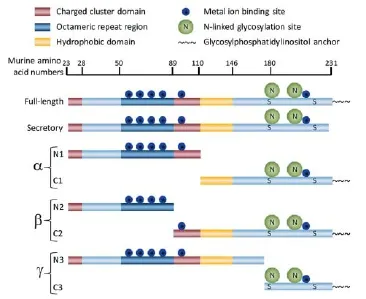
Alpha-(α-) and beta-(β-)cleavages of PrP were first characterized as constitutive processing events in both normal and diseased human brain twenty years ago (Chen et al., 1995). The cleavage sites and resultant fragments are shown schematically in Figure 1. The C-terminal fragments persist at detectible levels in cells post-cleavage, whereas the N-terminal fragments are likely secreted or released from cells and are detectible in the culture media. PrP cleavage differs depending upon cell type. In certain cells PrP can be over 50% α-cleaved suggesting that this processing may be part of its normal functioning (Lewis et al., 2009). The different properties and fates of the cleavage fragments further support that this cleavage is unlikely to represent a mere degradation step in the turnover of PrP but instead produces functional proteins. Indeed the different ratios across cell types may reflect the different functions of CNS cells. The β-cleavage event has traditionally been thought to be pathogenic as the relative amounts of cognate fragments are increased during prion disease (Chen et al., 1995). However, a cellular inability to undergo β-cleavage was found to result in a heightened susceptibility to cellular stress, which indicated that the produced N2/ C2 fragments were also likely to be functional (Watt et al., 2005). For both α- and β-cleavages the specific site of cleavage is “ragged” with these cleavages located either side of a charged cluster domain, defining which new peptide contains this basic domain.
Recently a further cleavage event has been extensively characterized (Lewis et al., 2015). Referred to as “gamma-cleavage”, this event occurs in the C-terminal structured domain and therefore produces fragments with very different features to both the α- and β-cleavages. A functional significance is yet to be assigned to this processing event but its presence in multiple cells and tissues, and in disease, suggests that the fragments produced are likely to exert cellular effects distinct to those produced by the other PrP constitutive processing events.
Our prior research has shown that the N2 fragment (and shorter fragments thereof that include the far N-terminal residues) display an anti-oxidant, neuroprotective function in response to the mild stress of serum starvation (Haigh et al., 2009a). This function required the N-terminal amino acids to be intact, including the structure conferred on the first charged cluster domain (residues 23-38) by the two proline residues at positions 26 and 28. Anti-oxidant function was further influenced by the octarepeat region and its copper-saturation, requiring a minimum of two copper molecules available for co-ordination into this site. N2 interaction with the cell surface required intact lipid rafts and heparan sulphate containing proteoglycans and if these were absent transduction of the protective effect was abolished. It was later shown that the cell surface engagement of N2 was also influenced by copper binding, directing the N2 internalisation pathway, which in turn permitted the specific activation of MEK1 in the absence of MEK2 or ERK1/2 activation (Haigh et al., 2015a). The outcome of the MEK1 activation was lower lysosomal and mitochondrial reactive oxygen species production. Therefore, two post-translational modifications of PrP, specifically β-cleavage and metal ion co-ordination, appear to co-operatively regulate and orchestrate N2 signalling, underscoring that the various permutations of post-translational modifications may determine PrP functional modulation. Further illustrating this, combinations of post-translational PrP modifications is not restricted to influencing MEK1 but has also been shown to alter other signalling pathways. For example, copper ion binding alters α-cleavage profiles as a function of membrane fluidity and lipid raft integrity and this correlates with downstream activation of the ERK1/2, p38 and JNK signalling pathways (Haigh et al., 2009b). Therefore, not only may cleavage be a function modifying event but the cellular consequences may be highly dependent upon the precise micro-environment context in which PrP exists.
Like the N2 cleavage fragment, N1 has also been shown to exert neuroprotective functions, counteracting staurosporine toxicity and hypoxia by reducing caspase-3 activation through modulating p53 protein levels and activity (Guillot-Sestier et al., 2009). Of interest, despite acting on different pathways, both N1 and N2 bind anionic synthetic lipid membranes at low pH. They integrate between the lipid head groups but do not significantly penetrate between the acyl tails and so the interactions are non-disruptive (Le Brun et al., 2014). The N1 peptide demonstrates a greater affinity for lipids than the N2. The lipid binding propensity of these peptides may function to sequester them, possibly to quench their function or alternatively to protect them from degradation and preserve their functional life-time. Additionally, lipid intercalation may serve to order membrane micro-domains for signal protein activation or to direct peptide trafficking, ensuring activation of specific signalling pathways. In anionic model membranes N1 and N2 peptide binding indeed results in alterations in lipid order (Le Brun et al., 2014). Despite N1 demonstrating a higher binding affinity for lipids, in a cellular context lipid changes appear to be influenced to a greater extent through N2 neuroprotective activity (Haigh et al., 2015b). Serum starvation of cells induces a number of changes in their lipid environment, including changes affecting phosphatidylserine and phosphatidic acid, with which both N1 and N2 interact. Through unresolved mechanisms, the N2 peptide (but not N1) was able to normalise these cellular changes during serum starvation. This could indicate that the extra poly-basic region alters engagement of membrane binding partners to such an extent that N1 and N2 exert their neuroprotective effects in different ways. A stronger binding affinity of N1 for membrane lipids may result in it being bound sufficiently tightly to prevent release for performing its function or ensure preferential binding to other partners to instigate transduction through a different signalling cascade.
Understanding the precise mechanisms underpinning the link between PrP functions and constitutive cleavage may prove valuable for understanding failing processes in disease and aging. Whilst C2 levels are increased during prion disease, possibly pathogenically (Chen et al., 1995), increased levels of C1 or PrP secretory cleavage have been shown to be protective against the uptake of prion infection (Lewis et al., 2009). The protective nature of C1 is thought to arise due to the α-cleavage site being located in the middle of a ‘toxic domain’ (amino acids at 106-126), which is thought to be important for efficiency of conversion into misfolded PrP. Whilst C1 may be protective in the context of prion disease, it has also been linked with signalling cellular death through p53 and caspase 3, in a pathway opposed by its counterpart N1 (Sunyach et al., 2007; Guillot-Sestier et al., 2009). No function has yet been found for C2, with an assumption that it is an inert substrate available for pathogenic misfolding during prion disease. With so many potential influences and different pathways to consider when attempting to discern functions of these fragments it could be a longtime before a definitive function, or lack thereof, can be assigned to this fragment.

Figure 1 Schematic representation of the prion protein (PrP) cleavage sites.
Beyond the causative role of PrP misfolding in prion diseases, cellular PrP has also been implicated in transducing the toxic signals that cause Alzheimer’s pathology. Of special interest, the N1 fragment has been shown to bind to the soluble beta-amyloid (Aβ) peptides associated with AD pathogenesis offering neuroprotection (Guillot-Sestier et al., 2012). N1 binding to soluble Aβ oligomers is thought to block their engagement with PrP at the cell surface and thereby prevent damaging signal transduction through fyn and tau. The N1/C1 cleavage event may be doubly protective as the important Aβ binding domain is thought to fall within the amino acids between the α- and β-cleavage sites. As a result, the membrane attached C1 fragment lacks the soluble Aβ-oligomer binding domain and so should be unable to transduce any toxic signals. The C2 fragment, however, contains this amino acid region and so the β-cleavage event may still be detrimental during Alzheimer’s disease, permitting toxic signalling to occur. The lack of the N-terminus when soluble Aβ oligomers bind C2 however, may also result in different cellular engagements thereby modulating signal transduction and neuronal outcome.
Understanding the influence of PrP cleavage on pathways regulating neuronal function are likely to not only provide significant information on the cellular failures that underscore or correlate with neurodegeneration but also produce insight into the role these cleavage fragments may have in the regenerative processes of the brain. Endogenous expression of PrP stimulates the activity of adult neural stem cells (Steele et al., 2006) yet the role of PrP cleavages within these cells remains to be determined. Clarifying the importance of PrP cleavage events and the resulting fragments, either as autocrine or paracrine intermediates, in neural stem cell growth and differentiation will enable us to judge whether there is potential to preserve, rescue or enhance regenerative processes by modifying PrP processing events.
In conclusion, there are many more regulatory influences both singly and in combination that must be considered before we can claim a thorough understanding of the functional roles of constitutive or inducible PrP cleavage within the cell. Nevertheless, cumulative data to date strongly suggests that constitutive PrP processing is not an irrelevant epiphenomenon of cellular turnover but regulated, precise and controlled events for specific ends. Cleavage profiles differ across different cell types (Lewis et al., 2009) and evidence so far suggests functional outcomes of cleavage are likely to differ and be context specific. The precise cellular locations of the cleavage events and of the resulting fragments, whether processing occurs at the cell surface and whether N-terminal fragments act in cis or trans, the enzymes controlling these events, the membrane micro-milieu, and dynamic rather than absolute cleavage levels may all potentially impact functional consequences. Until the various nuances of combined post-translational modifications of PrP coupled with cleavage events in different contexts and in different tissues are fully elucidated, PrP appears destined to remain an enigmatic “actor” playing in many apparent functional roles.
Cathryn L. Haigh*, Steven J. Collins*
Department of Medicine (Royal Melbourne Hospital), The University of Melbourne, Parkville, Victoria, Australia
*Correspondence to: Cathryn L. Haigh, Ph.D. or Steven J. Collins, Ph.D., chaigh@unimelb.edu.au or stevenjc@unimelb.edu.au.
Accepted: 2015-12-21
orcid: 0000-0001-7591-1149 (Cathryn L. Haigh) 0000-0002-5245-6611 (Steven J. Collins)
Chen SG, Teplow DB, Parchi P, Teller JK, Gambetti P, Autilio-Gambetti L (1995) Truncated forms of the human prion protein in normal brain and in prion diseases. J Biol Chem 270:19173-19180.
Guillot-Sestier MV, Sunyach C, Druon C, Scarzello S, Checler F (2009) The alpha-secretase-derived N-terminal product of cellular prion, N1, displays neuroprotective function in vitro and in vivo. J Biol Chem 284:35973-35986.
Guillot-Sestier MV, Sunyach C, Ferreira ST, Marzolo MP, Bauer C, Thevenet A, Checler F (2012) alpha-Secretase-derived fragment of cellular prion, N1, protects against monomeric and oligomeric amyloid beta (Abeta)-associated cell death. J Biol Chem 287:5021-5032.
Haigh CL, Marom SY, Collins SJ (2010) Copper, endoproteolytic processing of the prion protein and cell signalling. Front Biosci 15:1086-1104.
Haigh CL, McGlade AR, Collins SJ (2015a) MEK1 transduces the prion protein N2 fragment antioxidant effects. Cell Mol Life Sci 72:1613-1629.
Haigh CL, Tumpach C, Drew SC, Collins SJ (2015b) The prion protein N1 and N2 cleavage fragments bind to phosphatidylserine and phosphatidic acid; relevance to stress-protection responses. PLoS One 10:e0134680.
Haigh CL, Drew SC, Boland MP, Masters CL, Barnham KJ, Lawson VA, Collins SJ (2009a) Dominant roles of the polybasic proline motif and copper in the PrP23-89-mediated stress protection response. J Cell Sci 122:1518-1528.
Haigh CL, Lewis VA, Vella LJ, Masters CL, Hill AF, Lawson VA, Collins SJ (2009b) PrPC-related signal transduction is influenced by copper, membrane integrity and the alpha cleavage site. Cell Res 19:1062-1078.
Le Brun AP, Haigh CL, Drew SC, James M, Boland MP, Collins SJ (2014) Neutron reflectometry studies define prion protein N-terminal peptide membrane binding. Biophys J 107:2313-2324.
Lewis V, Johanssen VA, Crouch PJ, Klug GM, Hooper NM, Collins SJ (2015) Prion protein “gamma-cleavage”: characterizing a novel endoproteolytic processing event. Cell Mol Life Sci doi:10.1007/s00018-015-2022-z.
Lewis V, Hill AF, Haigh CL, Klug GM, Masters CL, Lawson VA, Collins SJ (2009) Increased proportions of C1 truncated prion protein protect against cellular M1000 prion infection. J Neuropathol Exp Neurol 68:1125-1135.
Nicolas O, Gavin R, del Rio JA (2009) New insights into cellular prion protein (PrPc) functions: the “ying and yang” of a relevant protein. Brain Res Rev 61:170-184.
Steele AD, Emsley JG, Ozdinler PH, Lindquist S, Macklis JD (2006) Prion protein (PrPc) positively regulates neural precursor proliferation during developmental and adult mammalian neurogenesis. Proc Natl Acad Sci U S A 103:3416-3421.
Sunyach C, Cisse MA, da Costa CA, Vincent B, Checler F (2007) The C-terminal products of cellular prion protein processing, C1 and C2, exert distinct influence on p53-dependent staurosporine-induced caspase-3 activation. J Biol Chem 282:1956-1963.
Watt NT, Taylor DR, Gillott A, Thomas DA, Perera WS, Hooper NM (2005) Reactive oxygen species-mediated beta-cleavage of the prion protein in the cellular response to oxidative stress. J Biol Chem 280:35914-35921.
10.4103/1673-5374.177726 http://www.nrronline.org/
How to cite this article: Haigh CL, Collins SJ (2016) Endoproteolytic cleavage as a molecular switch regulating and diversifying prion protein function. Neural Regen Res 11(2):238-239.
杂志排行
中国神经再生研究(英文版)的其它文章
- Tissue-type plasminogen activator is a modulator of the synaptic vesicle cycle
- Impaired consciousness caused by injury of the lower ascending reticular activating system: evaluation by diffusion tensor tractography
- Considering calcium-binding proteins in invertebrates: multi-functional proteins that shape neuronal growth
- Cardiovascular dysfunction following spinal cord injury
- Practical application of the neuroregenerative properties of ketamine: real world treatment experience
- Exergames: neuroplastic hypothesis about cognitive improvement and biological effects on physical function of institutionalized older persons
