Automatic counting of microglial cell activation and its applications
2016-12-01BeatrizGallegoColladoPablodeGracia
Beatriz I. Gallego Collado, Pablo de Gracia
1 Instituto de Investigaciones Oftalmológicas Ramón Castroviejo, Universidad Complutense de Madrid, Madrid, Spain2 Facultad de Óptica y Optometría, Departamento de Oftalmología y Otorrinolaringología, Universidad Complutense de Madrid, Madrid, Spain3 Midwestern University, Chicago College of Optometry, Downers Grove, IL, USA4 Department of Neurobiology, Barrow Neurological Institute, St. Joseph’s Hospital and Medical Center, Phoenix, AZ, USA
Automatic counting of microglial cell activation and its applications
Beatriz I. Gallego Collado1,2,*,#, Pablo de Gracia3,4,*,#
1 Instituto de Investigaciones Oftalmológicas Ramón Castroviejo, Universidad Complutense de Madrid, Madrid, Spain
2 Facultad de Óptica y Optometría, Departamento de Oftalmología y Otorrinolaringología, Universidad Complutense de Madrid, Madrid, Spain
3 Midwestern University, Chicago College of Optometry, Downers Grove, IL, USA
4 Department of Neurobiology, Barrow Neurological Institute, St. Joseph’s Hospital and Medical Center, Phoenix, AZ, USA
How to cite this article: Gallego BI, de Gracia P (2016) Automatic counting of microglial cell activation and its applications. Neural Regen Res 11(8)∶1212-1215.
Funding: This work was supported by the Science Foundation of Arizona through the Bisgrove Program to PdG, Grant Number∶ BSP 0529-13. BIG received funding from the Ophthalmological Network OFTARED (RD12-0034/0002) and the Institute of Health Carlos III. And also from the PN I+D+i 2008-2011, from the ISCIII-Subdireccion General de Redes y Centros de Investigación Cooperativa, from the European Programme FEDER, and from the project SAF2014-53779-R. BIG also received funding from the project∶ “The role of encapsulated NSAIDs in PLGA microparticles as a neuroprotective treatment” funded by the Spanish Ministry of Economy and Competitiveness.
Beatriz I. Gallego Collado, O.D., Ph.D. or Pablo de Gracia, O.D., Ph.D., F.A.A.O.,
bgallegocollado@gmail.com or pdegracia@midwestern.edu
Both of these two authors
contributed equally to this article.
orcid:
0000-0001-9864-3140
(Beatriz I. Gallego Collado) 0000-0003-4319-2797
(Pablo de Gracia)
Accepted: 2016-08-15
Glaucoma is a multifactorial optic neuropathy characterized by the damage and death of the retinal ganglion cells. This disease results in vision loss and blindness. Any vision loss resulting from the disease cannot be restored and nowadays there is no available cure for glaucoma; however an early detection and treatment, could offer neuronal protection and avoid later serious damages to the visual function. A full understanding of the etiology of the disease will still require the contribution of many scientific efforts. Glial activation has been observed in glaucoma, being microglial proliferation a hallmark in this neurodegenerative disease. A typical project studying these cellular changes involved in glaucoma often needs thousands of images - from several animals - covering different layers and regions of the retina. The gold standard to evaluate them is the manual count. This method requires a large amount of time from specialized personnel. It is a tedious process and prone to human error. We present here a new method to count microglial cells by using a computer algorithm. It counts in one hour the same number of images that a researcher counts in four weeks, with no loss of reliability.
glaucoma; glial cells; microglial cells; automatic counting; image processing; inner plexiform layer; outer plexiform layer; bilateral activation
Introduction
Vision in mammals begins at the retina, which is the innermost layer of the eye and part of the central nervous system (CNS). The retina comprises a high scaffold of complex neurons that transform light into nerve impulses, which propagate through the visual pathway to the brain where visual processing is completed.
Glaucoma is a chronic optic neuropathy characterized by neuronal death of retinal ganglion cells (RGCs). The disease is a prevalent visual pathology that leads to vision impairment (affecting > 60 million people worldwide) and is the second most frequent cause of irreversible blindness in the world (Quigley and Broman, 2006). Although age and ocular hypertension (OHT) constitute the major risk factors for the disease, the exact mechanisms involved in glaucoma pathophysiology are unknown. In some instances, the progress of the disease cannot be halted and, in others, major damage has already occurred by the time of diagnosis. Therefore, understanding the pathogenic mechanisms of glaucoma and developing new strategies for early diagnosis are paramount for improving the well-being of individuals suffering from glaucoma.
Glial Cell Activation in Glaucoma: the Good and the Bad
Activation of glial cells seems to play an important role in glaucomatous neurodegeneration. Glia are non-neuronal cells in the nervous system that support and protect neurons. Glia, especially microglial cells, are considered to be immune cells in the CNS (including the retina) and their activation after damage is crucial. Early, moderate, transient, well-controlled glial activation could be initially responsible for restoring damaged tissue. However, the sustained tissue stress that occurs in human glaucoma is associated with a chronic activation of glial cells-this hallmark of a harmful neuro-inflammatory process could lead to tissue damage. This concept supports the contention that, in glaucomatous neurodegeneration, glial cells could initiate an immune response that mayexacerbate the glaucomatous neurodegenerative injury (Tezel and the Fourth ARVO/Pfizer Ophthalmics Research Institute Conference Working Group, 2009).
Although gliotic processes are heterogeneous, common features are shared among them. The most important features of gliotic processes are morphologic and immunophenotypic changes, increase or de novo expression of certain molecules, production of pro- or anti-inflammatory molecules, and cell proliferation (Ramírez et al., 2015b; de Hoz et al., 2016).
Most research employs experimental unilateral glaucoma initiated in mice by an increase of intraocular pressure (IOP), with the contralateral normotensive eye used as a control. However, glaucomatous optic neuropathy is usually a bilateral disease, although asymmetric. Thus, the neuronal damage initially present in one eye eventually appears later in the contralateral eye (Ramírez et al., 2015b; de Hoz et al., 2016).
With this in mind, our recent work has focused on the role of bilateral glial activation observed in a unilateral OHT mouse model, as a possible mechanism for understanding early development and progression of glaucomatous neurodegeneration. A significant finding from our research is that bilateral retinal gliosis was observed in both hypertensive and contralateral normotensive untreated eyes; this result supports the concept that the eye contralateral to experimental glaucoma should not be used as an internal control. Briefly, in our study, hypertensive eyes exhibited neuronal damage, evidenced by the frequent presence of NF-200+immunostaining localized in the soma and primary dendrites of some RGCs; this indicated an impairment of these neurons. In hypertensive eyes, a gliotic phenomenon presence was characterized by i) non-proliferative glial fibrillary acidic protein (GFAP)+astrocytic gliosis with morphological changes (loss of cellular complexity); ii) an overall GFAP increase in astrocytes and Müller cells, which is a clear sign of glial activation; iii) proliferative gliosis of ionized calcium binding adaptor molecule 1 (Iba-1)+microglia, characterized by shrinkage of cell processes and displaced microglia between different retinal layers; and iv) the presence of new Iba-1+cell morphotypes (morphologically suggestive of cell migration from the bloodstream). More interesting, in the contralateral normotensive untreated eyes, despite the absence of evidence of RGC death, macroglial and microglial gliosis occurred, similar to the hypertensive eyes. To underline, in both hypertensive and contralateral normotensive untreated eyes, Iba-1+cells and GFAP+cells showed up-regulation of major histocompatibility complex class II (MHC-II) molecules immunostaining (Ramírez et al., 2010, 2015b; Gallego et al., 2012; de Hoz et al., 2013; Rojas et al., 2014). Under normal conditions, MHC-II expression is very low in the CNS, because it is required for antigen presentation to T cells; however, under nearly all inflammatory and neurodegenerative conditions, MHC-II expression significantly increases in reactive glia.
In light of these findings, and bearing in mind that glia constitute the immune cell population in the CNS, we suggested that an immune process was taking place in not only lasered eyes but also in contralateral retinas. Because we found no evidence of neuronal damage in contralateral retinas, we deduced that the glial response observed may represent an attempt to maintain homeostasis and protect retinal neurons from a stimulus that could come from the hypertensive eye and reach the contralateral retina by a hitherto unknown route. Possible mechanisms that would explain this bilateral eye communication include: i) a systemic hematic-immune involvement through a compromised blood-brain barrier in the hypertensive eyes, which has been found in glaucoma; ii) the propagation of signals into the opposite contralateral retina, passing through the optic chiasma; iii) some fibers from RGCs that cross the optic chiasm to reach the contralateral retina, known as retino-retinal projections; iv) a bilateral disruption of the anterior chamber associated immune deviation (ACAID), which has been reported with several unilateral eye injuries; or v) neurogenic mechanisms, which are also involved in the symmetrical spread of inflammation in rheumatoid arthritis (Ramírez et al., 2015b).
These results do not clarify whether glial activation precedes or is a consequence of neuronal damage in glaucoma. It is possible that before any neuronal damage occurs, some early inflammatory responses are involved in the onset or progression of the glaucomatous neurodegeneration.
We do know that ophthalmic diseases that affect retinal neurons share common pathophysiological features with cerebral neurodegenerative diseases. Thus, glial activation could be used in the development of new strategies for early diagnosis and treatment of neurodegenerative diseases, by controlling the development of neurodegeneration in the retina and also in other CNS locations.
Microglial Proliferation: Challenges in Quantitative Assessments
Microglial proliferation is a sign of gliosis and provides information about ongoing stress situations in the nervous system, including the retina (Ramírez et al., 2015a). This proliferation has been evaluated in quantitative studies of microglial cells using animal models of different eye diseases, and in other CNS conditions such as Alzheimer disease.
The analysis of large numbers of tissue samples is required to achieve decisive data of statistical significance in these studies. The manual method (a researcher counts cellson an image) is still considered the gold standard for quantitative assessments of microglial cells in the CNS. These manual processes, however, are time consuming, affected by the bias of the researcher, and prone to human error. In an attempt to overcome these shortcomings, our group recently developed an image processing algorithm in MATLAB that accurately and automatically identifies and quantifies mouse retinal microglial cells, in both naïve tissue and in a unilateral model of OHT exhibiting microgliotic processes (de Gracia et al., 2015).
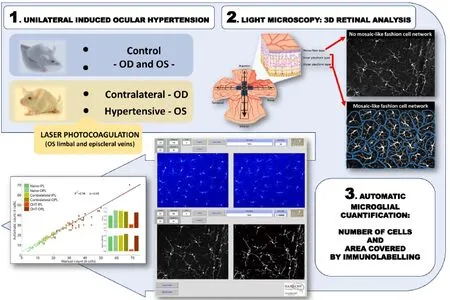
Figure 1 Illustration of the automatic retinal microglial cell quantification methodology.
Microglial cells are characterized by small cellular bodies from which emerge numerous, long, profusely ramified branches; these cells are distributed over the parenchyma of the nervous tissue, but without overlap of neighboring cells. These cellular features, which are also observed in the retina, allow visual identification of a single microglial cell and are the key to our algorithm, which automatically determines the number of microglial cells in the inner and outer plexiform layers of the retina (Ramírez et al., 2015a).
这是敲门歌,XX指出嫁姑娘家族的堂号。“堂号”是家族门户的代称,是家族文化重要的组成部分。如果出嫁姑娘姓黄,九寨黄姓的堂号是江夏堂,则会改成“江夏朝中招驸马”。这里用了借喻,“扬州琼花”代替嫁姑娘这件喜事;“荥阳城”代替女方的家。“扬州琼花”指《隋唐演义》中,扬州有一朵漂亮的牡丹花,杨广去看而花不开;但李世民去,花就开了,说明李世民才是花主,暗示他才是天子。所以这里就用“扬州琼花现”来表示要嫁的姑娘像扬州的花一样找到正主了。
These specific microglial cell features remain even during proliferative events; however, depending on particular characteristics of the tissue analyzed, microglial cells sometimes do not completely fulfil these criteria. This makes the process of cell recognition complex and not very reliable for both human and computational approaches. That complexity is noted on the nerve fiber layer of the retina, where Iba-1+cells adapt their somas and processes to the spaces within the fibers of the RGCs and the blood vessels. Nonetheless, instead of counting independent events in a research scenario, another quantitative approach is to calculate the area covered by specific immunolabeling, which produces an indirect measure of the number of cells in the tissue. An increase of the area covered by, for example, Iba-1 immunolabeling is also a mark of gliosis. This approach provided the key to developing another automatic tool in our algorithm that allows quantitative analysis of the area of the retina occupied by microglial cells (Figure 1).
One of the benefits of our algorithm is the interactive work interface, which was developed to supply researchers with a graphic visualization of the process and the ability to change some parameters of interest, such as cell distance and image threshold. As a result, and despite a complex mathematicalprogramming environment, this new algorithm is easy to use and does not require the user to have a programing background. Another advantage of our algorithm is that it allows researchers not only to work with a 2-dimensional image but also to study 3-dimensional volume (de Gracia et al., 2015). Because of the complex 3-dimensional spatial distribution of microglial cells in nervous tissue, this is a very useful feature.
With our new automated microglial cell quantification method, the time for counting a huge set of images can be radically reduced from weeks (manual procedure) to a few hours (computational analysis) without any statistically significant difference from results of a manual count by a human (gold standard). In addition, our results exhibited a good correlation not only in naïve tissues but also in highly proliferative gliotic states (de Gracia et al., 2015).
This algorithm has been developed for the quantification of microglial cells in retinal flat mount; however, due to the similarities of microglial cells within the CNS, this algorithm (once calibrated) will also facilitate quantitative tasks in other regions of the nervous system.
Microglial Analysis: Development of New Neuroprotective Therapies
Although it was already known that glaucoma is usually a bilateral but asymmetrically presenting disease, the contralateral eye in unilateral glaucoma models has been frequently used as an internal control. In our unilateral mouse model of laser induced OHT, the contralateral gliosis (bilateral reaction) may represent events linked with the initial steps of the glaucomatous neurodegeneration, previous to a neuronal death, and probably mediated by the ongoing inflammatory processes. Research about the events observed in the contralateral eyes in response to a unilateral model of glaucoma is sparse (Ramírez et al., 2015b); however, new studies focused on the implication of this activation of glial cells could provide a better understanding of glaucomatous pathophysiology. Research on steps prior to neurodegeneration in glaucoma, and therefore possible intervention points in the disease, has the potential to allow assessment and development of new neuroprotective therapies.
Microglial proliferation has been observed in our studies, both in the presence of neuronal death and also without it; thus, future quantitative microglial studies could assist in the detection of early neurodegeneration and establish signs or events related to neurodegeneration. Also, the number of microglial cells in tissue could be issued as an index of recovery after the application of neuroprotective therapies. A prodigious amount of research is needed to collect strong evidence to fully understand the etiology of glaucoma and to develop treatments. At this point in time, our algorithm provides researchers with a useful tool to perform quick and accurate microglial cell analysis on large data sets of images.
The algorithm will be provided at no cost to any researcher contacting the authors of this study (de Gracia et al., 2015).
Acknowledgments: The authors of this paper thanks the Neuroscience Publications office at Barrow Neurological Institute for their editorial and manuscript preparation assistance.
Conflicts of interest: None declared.
This work has been presented in the following meetings:
1. Salazar JJ, Gallego BI, Rojas B, Triviño A, Ramírez JM, de Gracia P (2013) A new automatic method for counting microglial cells in whole-mount mice retinas. Sociedad de Investigación de Retina de la Comunidad Valenciana (SIRCOVA). Co-patrocinio Association for Research in Vision and Ophthalmology (ARVO). Ophthal Res 50∶27-53.
2. de Hoz R, Gallego BI, Rojas B, Ramírez AI, Salazar JJ, Triviño A, de Gracia P, Ramírez JM (2013) A new automatic method for microglial-cell quantification in whole-mount mouse retinas. Joint Eu ropean Research Meeting in Ophthalmology and Vision. European As sociation for Vision and Eye Research (EVER). Acta Ophthalmol 89. 3. de Gracia P, Gallego BI (2012) A new automatic method for coun ting microglial cells in healthy and glaucomatous retinas. American Academy of Optometry meeting, American Academy of Optometry (AAO).
References
de Gracia P, Gallego BI, Rojas B, Ramírez AI, de Hoz R, Salazar JJ, Trivino A, Ramírez JM (2015) Automatic counting of microglial cells in healthy and glaucomatous mouse retinas. PLoS One 10:e0143278.
de Hoz R, Rojas B, Ramírez AI, Salazar JJ, Gallego BI, Trivino A, Ramírez JM (2016) Retinal macroglial responses in health and disease. Biomed Res Int 2016:2954721.
de Hoz R, Gallego BI, Ramírez AI, Rojas B, Salazar JJ, Valiente-Soriano FJ, Aviles-Trigueros M, Villegas-Perez MP, Vidal-Sanz M, Trivino A, Ramírez JM (2013) Rod-like microglia are restricted to eyes with laser-induced ocular hypertension but absent from the microglial changes in the contralateral untreated eye. PLoS One 8:e83733.
Gallego BI, Salazar JJ, de Hoz R, Rojas B, Ramírez AI, Salinas-Navarro M, Ortin-Martinez A, Valiente-Soriano FJ, Aviles-Trigueros M, Villegas-Perez MP, Vidal-Sanz M, Trivino A, Ramírez JM (2012) IOP induces upregulation of GFAP and MHC-II and microglia reactivity in mice retina contralateral to experimental glaucoma. J Neuroinflammation 9:92.
Quigley HA, Broman AT (2006) The number of people with glaucoma worldwide in 2010 and 2020. Br J Ophthalmol 90:262-267.
Ramírez AI, Rojas B, de Hoz R, Salazar JJ, Gallego BI, Triviño A, Ramírez JM (2015a) Microglia, inflammation, and glaucoma. Dover: SM Group Open Access eBooks ed.
Ramírez AI, Salazar JJ, de Hoz R, Rojas B, Gallego BI, Salobrar-Garcia E, Valiente-Soriano FJ, Trivino A, Ramírez JM (2015b) Macro- and microglial responses in the fellow eyes contralateral to glaucomatous eyes. Prog Brain Res 220:155-172.
Ramírez AI, Salazar JJ, de Hoz R, Rojas B, Gallego BI, Salinas-Navarro M, Alarcon-Martinez L, Ortin-Martinez A, Aviles-Trigueros M, Vidal-Sanz M, Trivino A, Ramírez JM (2010) Quantification of the effect of different levels of IOP in the astroglia of the rat retina ipsilateral and contralateral to experimental glaucoma. Invest Ophthalmol Vis Sci 51:5690-5696.
Rojas B, Gallego BI, Ramírez AI, Salazar JJ, de Hoz R, Valiente-Soriano FJ, Avilés-Trigueros M, Villegas Pérez MP, Vidal-Sanz M, Triviño A, Ramírez JM (2014) Microglia in mice retina contralateral to experimental glaucoma exhibit multiple signs of activation in all retinal layers: a detailed description. J Neuroinflammation 11:133.
Tezel G. the Fourth ARVO/Pfizer Ophthalmics Research Institute Conference Working Group (2009) The role of glia, mitochondria, and the immune system in glaucoma. Invest Ophthalmol Vis Sci 50:1001-1012.
10.4103/1673-5374.189166
*Correspondence to:
猜你喜欢
杂志排行

中国神经再生研究(英文版)的其它文章
- Secondary parkinsonism induced by hydrocephalus after subarachnoid and intraventricular hemorrhage
- Huangqi Guizhi Wuwu Decoction for treating diabetic peripheral neuropathy: a meta-analysis of 16 randomized controlled trials
- No synergism between bis(propyl)-cognitin and rasagiline on protecting dopaminergic neurons in Parkinson's disease mice
- Association between chromosomal aberration of COX8C and tethered spinal cord syndrome: arraybased comparative genomic hybridization analysis
- Rebuilding motor function of the spinal cord based on functional electrical stimulation
- Acellular allogeneic nerve grafting combined with bone marrow mesenchymal stem cell transplantation for the repair of long-segment sciatic nerve defects: biomechanics and validation of mathematical models