经皮肤入路鼻外侧截骨术在鼻整形中的应用
2016-07-18尉迟海深林金德陈小平郑翔宇万昌阳
尉迟海深, 林金德, 孙 露, 陈小平, 郑翔宇, 万昌阳
论著-鼻整形专题
经皮肤入路鼻外侧截骨术在鼻整形中的应用
尉迟海深, 林金德, 孙 露, 陈小平, 郑翔宇, 万昌阳
目的 探讨经皮肤入路鼻外侧截骨术在鼻整形中的应用效果。方法 对12例鼻外形不佳者行皮肤入路鼻外侧截骨术,于双侧鼻面沟中部做一长约2 mm的皮肤切口,通过此切口插入一锋利的宽为2 mm的截骨刀直达骨膜下,沿双侧鼻面沟设计的截骨线每隔2 mm间断截断上颌骨额突。完成截骨后,用指力向内侧挤压鼻骨,造成青枝骨折,重塑理想的骨性鼻锥。7例患者同时行假体隆鼻、鼻中隔延长等手术。结果 所有患者术后骨性鼻锥外形均得到明显改善。术后早期眶周淤血、水肿等症状较轻,随访2~9个月未发生通气功能异常,双侧鼻骨未发生明显的不对称,皮肤切口瘢痕不明显,未发生色素沉着等并发症。结论 采用经皮肤入路鼻外侧截骨术重塑骨性鼻锥,易于操作,方法精确、安全,效果确切,术后并发症较少,是一种值得推广的手术方法。
鼻整形术; 鼻外侧截骨术; 皮肤入路; 骨性鼻锥
自2014年5月至2016 年1月,南京医科大学附属友谊整形外科医院整形外科采用经皮肤入路鼻外侧截骨术矫正鼻部骨性畸形患者12例,手术效果良好。现报道如下。
1 临床资料
本组共12例患者。均为女性;年龄23~31岁,平均27岁。均为先天性宽鼻畸形,其中5例行经皮肤鼻外侧截骨术单纯纠正宽鼻,7例行经皮肤鼻外侧截骨术+假体隆鼻、鼻中隔延长和鼻翼缩小等综合鼻整形手术纠正鼻外形。
2 手术方法
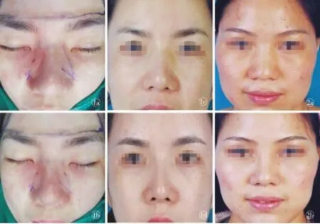
采用全身麻醉。如果要同时行隆鼻、鼻中隔延长、鼻尖成形术等其他鼻部手术,一般在其他鼻部手术完成之后再行截骨,以保证截骨塑形后的稳定性。手术要点:⑴沿鼻面沟设计截骨线,上至内眦连线。以亚甲蓝于面部标记。⑵以1%利多卡因+1∶10万肾上腺素溶液沿双侧鼻面沟行局部浸润麻醉10 min。⑶在设计线的中部做一约2 mm的皮肤切口,通过此切口插入宽为2 mm的截骨刀直达骨膜下,用锤子敲打截骨刀尾部,当感到一明显的突破感和截骨声音改变时,表示此点的骨质已被穿透。沿设计线截断上部和下部的上颌骨额突,每当截骨刀移到下一个截骨点时,需要确认截骨刀尖端所接触的骨质位于设计的截骨线的正下方,以确保严格按照预设的截骨线截骨,避免截骨出现偏移,导致骨锥的继发畸形,每个截骨点之间保持2 mm的正常骨质。截骨在骨膜下进行,这样可以避免损伤内眦动脉,减少术中出血及术后眼周淤血。截骨线始于梨状孔的下鼻甲前缘处,向上达内眦连线水平。用同样方法完成对侧截骨(图1a,b)。⑷当完成两侧截骨后,用手指将两侧鼻骨向内侧挤压,造成青枝骨折,将鼻骨移动到合适位置。注意避免手指的过度用力,如果轻力不能压断骨质,则需沿原截骨线再行多个穿刺点,缩窄穿刺点间的距离;如果骨质过厚过硬,则需将截骨点连接起来。⑸鼻背先用胶布固定,再用鼻外夹板固定。鼻外夹板在术后第10天去除。如单纯行鼻外侧截骨术,可以不使用抗生素。如联合隆鼻或鼻中隔延长术,则术后预防性使用抗生素72 h。3~4周避免擤鼻涕及剧烈运动。
3 结果
本组共12例患者。因联合其他手术,所有患者住院期间(3 d左右)眶周水肿和淤斑等表现明显。术后随访2~9个月,所有患者术后均恢复良好,未发生通气功能异常,无明显的双侧鼻骨不对称、皮肤穿刺点瘢痕增生、色素沉着等并发症发生。
4 典型病例
例1,患者女性,25岁。单纯行经皮肤入路鼻外侧截骨术,术中损伤小,眶周肿胀程度轻。术后9个月,切口瘢痕不明显(图1c,d)。
例2,患者女性,31岁。因鼻背低平、宽鼻畸形行“假体隆鼻+鼻中隔延长+宽鼻畸形矫正术”。经皮肤入路行鼻外侧截骨术矫正宽鼻畸形,术后鼻背缩窄明显。术后2个月,鼻外形满意(图2)。皮肤切口少许瘢痕,颜色稍红。

图1 单纯经皮肤入路鼻外侧截骨术前后对比 a.完成右侧截骨 b.完成双侧截骨 c.术前 d.术后9个月 图2 假体隆鼻+鼻中隔延长+宽鼻畸形矫正术前后对比 a.术前 b.术后2个月
Fig 1 Comparison between preview and postview of transcutaneous lateral nasal osteotomy. a.complete image of right side osteotomy. b.complete image of bilateral osteotomy. c.preview. d.postview at 9 months. Fig 2 Comparison between preview and postview of augmentation rhinoplasty, septal extension and correction of wide nasal deformity. a.preview. b.postview at 2 months.
5 讨论
鼻外侧截骨术是矫正鼻骨性支架畸形的必需手术步骤之一[1-6]。传统的鼻外侧截骨术是通过鼻内或口内入路,分离的软组织较多,充分显露鼻骨和部分上颌骨额突后,将截骨线周围的骨膜完全剥离,再用宽截骨刀连续截断上颌骨额突,重塑鼻骨性支架。由于盲视操作,这种传统的截骨术损伤较大,截骨准确性和可控性较差。手术效果更多地依赖术者的经验,截骨可能不精确,不能达到理想的手术效果。
1984年,CN Ford等通过尸体研究比较鼻内入路连续截骨与间断截骨术的效果,再用CT扫描,发现间断截骨能保留骨膜与骨片的连接,减少了死腔的形成,减轻了对气道的不良影响,并增加了骨性鼻拱的稳定性。1993年,M Goldfard等在其临床试验中回顾了100多例行鼻外侧间断穿刺截骨的患者,在2年的随访期内,发现术后淤血、水肿轻微且发生率很低。
1961年,CR Straatsma介绍了经皮肤入路鼻外侧截骨术,具有损伤轻,不易在鼻背基底形成阶梯状畸形等优点。1997年,RJ Rohrich等研究截骨方法和鼻内损伤的关系,在新鲜尸体中发现,经皮肤入路截骨术鼻黏膜损伤程度比鼻内入路连续鼻外侧截骨术轻。
理想的鼻外侧截骨术应该是可重复,效果可控,易于操作,创伤小,恢复快,并发症少[7-14]。经皮肤入路鼻外侧截骨术具有操作简单、截骨精确等优点。从解剖学看,内眦韧带通过泪囊,大范围剥离骨膜可能损伤内眦韧带;内眦动脉横跨截骨线,传统方法截骨容易损伤内眦血管,不剥离骨膜的间断截骨大大减少了以上情况发生的可能性,由此导致的皮下出血和肿胀也相应减少。另外,经皮肤入路鼻外侧截骨术的青枝骨折更加稳定,减少死腔形成,对气道的影响更小。因为相邻截骨点之间的骨膜是不剥离的,保留了绝大部分骨膜与骨的贴附,能作为支撑力量增加骨折后骨块的稳定性。
如果鼻骨远端至内眦连线长度小于1 cm,不建议截骨,因为这会导致侧鼻软骨的塌陷,引起鼻部畸形和鼻气道的阻塞。对老年患者、戴较重眼镜和皮肤层较厚患者应慎重截骨;非高加索人种如果鼻背过低或过宽,则不建议截骨。本术式尽管在两侧鼻面沟处各遗留长约2 mm的皮肤切口,即使不加缝合,切口也不会有明显的瘢痕。
综上所述,经皮肤入路鼻外侧截骨术疗效肯定,能精确可控地截骨,所引起的鼻内创伤较轻。由于大部分骨膜不被剥离,减少了死腔形成和对气道的影响,并增加了鼻骨性支架的稳定性,是鼻骨截骨整形术的一种理想选择。
[1] Tirelli G, Tofanelli M, Bullo F, et al. External osteotomy in rhinoplasty: Piezosurgery vs osteotome[J]. Am J Otolaryngol, 2015,36(5):666-671.
[2] Ghazipour A, Alani N, Ghavami Lahiji S, et al. Buccal sulcus versus intranasal approach for postoperative periorbital oedema and ecchymosis in lateral nasal osteotomy[J]. J Craniomaxillofac Surg, 2014,42(7):1456-1459.
[3] Caglar E, Celebi S, Topak M, et al. How can periorbital oedema and ecchymose be reduced in rhinoplasty?[J]. Eur Arch Otorhinolaryngol, 2016,273(9):2549-2554.
[4] Azizzadeh B, Reilly M. Dorsal hump reduction and osteotomies[J]. Clin Plast Surg, 2016,43(1):47-58.
[5] Bali ZU, Ahmedov A, Aydemir L, et al. A novel method to prevent complications of nasal osteotomy: matress suture which traverses lateral walls and septum[J]. Kulak Burun Bogaz Ihtis Derg, 2015,25(6):324-328.
[6] Simsek G, Demirtas E. Comparison of surgical outcomes and patient satisfaction after 2 different rhinoplastytechniques[J]. J Craniofac Surg, 2014,25(4):1284-1286.
[7] Cakr B, Finocchi V, Tambasco D, et al. Osteoectomy in rhinoplasty: a new concept in nasal bones repositioning[J]. Ann Plast Surg, 2016,76(6):622-628.
[8] Cochran CS, Roostaeian J. Use of the ultrasonic bone aspirator for lateral osteotomies in rhinoplasty[J]. Plast Reconstr Surg, 2013,132(6):1430-1433.
[9] Gabra N, Rahal A, Ahmarani C, et al. Nasal osteotomies: a cadaveric study of fracture lines[J]. JAMA Facial Plast Surg, 2014,16(4):268-271.
[10] Ghassemi A, Ayoub A, Modabber A, et al. Lateral nasal osteotomy: a comparative study between the use of osteotome and a diamond surgical burr-a cadaver study[J]. Head Face Med, 2013,9:41.
[11] Rho BI, Lee IH, Park ES, et al. Visible perforating lateral osteotomy: internal perforating technique with wide periosteal dissection[J]. Arch Plast Surg, 2016,43(1):88-92.
[12] Ilhan AE, Cengiz B, Caypinar Eser B, et al. Double-blind comparison of ultrasonic and conventional osteotomy in terms of early postoperative edema and ecchymosis[J]. Aesthet Surg J, 2016,36(4):390-401.
[13] Ghassemi A, Prescher A, Talebzadeh M, et al. Osteotomy of the nasal wall using a newly designed piezo scalpel-a cadaver study[J]. J Oral Maxillofac Surg, 2013,71(12):2155.e1-e6.
[14] Al Arfaj AM. The use of nasal packing post rhinoplasty: does it increase periorbital ecchymosis? a prospective study[J]. J Otolaryngol Head Neck Surg, 2015,44:22.
Application of transcutaneous lateral nasal osteotomy in rhinoplasty
YU-CHIHai-shen,LINJin-de,SUNLu,CHENXiao-ping,ZHENGXiang-yu,WANChang-yang.
(DepartmentofPlasticSurgery,FriendshipPlasticSurgeryHospitalofNanjingMedicalUniversity,Nanjing210009,China)Correspondingauthor:LINJin-de,Email:hzljd@sohu.com
Objective To investigate the effect of transcutaneous lateral nasal osteotomy applied in rhinoplasty. Methods Twelve patients with poor nasal shape underwent transcutaneous lateral nasal osteotomy. Using a 2 mm sharp osteotome, a 2 mm incision was made at the middle of the nasofacial junction to the periosteum. An intermittent cut every 2 mm following the cutting line designed along the bilateral nasofacial sulcus was made to the processus frontalis. Subsequently, greenstick fractures of nasal bone were induced by gentle force with fingers to reform the ideal bony nose cone. Seven patients were also treated with other procedures such as implant augmentation and septal extension. Results All patients received obviously improved shape of nasal bony cone after surgery. Early postoperative periorbital edema and congestion were mild. No instances of abnormal ventilation, bilateral nasal bone asymmetry, or pigmentation were found during follow-up for 2 to 9 months, and the skin incision scars were concealed. Conclusion Transcutaneous lateral nasal osteotomy for reshaping nasal bony cone is simpleand convenient for the surgeon, resulting in accurate and safe osteotomy. The obvious effectiveness and few complications make this a procedure worthy of wider application.
Rhinoplasty; Lateral nasal osteotomy; Transcutaneous; Nasal bony cone
210009 江苏 南京,南京医科大学附属友谊整形外科医院 整形外科
尉迟海深(1988-),男,河南南阳人,硕士.
林金德,210009,南京医科大学附属友谊整形外科医院 整形外科,电子信箱:hzljd@sohu.com
10.3969/j.issn.1673-7040.2016.10.004
R622
A
1673-7040(2016)10-0589-03
2016-06-16)
