426例甲状腺结节的临床回顾性分析
2016-07-11王文超杨丽张春霞
王文超 杨丽 张春霞

[摘要] 目的 探讨与甲状腺结节良、恶性鉴别相关的临床因素。 方法 回顾性分析2009年1月~2014年10月北京市顺义区医院收治的426例甲状腺结节患者的临床资料。 结果 本组资料中,良性病变382例(89.67%),恶性病变44例(10.33%);单因素分析结果:患者为男性、年龄≤20岁、位于右叶的病灶及伴钙化的甲状腺结节恶性可能性大(P<0.05),TSH水平对甲状腺结节性质的鉴别无统计学意义(P>0.05);多因素分析结果:患者为男性、年龄≤20岁、位于右叶的病灶及伴钙化者患甲状腺恶性肿瘤的可能性大(P<0.05)。 结论 患者性别、年龄、病灶位置及是否伴钙化对甲状腺结节病变性质的鉴别具有重要临床意义。
[关键词] 甲状腺结节;良性;恶性;钙化
[中图分类号] R736.1 [文献标识码] A [文章编号] 1674-4721(2016)03(b)-0046-03
[Abstract] Objective To investigate the relationship between the clinical factors and the character of the thyroid nodules. Methods The clinical data of 426 patients with thyroid nodules who were treated in Shunyi Hospital were retrospectively analyzed. Results In this study,382 cases (89.67%) were benign and 44 cases (10.33%) were malignant.Univariate analysis:The possibility of suffering thyroid cancer increased when the patient with thyroid nodules was male,younger than 20 years of age,the nodules located in the right lobe,or when the patient was accompanied by calcification(P<0.05).The level of thyroid stimulating hormone was not correlated with diagnosis of benign and malignant lesions of the nodules (P>0.05).Multivariate analysis:The patient with thyroid nodules was male,younger than 20 years of age,the nodules located in the right lobe,or when the patient was accompanied by calcification had a higher chance of suffering from thyroid cancer. Conclusion For the patient presenting with thyroid nodules,the sex and the age of patient,the locations of the thyroid nodules and the accompanying by calcification have important clinical significance in diagnosis of benign and malignant lesions of thyroid nodules.
[Key words] Thyroid Nodules;Benign;Malignant;Calcification甲状腺结节是甲状腺外科门诊日常患者就诊最常见的主诉,其发病率呈逐年上升的趋势,成人中其发病率达3%~7%,尤其以女性多见[1]。甲状腺结节以良性病变为主,但恶性病变仍占有很大比例,占甲状腺结节的5%~7%[2]。如何结合患者的临床因素提高对甲状腺结节性质判断的准确性具有重要意义。本文通过对426例甲状腺结节患者的临床资料进行回顾性分析,探讨与甲状腺结节性质鉴别相关的临床因素与诊断问题。
1 资料与方法
1.1 一般资料
收集2009年1月~2014年10月于北京市顺义区医院就诊并行手术治疗的426例甲状腺结节患者的临床资料,其中男80例,女346例,男女比例为1∶4.33,年龄18~84岁,中位年龄49.5岁。病灶位于甲状腺左叶100例,右叶139例,双侧179例,峡部8例。
1.2 辅助检查
术前均行血清促甲状腺素(thyroid stimulating hormone,TSH)水平检测及甲状腺超声检查,其中TSH低于正常水平70例,正常或高于正常水平356例;超声检查提示甲状腺单发结节175例,多发结节202例,单发甲状腺结节伴钙化者20例,多发结节伴钙化29例。
1.3 石蜡病理结果
良性病变382例,其中结节性甲状腺肿187例,腺瘤191例,甲状腺炎4例;恶性病变44例,其中乳头状癌42例,滤泡状癌1例,髓样癌1例。
1.4统计学方法
采用SPSS 17.0统计学软件对数据进行处理,采用χ2检验从中筛选对病变性质影响有统计学意义的临床因素作为自变量,运用逐步Logistic回归分析法进行多因素分析,以P<0.05为差异有统计学意义。
2 结果
2.1甲状腺结节病变性质与临床因素的关系(单因素分析)
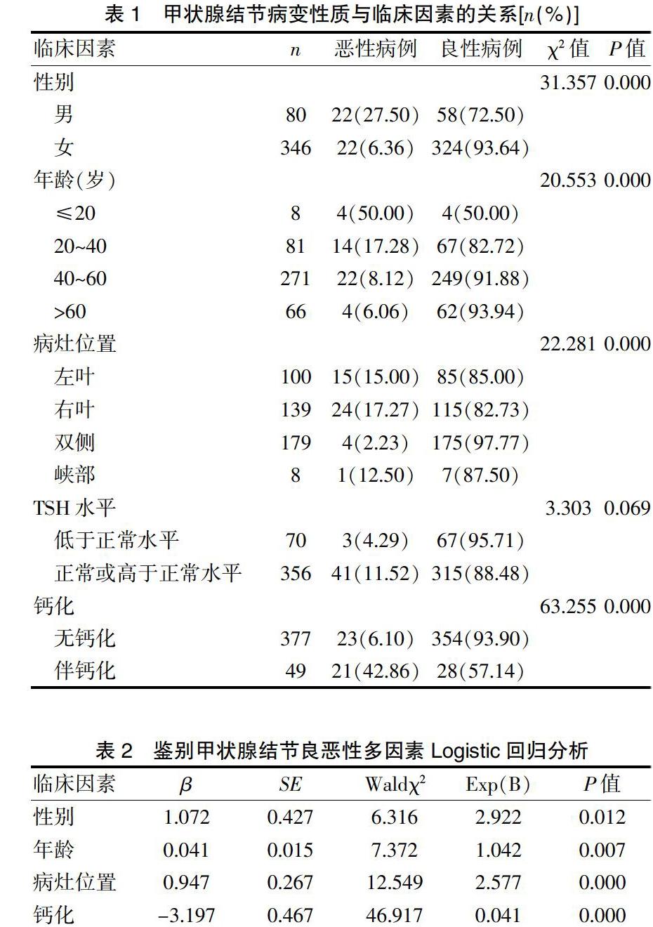
通过比较分析得出患者为男性、年纪≤20岁、位于右叶的病灶及伴钙化的甲状腺结节恶性可能性大,TSH水平对甲状腺结节性质的鉴别差异无统计学意义意义(P>0.05)(表1)。
2.2 甲状腺结节病变性质与临床因素的关系(多因素分析)
通过单因素分析,筛出对甲状腺结节性质影响的四组变量,即患者性别、年龄、病灶位置及是否伴钙化,差异有统计学意义(P<0.05)(表2)。
3 讨论
目前公认的对于甲状腺结节良、恶性鉴别有重要意义的临床因素包括患者年龄、性别、家族史、超声表现、颈部放射线照射史等,尤其是儿童时期的放射线接触[1]。本研究主要对患者性别、年龄、病灶位置、血清TSH水平及是否伴钙化与甲状腺结节性质的关系进行研究。
研究表明,女性甲状腺结节发病率高于男性,但男性患者中甲状腺结节恶性率更高[3],其中滤泡状癌在男性患者中发病率高于女性患者[4]。本研究的结果中女性患者占全部病例的81.22%,其恶性率为6.36%,男性患者中甲状腺结节的恶性率为27.50%,明显高于女性患者,与上述报道结论相符,提示对于男性甲状腺结节的患者应该予以足够的重视。
年龄作为鉴别甲状腺结节性质的重要因素之一,近年来有学者进行了大量的研究调查,结果显示,年龄<55岁的患者更易发生甲状腺癌,且年龄越小甲状腺结节的恶性率越高[3]。在成年人及儿童中,甲状腺结节的恶性率分别为5%和25%[5-7],本研究中年龄≤20岁组中甲状腺结节的恶性率最高,年龄越大,甲状腺结节的恶性率越低,对于年轻患者应予以足够重视,但对于高龄的患者,应结合其性别、临床表现及超声等结果对甲状腺结节性质进行综合判断。
近年来对双侧同时发生的甲状腺癌研究较多,据报道,双侧同时发生的甲状腺癌占总甲状腺癌的10.9%[8]。本组资料中,双侧甲状腺癌均为同时发生,其发病率占总甲状腺癌的9.09%(4/44),相对于单侧及峡部的甲状腺结节,双侧甲状腺结节的恶性率最低,考虑双侧甲状腺肿物多为多发结节,病灶弥漫散在,结节性甲状腺肿的发病率高,发现甲状腺结节后患者的手术意愿较高,早期即行手术治疗,手术适应证范围相对较宽所致。既往认为甲状腺的单发结节相对于多发结节的恶性率高[9],但亦有学者提出反对意见,如Mihailescu等[10]认为甲状腺结节的良恶性与结节大小和数目无关。对于甲状腺结节的病灶位置与其病变性质的研究目前较少,且无定论[11],本研究中的右叶甲状腺结节发病率相对高于左叶及峡部,且其恶性率也最高,考虑与甲状腺右叶体积较大有关,与Gessl等[12]报道结果相近。
血清TSH作为预测甲状腺癌的独立指标之一,随着血清TSH水平的升高,甲状腺结节的恶性可能性越大[13]。本研究中血清TSH对于甲状腺结节的良恶性影响差异无统计学意义,考虑与本研究中样本量小及对于TSH水平未进行详细分组研究有关,尚需大样本量的后续研究。
超声下提示甲状腺恶性肿瘤的征象包括:微钙化、边缘不规则、内部血流丰富、伴有肿大淋巴结及高径>宽径等[4],其中以微钙化的研究最多,是目前公认的甲状腺癌的重要指标之一,而粗大或弧形钙化被认为是良性病变。有学者提出伴有微钙化的甲状腺结节的恶性发病率明显高于不伴钙化者[14]。本组资料中,甲状腺结节伴钙化患者的恶性率明显高于不伴钙化者,但亦有学者持反对意见,认为结节性甲状腺肿或甲状腺腺瘤也可在影像学上表现为微钙化,而粗大或弧形钙化也可以出现在甲状腺癌的影像学表现中,目前通过超声检查并不能对钙化的良、恶性进行鉴别[15],支持此观点的学者研究提示,恶性钙化的应为砂砾样钙化,但并非所有微钙化均为砂砾样钙化。近年来超声弹性成像技术在临床超声中逐步推广,使得超声对于甲状腺结良恶性诊断的准确性有了很大提高[16],对于钙化性质的鉴别有赖于超声技术的发展及超声科医师水平的提高,进而提高对其诊断的准确性。
综上所述,甲状腺结节为最常见的临床疾病,其性质的判断需结合患者性别、年龄、病灶位置及超声是否伴有钙化等因素进行综合考虑,对于患者血清的TSH水平是否可作为预测甲状腺结节良、恶性的指标仍有待临床研究证明。
[参考文献]
[1] Syrenicz A,Koziolek M,Ciechanowicz A,et al.New insights into the diagnosis of nodular goiter[J].Thyroid Res,2014,7:6.
[2] Gharib H,Papini E,Paschke R,et al.American association of clinical endocrinologists,associazione medici endocrinologi,and european thyroid association medical guidelines for clinical practice for the diagnosis and management of thyroid nodules:executive summary of recommendations[J].J Endocrinol Invest,2010,33(5 Suppl):51-56.
[3] Azizi G,Malchoff CD.Autoimmune thyroid disease:a risk factor for thyroid cancer[J].Endocr Pract,2011,17(2):201-209.
[4] Nachiappan AC,Metwalli ZA,Hailey BS,et al.The thyroid:review of imaging features and biopsy techniques with radiologic-pathologic correlation[J].Radiographics,2014,34(2):276-293.
[5] Wiersinga WM.Management of thyroid nodules in children and adolescents[J].Hormones (Athens),2007,6(3):194-199.
[6] Osipoff JN,Wilson TA.Consultation with the specialist:thyroid nodules[J].Pediatr Rev,2012,33(2):75-81.
[7] Bas VN,Aycan Z,Cetinkaya S,et al.Thyroid nodules in children and adolescents:a single institution′s experience[J].J Pediatr Endocrinol Metab,2012,25(7-8):633-638.
[8] 陈振宇,吴毅.双侧甲状腺癌的临床新特点[J].中国实用外科杂志,2012,32(1):77-79.
[9] Rago T,Fiore E,Scutari M,et al.Male sex,single nodularity,and young age are associated with the risk of finding a papillary thyroid cancer on fine-needle aspiration cytology in a large series of patients with nodular thyroid disease[J].Eur J Endocrinol,2010,162(4):763-770.
[10] Mihailescu DV,Schneider AB.Size,number,and distribution of thyroid nodules and the risk of malignancy in radiation-exposed patients who underwent surgery[J].J Clin Endocrinol Metab,2008,93(6):2188-2193.
[11] Campennì A,Giovanella L,Siracusa M,et al.Is malignant nodule topography an additional risk factor for metastatic disease in low-risk differentiated thyroid cancer?[J].Thyroid,2014,24(11):1607-1611.
[12] Gessl A,Raber W,Staudenherz A,et al.Higher frequency of thyroid tumors in the right lobe[J].Endocr Pathol,2010, 21(3):186-189.
[13] Boelaert K.The association between serum TSH concentration and thyroid cancer[J].Endocr Relat Cancer,2009,16(4):1065-107.
[14] 矫杰,周迎生,陈宝玥,等.甲状腺结节伴乳头状癌临床特征及恶性风险因素分析[J].中华实用诊断与治疗杂志,2015,29(1):53-55.
[15] 吴毅.关于甲状腺结节诊断和治疗的若干思考[J].中国实用外科杂志,2010,30(10):821-823.
[16] 王伟,金正吉,唐波,等.超声及超声弹性成像诊断甲状腺结节的良恶性[J].中华实用诊断与治疗杂志,2013, 27(5):467-469.
