正常青年人下颌管全长三维走向及下颌骨形态的锥形束CT测量
2016-07-08绳兰兰曲卫国李阳屈振宇岳柏王吉大连市口腔医院口腔颌面外科放射科大连60
绳兰兰 曲卫国 李阳 屈振宇 岳柏 王吉.大连市口腔医院口腔颌面外科;.放射科,大连 60
正常青年人下颌管全长三维走向及下颌骨形态的锥形束CT测量
绳兰兰1曲卫国1李阳1屈振宇2岳柏1王吉1
1.大连市口腔医院口腔颌面外科;2.放射科,大连 116021
[摘要]目的 采用锥形束CT(CBCT)分析正常青年人下颌管在下颌骨内的三维位置以及下颌骨的形态特征,为临床下颌骨手术提供解剖学依据。方法 对29例个别正常�进行CBCT扫描,用InVivo 5软件对下颌骨进行三维重建,定位标记点,测量下颌骨形态以及下颌管在其内的三维走行。采用SPSS 17.0软件对测量值进行统计分析。结果下颌管舌侧骨皮质厚度明显较颊侧骨皮质薄。下颌管到颊侧骨皮质的距离从近中到远中逐渐增加,到舌侧骨皮质、牙槽嵴顶的距离从近中到远中逐渐减小,到下颌下缘的距离在第一磨牙处最小,第二前磨牙处最大。下颌体截面高度、宽度、皮质骨厚度左右侧无统计学差异,从中线至远中,下颌体截面高度、舌侧下1/3皮质骨厚度逐渐减小,上截面宽度、唇/颊侧上1/3皮质骨厚度逐渐增大。部分测量项目性别间有统计学差异。结论 下颌管入下颌孔后渐渐远离舌侧而向颊侧靠近,然后又逐渐远离偏向舌侧,但其总体走行还是靠近舌侧。男性下颌骨较女性更坚厚。CBCT能精确地显示下颌神经管的走行及其与周边结构的关系。
[关键词]下颌管; 下颌骨; 锥形束CT
对下颌管位置和走向的准确判断,有助于下颌骨手术(如牙种植术、囊肿刮治术、根尖外科手术等)时减少对下牙槽神经的损伤。本研究采用锥形束CT(cone beam computed tomography,CBCT)分析正常青年人下颌管在下颌骨内的三维位置以及下颌骨的形态特征,为临床下颌骨手术时提供解剖学依据。
1 材料和方法
1.1 样本的选择
随机选择2015年1月—2015年4月于大连市口腔医院颌面外科就诊的29例个别正常为研究对象。29例中,男17例,女12例;年龄19~30岁,平均年龄24.48岁。纳入标准:下颌骨无发育异常、无牙列缺损、无牙周病、无炎症、无颌骨囊肿及肿瘤,且无下颌骨外伤史。排除标准:存在能影响下颌管位置及形态的各种疾病。
1.2 CBCT扫描和图像处理
所有研究对象均在牙尖交错位使用同一CBCT机(Kavo 3D eXam,Imaging Sciences International公司,美国)接受检查。受检者取坐位,头直立,表情自然,眶耳平面与地面平行,自然咬合。投照角度为单次360°旋转扫描,扫描野16.5 cm×13.5 cm,电压120 kV、电流5 mA,层厚0.2 mm,扫描时间14.7 s。使用三维重建测量软件InVivo 5进行三维重建图像处理和测量研究。
1.3 测量
1.3.1 下颌管的测量 1)下颌升支部下颌管的位置。将通过下颌管最先形成的平面做为0平面,向下每5 mm作为一个测量平面,依次为1、2、3、4、5平面,在每一平面测量:下颌管内径(ID),通过下颌管中心的下颌骨厚度(LB),该处颊舌侧骨皮质的厚度(BC、LC),下颌管外侧壁到颊舌侧骨皮质之间的距离即颊舌侧骨髓腔的宽度(BP、LP)(图1)。2)下颌骨体部下颌管的空间定位。调整牙齿远中截面与下颌牙弓垂直,分别于第二磨牙、第一磨牙、第二前磨牙的远中面测量下颌管内缘距下颌骨各表面(牙槽嵴顶、下颌下缘、颊侧骨板、舌侧骨板)的距离(L1~L4,图2)。
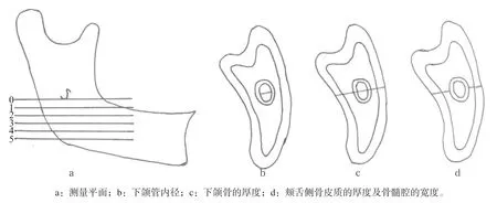
图 1 下颌升支的测量平面及测量项目Fig 1 The measurement plans and projects of mandibular ramus
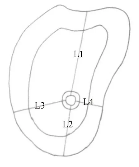
图 2 下颌骨体部下颌管内缘距各表面的距离测量 Fig 2 The distance from the inner edge of mandibular canal to the surface of body
1.3.2 下颌骨的测量 1)下颌孔的位置。测量项目:下颌孔中心到下颌切迹的距离(M1),下颌孔中心到下颌下缘的最小距离(M2),下颌孔中心到升支前缘的最小距离(M3),下颌孔中心到升支后缘的最小距离(M4)(图3)。2)下颌体截面形态。采用Swasty等[1]的测量方法,从左下颌第二磨牙远中到右下颌第二磨牙远中截取13个截面(图4),分别测量下颌体各截面的高度、宽度以及皮质骨厚度(图5)。
每个项目测量2次,取平均值,每次测量在相同条件下由同一位医生在连续时间内完成。正式测量前研究者进行CBCT影像学方面的专业培训,且预试验中2次测量结果具有较高的一致性。
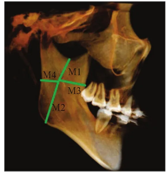
图 3 下颌孔在下颌升支的位置测量Fig 3 Measurement of the position of mandibular foramen in ramus
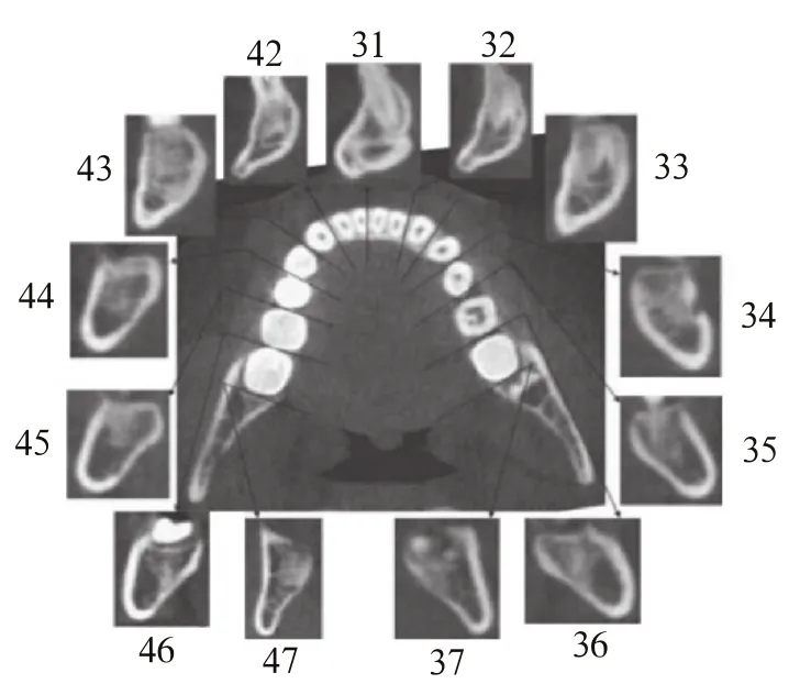
图 4 下颌体13个截面的形态Fig 4 The morphology of 13 cross-sections of mandible body
1.4 统计学分析
采用SPSS 17.0软件进行统计学分析。采用独立样本t检验进行比较。
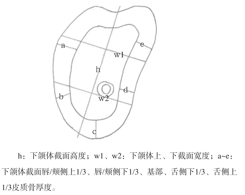
图 5 下颌体截面测量项目Fig 5 The measurement projects of cross-section of mandible body
2 结果
2.1 下颌管在下颌骨内的三维走行
2.1.1 下颌升支部下颌管的位置 下颌升支各层面下颌管测量分析见表1。下颌管舌侧骨皮质厚度无论在哪一层均明显较颊侧骨皮质薄,BC从1平面到5平面都逐渐增厚。统计分析表明,LC、LP分别在4平面、2平面时不同性别间有统计学差异(P<0.05),ID在3平面时不同性别间有统计学差异(P<0.01)。
表 1 下颌升支各层面下颌管测量分析Tab 1 Analysis of measured values at various levels in ramus mm,±s

表 1 下颌升支各层面下颌管测量分析Tab 1 Analysis of measured values at various levels in ramus mm,±s
注:不同性别间相比,*P<0.05,**P<0.01。
测量项目 ID LB BC LC BP LP 0平面 男 2.60±0.40 10.10±1.18 2.20±0.27 1.09±0.32 3.32±1.45 0.00±0.00 女2.51±0.20 9.77±1.10 2.16±0.28 1.15±0.38 3.26±0.98 0.00±0.00 1平面 男 2.45±0.34 9.57±1.89 2.14±0.37 1.21±0.24 3.12±1.18 0.02±0.09 女2.40±0.06 9.34±1.16 2.05±0.32 1.23±0.39 2.77±1.37 0.02±0.05 2平面 男 2.47±0.25 10.28±1.53 2.29±0.25 1.54±0.39 2.52±1.41 0.14±0.32* 女2.36±0.28 9.59±1.45 2.18±0.27 1.49±0.42 2.31±1.31 0.00±0.00 3平面 男 2.55±0.18** 11.52±1.88 2.40±0.31 1.67±0.27 2.45±1.79 0.57±0.56 女2.38±0.21 10.85±2.07 2.26±0.31 1.68±0.31 2.18±1.60 0.29±0.52 4平面 男 2.46±0.20 12.14±2.09 2.83±0.69 1.62±0.23* 2.85±1.74 0.70±0.70 女2.45±0.19 11.49±2.21 2.76±0.92 1.77±0.23 2.41±1.16 0.36±0.51 5平面 男 2.43±0.30 13.66±2.09 2.97±0.75 1.60±0.30 4.06±1.97 1.00±0.73 女2.36±0.28 12.60±2.55 2.82±0.80 1.76±0.36 3.27±1.98 0.74±0.52合计 2.46±0.06 10.96±1.38 2.46±0.33 1.48±0.26 2.91±0.55 0.33±0.35
2.1.2 下颌骨体部下颌管的空间定位 下颌骨体部下颌管距各表面的距离见表2。下颌管到颊侧骨皮质的距离从近中到远中逐渐增加,到舌侧骨皮质、牙槽嵴顶的距离从近中到远中逐渐减小,到下颌下缘的距离在第一磨牙处最小,第二前磨牙处最大。统计分析表明:L1>L2>L3>L4,均具有统计学差异(P< 0.05);不同性别间的L1具有统计学差异,男性大于女性(P<0.01)。

表 2 下颌骨体部下颌管距各表面的距离Tab 2 The distance from mandibular canal to the surface of mandibular body mm, x±s
2.2 下颌孔的位置
下颌孔的位置分析见表3。左右侧M1、M2、M3、M4之间均无统计学差异(P>0.05);除M3外,其他项目不同性别间均具有统计学差异,男性大于女性(P<0.01)。

表 3 下颌孔的位置Tab 3 The position of mandibular foramen mm, x±s
2.3 下颌体截面形态
下颌体截面高度、宽度及皮质骨厚度各测量值左右侧之间均无统计学差异(P>0.05),故两侧合并一起处理。
2.3.1 下颌体截面高度 下颌体截面高度见表4。从中线至远中,男性与女性的下颌体截面高度均有减小的趋势,牙弓前段明显大于牙弓后段,男性的下颌体高度大于女性(P<0.01)。

表 4 下颌体截面高度Tab 4 The height of mandibular body cross sections mm, x±s
2.3.2 下颌体截面宽度 下颌体截面宽度见表5。上截面宽度从中线至远中均逐渐增大,在侧切牙、尖牙、第一前磨牙处男女差异具有统计学意义(P< 0.05);下截面宽度从中线至前磨牙段逐渐减小,至磨牙段又逐渐增大,在侧切牙、尖牙处男女差异具有统计学意义(P<0.05)。

表 5 下颌体截面宽度Tab 5 The width of mandibular body cross sections mm, x±s
2.3.3 下颌骨体截面皮质骨厚度 下颌骨体截面皮质骨厚度分析见表6。唇/颊侧上1/3皮质骨厚度从中线至远中逐渐增加,在侧切牙至第二前磨牙处性别差异有统计学意义(P<0.05);唇/颊下1/3皮质骨厚度从中线至尖牙段逐渐减小,从第一前磨牙至远中逐渐增大,在正中联合部、侧切牙、尖牙处男女差异有统计学意义(P<0.05);下颌骨基部皮质骨厚度在正中联合部、尖牙、第一前磨牙处男女差异有统计学意义(P<0.05);舌侧上1/3皮质骨厚度从中线至第一前磨牙段逐渐增大,至远中逐渐减小,在第一前磨牙、第二前磨牙、第二磨牙处男女差异有统计学意义(P<0.01);舌侧下1/3厚度从中线至远中逐渐减小,在正中联合部男女差异有统计学意义(P<0.05)。
表 6 下颌体截面皮质骨厚度Tab 6 The thickness of the cortical bone of mandibular body cross sections mm,±s
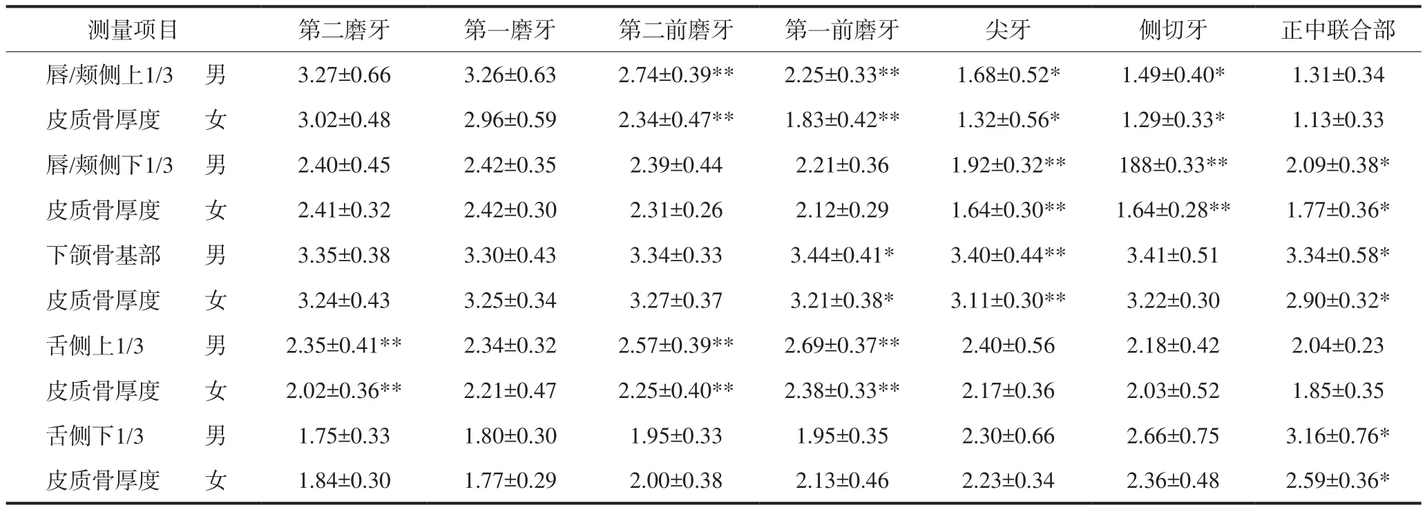
表 6 下颌体截面皮质骨厚度Tab 6 The thickness of the cortical bone of mandibular body cross sections mm,±s
注:不同性别间相比,*P<0.05,**P<0.01。
测量项目 第二磨牙 第一磨牙 第二前磨牙 第一前磨牙 尖牙 侧切牙 正中联合部唇/颊侧上1/3 男 3.27±0.66 3.26±0.63 2.74±0.39** 2.25±0.33** 1.68±0.52* 1.49±0.40* 1.31±0.34皮质骨厚度 女 3.02±0.48 2.96±0.59 2.34±0.47** 1.83±0.42** 1.32±0.56* 1.29±0.33* 1.13±0.33 唇/颊侧下1/3 男 2.40±0.45 2.42±0.35 2.39±0.44 2.21±0.36 1.92±0.32** 188±0.33** 2.09±0.38*皮质骨厚度 女 2.41±0.32 2.42±0.30 2.31±0.26 2.12±0.29 1.64±0.30** 1.64±0.28** 1.77±0.36*下颌骨基部 男 3.35±0.38 3.30±0.43 3.34±0.33 3.44±0.41* 3.40±0.44** 3.41±0.51 3.34±0.58*皮质骨厚度 女 3.24±0.43 3.25±0.34 3.27±0.37 3.21±0.38* 3.11±0.30** 3.22±0.30 2.90±0.32*舌侧上1/3 男 2.35±0.41** 2.34±0.32 2.57±0.39** 2.69±0.37** 2.40±0.56 2.18±0.42 2.04±0.23皮质骨厚度 女 2.02±0.36** 2.21±0.47 2.25±0.40** 2.38±0.33** 2.17±0.36 2.03±0.52 1.85±0.35舌侧下1/3 男 1.75±0.33 1.80±0.30 1.95±0.33 1.95±0.35 2.30±0.66 2.66±0.75 3.16±0.76*皮质骨厚度 女 1.84±0.30 1.77±0.29 2.00±0.38 2.13±0.46 2.23±0.34 2.36±0.48 2.59±0.36*
3 讨论
3.1 下颌骨、下颌管的研究背景及测量方法
下颌管的位置和走向一直是口腔颌面外科关注的重点之一,以往对下颌管的研究大多在离体的下颌骨上进行,采用游标卡尺进行测量研究,这种方法易受到样本量以及年龄的限制。二维平片有放大、扭曲以及不清晰等缺点,且存在放大率问题。三维CT重建,在计算机上可以任意角度进行整体或局部三维显示,但其放射量大、成本高而无法在临床常规使用。CBCT则有许多优势:通过冠状面、矢状面、轴位三维联动,实现了更清晰的立体图像观察,精度高,与被投照物之间比例为1︰1,可以进行实际测量;具有辐射量小、成像迅速、费用相对低得多等优势。本研究采用CBCT对研究对象进行三维测量,克服了二维图像的局限性,通过定量测量为临床诊断、治疗提供了更为详尽、准确的数据。
3.2 下颌管的空间位置分析
准确定位下颌孔,直接关系到下牙槽神经的麻醉效果。本文测得下颌孔到下颌切迹和下颌下缘的距离分别是18.51、29.12 mm,说明下颌孔位于下颌管内面中央偏上。下颌孔到下颌支前缘、后缘的距离分别是19.89、13.71 mm,说明下颌孔位于下颌支内面中央偏后。下牙槽神经阻滞麻醉时要掌握好进针的角度和深度,否则很容易将麻醉剂注射到下颌体后缘或者下颌切迹处,达不到麻醉效果。
从下颌孔到下颌骨下缘,下颌管内径变化不大,下颌骨厚度增加,颊舌侧骨皮质也逐渐增厚,舌侧骨髓腔的宽度是从无到有的逐渐递增趋势,在每一层中,颊侧骨髓腔的宽度均大于舌侧。舌侧骨皮质厚度为1.48 mm,比冉炜等[2]的研究结果(1.22 mm)稍大,可能是因研究对象及测量方法不同所致。研究结果提示,行下颌升支部手术时舌侧切骨深度不应超过2 mm,以免损伤下牙槽神经血管束。
从下颌孔至下颌骨体部,下颌管距离升支前缘较后缘近,至舌侧骨板的距离较颊侧骨板小,下颌管上缘至牙槽嵴顶的距离较下颌管下缘至下颌骨下缘的距离大。这与皮昕[3]的研究结论一致。下颌管入下颌孔后,先紧贴着舌侧骨板沿升支行向前下,然后逐渐离开贴附的舌侧骨板向颊侧移动,抵达颏孔下方时即折转向上向外出颏孔,略呈“S”形走行。de Oliveira Júnior等[4]报道,下颌骨体部下颌管到颊侧骨壁距离为6.10 mm,到舌侧骨壁距离为3.98 mm,到牙槽嵴顶距离为16.98 mm,本研究的结果分别为7.05、3.66、17.09 mm,与其基本相符。
下牙槽神经与下牙槽血管共同包裹于一薄层的骨壳内,下牙槽血管有66.7%位于下牙槽神经外上方[5],在临床上进行下颌骨相关手术操作时,若出现出血应立即停止操作,防止损伤下牙槽神经。本研究中,下颌管距牙槽嵴顶的距离由第二磨牙到第二前磨牙处逐渐增加,最小值为14.49 mm,由于多数学者[6-7]建议下牙种植体尖端与下颌管的安全距离为2 mm,因此在下颌第二磨牙区种植牙时种牙深度尽量小于12 mm,此结果与范锡印等[8]、冯文颢等[5]的研究结果(最小距离9.5~12.0 mm)相符。
本研究还发现,下颌管距牙槽嵴顶的距离性别间有统计学差异,男性平均为18.24 mm,比女性大2.79 mm。胥爱文等[9]螺旋CT扫描发现,男、女下颌管在下颌骨内的位置无显著差异。本研究结果与其不同,可能是由于其选用的螺旋CT扫描是由二维切片堆积而成,而本研究采用牙科专用CBCT进行扫描,InVivo 5软件进行图像处理和测量,有效地避免误差。在手术时,应结合影像学检查确定下颌管解剖定位,减少并发症的发生。
3.3 下颌体截面形态分析
3.3.1 下颌体截面高度、宽度的特点及性别差异 从中线至远中,男性与女性下颌骨体截面高度均有减小的趋势,在牙弓前段明显大于牙弓后段,此变化在前磨牙段开始明显,且男性变化比女性更明显。下颌体上截面宽度从中线至远中均逐渐增大;下截面宽度从中线至前磨牙段逐渐减小,至磨牙段又逐渐增大。下颌体截面高度、宽度的特点与其椭圆形结构相一致,van Eijden[10]研究发现,下颌体是一个垂直向大于横向的椭圆形,这一特点有利于增强下颌抵抗相对大的矢状向弯曲和剪切力。
3.3.2 下颌体截面皮质骨厚度的特点及差异 下颌体截面皮质骨厚度各测量值左右侧无统计学差异,其三维形态左右基本对称,但部分测量值性别差异显著,尤其是下颌骨基部皮质骨厚度。
皮质骨厚度在颌面部肌肉作用下可以发生相应的变化,咀嚼时磨牙受到咬肌的直接压力,为了抵抗此压力,磨牙颊侧皮质骨厚度适应性增厚[10]。下颌体上下方皮质骨厚度与其抵抗弯曲的能力有关。Schwartz-Dabney等[11]发现,下颌骨在受到垂直向弯曲时,其联合部舌侧皮质骨受到的张力最大,而皮质骨厚度与作用力的大小相适应,无论是张力还是压力,均可在力作用下发生变形。下颌磨牙后区舌侧骨板较颊侧薄,临床上拔除下颌后牙时宜多向舌侧摇动,从舌向拔出,当用劈开法拔除阻生第三磨牙时用力不应过猛,否则会导致舌侧骨板的劈裂。
综上,正常人下颌管在下颌骨内的位置变异较大,形态也不尽相同,性别差异明显,在植骨及种植等手术前应仔细检查,尽量减小神经损伤的风险。
[参考文献]
[1] Swasty D, Lee J, Huang JC, et al. Cross-sectional human mandibular morphology as assessed in vivo by cone-beam computed tomography in patients with different vertical facial dimensions[J]. Am J Orthod Dentofacial Orthop, 2011, 139(4 Suppl):e377-e389.
[2] 冉炜, 郭冰, 陈松龄, 等. 下颌神经管全长三维走向的测量及其临床意义[J]. 解剖学研究, 2002, 24(2):116-118,155. Ran W, Guo B, Chen SL, et al. Three dimensional survey of trend of whole inferior alveolar nerve canal and its relation to adjacent structures and its clinical significance[J]. Anat Res, 2002, 24(2):116-118,155.
[3] 皮昕. 下颌骨矢状劈开截骨术中下颌管的应用解剖[J]. 临床口腔医学杂志, 1986, 2(1):16-18. Pi X. The applied anatomy of mandibular canal in mandibular sagittal split osteotomy[J]. J Clin Stomatol, 1986, 2 (1):16-18.
[4] de Oliveira Júnior MR, Saud AL, Fonseca DR, et al. Morphometrical analysis of the human mandibular canal: a CT investigation[J]. Surg Radiol Anat, 2011, 33(4):345-352.
[5] 冯文颢, 杨能瑞, 纪荣明, 等. 下颌管及下牙槽神经的应用解剖[J]. 解剖学杂志, 2014, 37(1):75-78. Feng WH, Yang NR, Ji RM, et al. Applied anatomy of mandibular canal and inferior alveolar nerve[J]. Chin J Anat, 2014, 37(1):75-78.
[6] Etoz OA, Er N, Demirbas AE. Is supraperiosteal infiltration anesthesia safe enough to prevent inferior alveolar nerve during posterior mandibular implant surgery[J]. Med Oral Patol Oral Cir Bucal, 2011, 16(3):e386-e389.
[7] Onur B, Dilek OC. Assessment of the risk of perforation of the mandibular canal by implant drill using density and thickness parameters[J]. Gerodontology, 2011, 28(3):213-220.
[8] 范锡印, 苗莹莹, 付升旗, 等. 下颌管的应用解剖及临床意义[J]. 中华解剖与临床杂志, 2011, 16(5):396-399. Fan XY, Miao YY, Fu SQ, et al. The applied anatomy and clinical signifiance of mandibular canal[J]. Chin J Anat Clin, 2011, 16(5):396-399.
[9] 胥爱文, 张利, 王君琛, 等. 螺旋CT三维成像对下颌神经管走向的研究[J]. 现代口腔医学杂志, 2011, 25(1):12-15. Xu AW, Zhang L, Wang JC, et al. Study of the course of the mandibular canal by spiral CT 3-D imaging[J]. J Modern Stomatol, 2011, 25(1):12-15.
[10] van Eijden TM. Biomechanics of the mandible[J]. Crit Rev Oral Biol Med, 2000, 11(1):123-136.
[11] Schwartz-Dabney CL, Dechow PC. Variations in cortical material properties throughout the human dentate mandible [J]. Am J Phys Anthropol, 2003, 120(3):252-277.
(本文采编 石冰)
[中图分类号]R 322.4+1
[文献标志码]A [doi] 10.7518/hxkq.2016.02.010
[收稿日期]2015-06-15; [修回日期] 2015-10-20
[作者简介]绳兰兰,硕士,E-mail:shenglanlan2010@163.com
[通信作者]曲卫国,主任医师,硕士,E-mail:qwg2002521@163.com
Three-dimensional survey of the whole mandibular canal and mandibular morphology by cone beam computed tomo-graphy in normal young people
Sheng Lanlan1, Qu Weiguo1, Li Yang1, Qu Zhenyu2, Yue Bai1, Wang Ji1. (1. Dept. of Oral and Maxillofacial Surgery, Dalian Stomatological Hospital, Dalian 116021, China; 2. Dept. of Radiology, Dalian Stomatological Hospital, Dalian 116021, China)
Correspondence: Qu Weiguo, E-mail: qwg2002521@163.com.
[Abstract]Objective This research aimed to analyze the three-dimensional position of mandibular canal (MC) and mandibular morphology of normal young males and females by using data from cone beam computed tomography (CBCT), as well as to provide an anatomical basis for clinical surgery of the mandible. Methods Normal occlusion and CBCT scans of 29 normal young people were conducted. InVivo 5 software was used to reconstruct the mandible, anchor the points, and measure the jaw shape and three-dimensional course of MC. All measurements were analyzed with SSPS 17.0 software. Results The MC lingual bone cortex was thinner than the MC buccal bone cortex, and the distance of the MC to the buccal bone cortex gradually increased. However, the distance of the MC to the tongue bone cortex and alveolar crest gradually decreased from proximal to distal. In addition, the distance of the MC to the mandibular lower margin was minimal at the first molar and reached the maximum at the second premolar. No significant difference was observed among the heights, widths, and thicknesses of the left and right sides of the cortical bone of the mandibular body cross sections. From the midline to the farthest point, the height and lower one-third thickness of the lingual cortical bone of the mandibular body cross sections gradually decreased, whereas the width of the upper cross section and upper one-third thickness of the buccal cortical bone gradually increased. Significant difference was observed in some measured values. Conclusion After MC enter into the mandibular foramen, it moved away from the lingual to the buccal bone but gradually returned to the lingual bone; its general course is closer to the lingual bone. The mandibles of males are thicker than those of females. CBCT can accurately display the course of MC and its relationship with the surrounding structures.
[Key words]mandibular canal; mandible; cone beam computed tomography
