RNA干扰下调YAP基因对甲状腺癌细胞TPC-1功能的影响
2016-07-07杨卫国花开尧金佳利
杨卫国, 花开尧, 金佳利, 房 林
(1. 同济大学附属第十人民医院甲状腺乳腺外科,上海 200072; 2. 同济大学医学院,上海 200092)
·基础研究·
RNA干扰下调YAP基因对甲状腺癌细胞TPC-1功能的影响
杨卫国1, 花开尧1, 金佳利2, 房 林1
(1. 同济大学附属第十人民医院甲状腺乳腺外科,上海 200072; 2. 同济大学医学院,上海 200092)
目的 检测siRNA沉默Yes相关蛋白(Yes associated protein,YAP)后的人甲状腺癌TPC-1细胞功能变化。方法 应用阳离子脂质体转染试剂LipofectamineTM2000将靶向沉默YAP基因的siRNA序列转染至甲状腺癌TPC-1细胞,应用qRT-PCR、Western印迹法检测转染后TPC-1细胞YAP基因及蛋白表达,MTT、平板克隆实验、Transwell小室、划痕实验和流式细胞术分别检测转染对细胞增殖、侵袭、迁移和细胞周期及凋亡的影响。结果 转染siRNA后,YAP mRNA和蛋白表达明显下降(P<0.05);TPC-1细胞的增殖、侵袭、迁移能力受到抑制,细胞周期G0/G1阻滞,细胞凋亡未明显上升。结论 抑制甲状腺癌细胞YAP表达可有效降低细胞的增殖、迁移和侵袭能力,改变细胞周期分布,但对细胞凋亡无明显影响。
甲状腺肿瘤; Yes相关蛋白; siRNA; 细胞功能
YAP基因位于Hippo信号通路下游,是一重要的转录共激活因子[1]和癌基因,在乳腺、肺、膀胱等癌中高表达[2-3]。研究[4]显示,YAP基因在甲状腺乳头状癌和未分化癌中高表达且与肿瘤增殖正相关。近年来,siRNA技术引起重视[5]。本研究观察应用siRNA敲除YAP基因后甲状腺癌TPC-1细胞功能的变化,以探讨其作为甲状腺癌基因治疗的可行性。
1 材料与方法
1.1 材料
人甲状腺癌TPC-1细胞购自中国科学院上海生命科学研究院;DMEM培养基、胎牛血清购自美国Gibco公司;LipofectamineTM2000购自美国Invitrogen公司;Annexin-V/PI凋亡检测试剂盒购自BD Pharmingen公司;YAP抗体购自Cell Signaling Technology公司。在NCBI上Genbank中检索YAP基因的全部碱基序列,采用在线RNA干扰设计软件并根据siRNA设计原则设计干扰位点,siRNA由广州市锐博生物科技有限公司合成。靶向沉默YAP基因的siRNA顺义链: 5′-GCAUCUUCGACAGUC-UUCUTT-3′;反义链为: 5′-AGAAGACUGUCGA-AGAUGCTT-3′。以siRNA NC序列作为与人类基因组序列无任何匹配的阴性对照,siRNA NC顺义链为: 5′-UUCUCCGAACGUGUCACGUTT-3′;反义链为: 5′-ACGUGACACGUUCGGAGAA-TT-3′。
1.2 方法
1.2.1 细胞培养及TPC-1细胞转染 人甲状腺癌TPC-1细胞培养于含10%澳洲胎牛血清及 100U/ml 双抗DMEM培养液中,置于37℃、5% CO2、100%湿度培养箱中培养,3~4d更换培养基1次。选取活力较好的指数生长期细胞进行实验,细胞计数后以每孔1×10⁵个TPC-1细胞分种于6孔培养板中。当细胞密度达40%~50%后,应用阳离子脂质载体LipofectamineTM2000将siRNA转染入TPC-1细胞,每孔siRNA终浓度为 50nmol/L,同时转染siRNA NC作为阴性对照,而未做任何处理组细胞作为空白对照组。
1.2.2 qRT-PCR检测 总RNA提取试剂(TRIzol reagent)提取各实验组TPC-1细胞总RNA,紫外分光光度计准确定量。将提取的总RNA进行反转录,获取cDNA后进行实时定量PCR反应。反应条件为95℃变性30s,57℃退火 1min,72℃延伸 1min,循环30次,最后72℃温育10min。qRT-PCR引物YAP基因顺义链为: 5′-ACCCACAGCTCAG-CATCTTCG-3′,反义链为: 5′-TGGCTTGTTCCC-ATCCATCAG-3′;β-actin基因顺义链为: 5′-CGT-CTTCCCCTC CATCGT-3′,反义链为: 5′-GAAG-GTGTGGTGCCAGATTT-3′。
1.2.3 Western印迹法检测 转染后72h收集细胞,用RIPA细胞裂解液裂解TPC-1细胞并全程于低温下提取总蛋白,BCA蛋白定量试剂盒测定蛋白浓度。每孔加蛋白样品20μg,用10%SDS-PAGE凝胶进行电泳,转膜、封闭,按1∶800稀释一抗孵育,于4℃冰箱放置过夜。TBST洗膜三次,每次约 10min,按1∶1 500稀释二抗孵育50min,TBST再次洗膜3次,每次约10min。上机操作,通过Odyssey荧光成像系统扫描并进行蛋白条带灰度分析。
1.2.4 四甲基偶氮唑蓝(MTT)检测 实验取对数生长期的TPC-1细胞,按2000个/孔接种于96孔板,每孔200μl,边缘加200μl磷酸盐缓冲液(PBS),在培养箱中培养12h待细胞贴壁完全后转染siRNA,转染浓度为50nmol/L。转染后细胞培养4d,检测时间点分别为24、48、72和96h。每个检测时间点待侧孔每孔加入MTT溶液(5mg/ml)20μl,继续培养4h后终止培养,小心吸去孔内培养上清液,每孔加入150μl DMSO,均匀振荡10min,使结晶物充分溶解。酶联免疫检测仪上选择 490nm 波长,测定各孔吸光度值(D490),记录结果并绘制细胞生长曲线。
1.2.5 克隆形成实验 将各实验组细胞消化,重悬后计数,按500个/孔接种于6孔板,十字形晃动使细胞在培养基中分散均匀,培养箱中培养8~10d,期间换全培养基一次。当观察发现肉眼可见的克隆时终止培养,弃上清液,PBS浸洗2次,95%乙醇固定10min后用0.1%结晶紫染色10min,蒸馏水冲洗3次,风干后在显微镜下选取具有代表性的视野拍照。
1.2.6 划痕试验 取指数生长期状态良好的TPC-1细胞,按1×10⁵个/孔接种于96孔板,培养12h待细胞贴壁牢固后转染siRNA,将全培养基换为含2%澳洲胎牛血清的DMEM后继续培养,当细胞长满6孔板后,用枪头在细胞表面笔直划一条直线,PBS清洗2次去除脱落细胞,放入培养箱中继续培养,12、24h后镜下观察各组划痕的愈合情况,显微镜下选取具有代表性的视野拍照。
1.2.7 Transwell侵袭实验 将已转染的状态良好的TPC-1细胞消化后离心,细胞计数后加入一定量的培养基,调整细胞密度为5×10⁵个/ml,取 0.2ml 接种于Transwell小室内,将小室置于24孔板内,上室为含2%灭活血清的培养基,下室为含15%灭活血清的培养基。培养箱内培养24h,用消毒棉签缓慢擦除上层小室内细胞,95%乙醇固定,0.1%结晶紫染色,蒸馏水浸洗3次,风干后荧光倒置显微镜200倍视野下拍照。
1.2.8 细胞周期和凋亡测定 将已转染的状态良好TPC-1细胞消化后离心,一部分用预冷的PBS洗涤3次后离心,加入预冷的70%乙醇固定过夜,再次离心并洗涤后用含RNase及碘化丙啶(PI)的染色液室温下避光孵育30min,流式细胞仪分析细胞周期。另一部分加500μl的结合缓冲液重悬细胞,再加1μl Annexin V-PI荧光染料充分混匀,室温下避光孵育20min,1h内进行流式细胞仪检测,计算细胞凋亡率。
1.3 统计学处理

2 结 果
2.1 qRT-PCR和Western印迹法检测siRNA转染后YAP基因和蛋白的表达
qRT-PCR和Western印迹法结果显示,与NC组和空白对照组相比,siRNA处理组TPC-1细胞的YAP mRNA和YAP蛋白表达均受到显著抑制(P<0.05),见图1。
2.2 MTT检测和平板克隆实验检测siRNA转染后细胞增殖、迁移和侵袭能力的变化
MTT测定和平板克隆实验显示: 随时间延长,空白组与NC组细胞增殖能力和克隆数无差异,YAP-siRNA转染组细胞增殖能力和克隆数则明显减缓或减少(P<0.05),见图2;划痕和侵袭实验显示: 与空白对照组和NC组比较,siRNA-YAP组细胞愈合速度较慢,Transwell小室下层细胞数减少(均P<0.05),见图3;表明siRNA沉默YAP基因可抑制TPC-1细胞的增殖、迁移和侵袭能力。
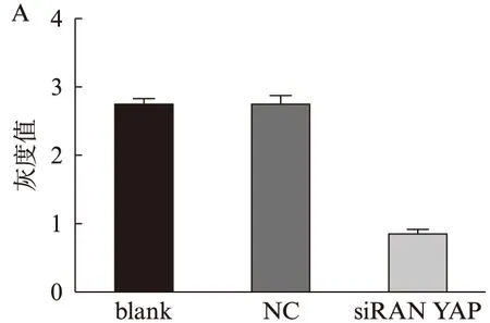

图1 转染siRNA后的甲状腺癌TPC-1细胞YAP mRNA和蛋白表达Fig.1 Relative expression of YAP mRNAand protein in TPC-1 thyroid cancer cells after transfected with siRNA demonstrated by qRT-PCR and Western blottingA: qRT-PCR;B: Western印迹法
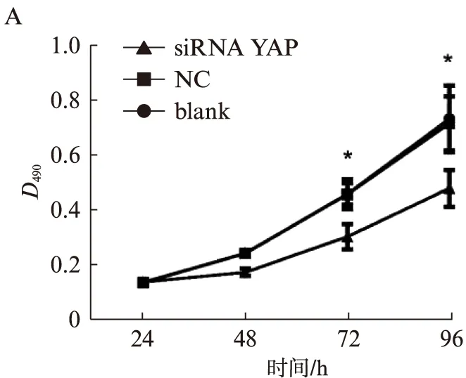
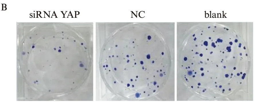
图2 MTT和平板克隆形成实验检测转染siRNA后TPC-1细胞增殖能力Fig.2 Proliferation of TPC-1 cells demonstrated by MTT assay and colony formation assayA: MTT;B: 平板克隆形成实验
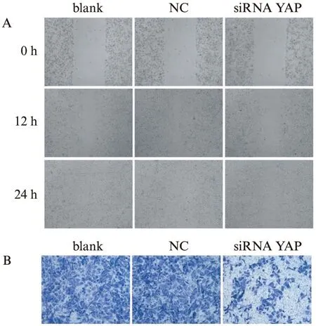
图3 划痕试验和Transwell侵袭实验检测转染siRNA后TPC-1细胞迁移、侵袭能力Fig.3 The migration and invasion of TPC-1 cellsdemonstrated by Cell scratch assay and Transwell assayA: 划痕试验;B: Transwell侵袭实验
2.3 流式细胞仪检测siRNA转染后细胞周期的变化
与空白对照组(53.60±0.73)%和NC组(55.27±2.10)%相比,siRNA-YAP组G0/G1期细胞数(64.40±1.69)%明显上升(P<0.01),S期和G2/M期细胞明显下降,但细胞凋亡率则无差异,表明siRNA沉默YAP可使TPC-1细胞出现G0/G1期阻滞,但对细胞凋亡无影响(表1)。流式细胞仪检测所示,siRNA干扰YAP基因后TPC-1细胞周期受到明显影响,见图4。
表1 流式细胞仪检测blank、NC、siRNA组TPC-1细胞周期分布
Tab.1 Cell cycle distribution detected by Flow cytometry in TPC-1 cell line (%)

组别G0/G1G2MSBlank53.60±0.7330.40±1.1016.00±1.00NC55.27±2.1029.00±1.0015.73±1.00siRNA64.40±1.6924.00±1.0011.60±1.00
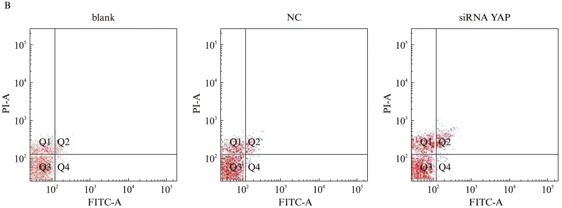
图4 流式细胞仪检测的TPC-1细胞周期分布和凋亡Fig.4 Cell cycle distribution and apoptosis in TPC-1 cells shown by flow cytometryA: 细胞周期分布;B: 凋亡;图B中右下象限AnnexinV(+)PI(-)代表早期凋亡细胞,右上象限AnnexinV(+)PI(+)代表晚期凋亡细胞或死亡细胞
3 讨 论
甲状腺癌的发生是多因素共同作用的结果。利用siRNA沉默相关关键基因治疗恶性肿瘤已取得一定的进展[6]。
Hippo通路最早在果蝇体内被发现,主要通过调节细胞增殖和凋亡调控器官发育,近年不少研究表明与肿瘤发病密切相关[7]。YAP作为Hippo通路的主要效应因子,其上游的转录因子如Mast1/2、Ww45、Lats1/2、Mob1等通过磷酸化和促进细胞质/细胞核转位对YAP起负性调节作用。这些调控因子的表达缺失能使YAP过表达,导致其下游的转录因子cyclinE、DIAP1、TEAD1、TEAD2等表达增加,从而促进细胞增殖,抑制细胞凋亡[8]。研究[9-12]报道,YAP基因在结肠癌、肝癌、肺癌、宫颈癌、膀胱癌等过表达。Tschaharganeh等[13]证实,肝癌细胞YAP能上调Notch信号通路的配体Jagged-1(Jag-1)、激活Notch信号通路,从而调控肝癌细胞的增殖、迁移、侵袭。
本实验通过siRNA下调甲状腺癌TPC-1细胞YAP表达,以观察细胞功能的变化。转染siRNA-YAP后,mRNA和蛋白水平YAP表达均被显著抑制;随着YAP下调,TPC-1细胞的增殖、迁移、侵袭也均受到抑制,细胞周期阻滞在G1期,但对细胞凋亡无明显作用。本实验只是应用阳离子脂质体转染试剂介导的YAP特异性siRNA的甲状腺癌细胞瞬时转染,对基因抑制状态不稳定;且实验只在甲状腺乳头状癌细胞TPC-1中进行验证,也未进行体内实验,因此相关的研究有待进一步进行。
综上所述,siRNA能特异性沉默人甲状腺癌TPC-1细胞YAP基因,下调其mRNA及蛋白表达水平,抑制癌细胞的增殖、迁移和侵袭,为甲状腺癌的基因治疗提供了新的实验依据。
[1] Dong J, Feldmann G, Huang J, et al. Elucidation of a universal size-control mechanism in Drosophila and mammals[J]. Cell, 2007, 130(6): 1120-1133.
[2] Liu JY, Li YH, Lin HX, et al. Overexpression of YAP 1 contributes to progressive features and poor prognosis of human urothelial carcinoma of the bladder[J]. BMC Cancer, 2013,13: 349.
[3] Yeung B, Yu J, Yang X. Roles of the Hippo pathway in lung development and tumorigenesis[J]. Int J Cancer, 2016,138(3): 533-539.
[4] Lee SE, Lee JU, Lee MH, et al. RAF kinase inhi-bitor-independent constitutive activation of Yes-associated protein 1 promotes tumor progression in thyroid cancer[J]. Oncogenesis, 2013,2: e55.
[5] Størvold GL, Andersen TI, Perou CM, et al. siRNA: a potential tool for future breast cancer therapy?[J]. Crit Rev Oncog, 2006,12(1-2): 127-150.
[6] Datta M, Schwartz GG. Calcium and vitamin D supplementation and loss of bonemineral density in women undergoing breast cancer therapy[J]. Crit Rev Oncol Hematol, 2013,88(3): 613-624.
[7] Shi P, Feng J, Chen C. Hippo pathway in mammary gland development and breast cancer[J]. Acta Biochim Biophys Sin (Shanghai), 2015,47(1): 53-59.
[8] Ma Y, Yang Y, Wang F, et al. Hippo-YAP signaling pathway: a new paradigm for cancer therapy[J]. Int J Cancer, 2015,137(10): 2275-2286.
[9] Wang L, Shi S, Guo Z, et al. Overexpression of YAP and TAZ is an independent predictor of prognosis in colorectal cancer and related to the proliferation and metastasis of colon cancer cells[J]. PLoS One, 2013,8(6): e65539.
[10] Perra A, Kowalik MA, Ghiso E, et al. YAP activation is an early event and a potential therapeutic target in liver cancer development[J]. J Hepatol, 2014,61(5): 1088-1096.
[11] Wang Y, Dong Q, Zhang Q, et al. Overexpression of yes-associated protein contributes to progression and poor prognosis of non-small-cell lung cancer[J]. Cancer Sci, 2010,101(5): 1279-1285.
[12] Liu T, Liu Y, Gao H, et al. Clinical significance of yes-associated protein overexpression in cervical carcinoma: the differential effects based on histotypes[J]. Int J Gynecol Cancer, 2013,23(4): 735-742.
[13] Tschaharganeh DF, Chen X, Latzko P, et al. Yes-associated protein up-regulates Jagged-1 and activates the Notch pathway in human hepatocellular carcinoma[J]. Gastroenterology, 2013,144(7): 1530-1542.
Effect of RNA interference targeting YAP gene on thyroid cancer TPC-1 cells
YANGWei-guo1,HUAKai-yao1,JINJia-li2,FANGLin1
(1. Dept. of Breast and Thyroid Surgery, Tenth People’s Hospital, Tongji University, Shanghai 200072, China;2. Medical College, Tongji University, Shanghai 200092, China)
Objective To investigate the effect of small interfering RNA (siRNA) targeting Yes associated protein (YAP) gene on thyroid cancer TPC-1 cells. Methods siRNA targeting YAP gene was transfected in thyroid cancer TPC-1 cells with LipofectamineTM2000. qRT-PCR and Western blotting were conducted to determine YAP suppression at mRNA and protein levels by siRNA. MTT assay, colony formation assay, Transwell migration assay wound healing assay and flow cytometry were used to study the effects of siRNA on cell proliferation, migration, cell cycle and apoptosis, respectively. Results Expression of YAP was suppressed after interference of YAP siRNA (P<0.05), proliferation, migration and invasion of TPC-1 cells were inhibited; cells were arrested at G0/G1phase, but cellular apoptosis rate was not significantly changed. Conclusion The results indicate that YAP gene exerts a vital role in biological behaviors of thyroid cancer TPC-1 cells.
thyroid cancer; Yes associated protein; siRNA; cellular function
10.16118/j.1008-0392.2016.01.006
2015-10-12
国家自然科学基金(81272240)
杨卫国(1981—),男,硕士研究生.E-mail: changning021@163.com
房 林.E-mail: fanglin_f@126.com
R 736.1
A
1008-0392(2016)01-0028-05