VEGF, HIF-1α and PEDF expression in the retina of streptozotocin-induced diabetics rats treated with ozone
2016-02-27TingYuXieCuiLiZhangXueYiChen
Ting-Yu Xie, Cui-Li Zhang, Xue-Yi Chen
Foundation item:Supported by Natural Scientific Research Fund of the Xinjiang Uygur Autonomous Region in China (No.2014211C046)
Department of Ophthalmology, the First Affiliated Hospital of Xinjiang Medical University,Urumchi 830011 , Xinjiang Uygur Autonomous Region, China
Correspondence to:Xue-Yi Chen. Department of Ophthalmology, the First Affiliated Hospital of Xinjiang Medical University, Urumchi 830011, Xinjiang Uygur Autonomous Region, China. xtygood@126.com
Received: 2014-11-25 Accepted: 2015-08-20
VEGF, HIF-1α and PEDF expression in the retina of streptozotocin-induced diabetics rats treated with ozone
Ting-Yu Xie, Cui-Li Zhang, Xue-Yi Chen

Foundation item:Supported by Natural Scientific Research Fund of the Xinjiang Uygur Autonomous Region in China (No.2014211C046)
Department of Ophthalmology, the First Affiliated Hospital of Xinjiang Medical University,Urumchi 830011 , Xinjiang Uygur Autonomous Region, China
Correspondence to:Xue-Yi Chen. Department of Ophthalmology, the First Affiliated Hospital of Xinjiang Medical University, Urumchi 830011, Xinjiang Uygur Autonomous Region, China. xtygood@126.com
Received: 2014-11-25Accepted: 2015-08-20
引用:谢婷玉, 张萃丽, 陈雪艺. 臭氧对STZ诱导的糖尿病大鼠视网膜表达VEGF, HIF-1α与PEDF的影响.国际眼科杂志2016;16(2):195-200
Abstract
•AIM:To study vascular endothelial growth factor (VEGF), pigment epithelium derived factor (PEDF) and hypoxia inducible factor-1α (HIF-1α) expression in retinal and serum in ozone-treated streptozotocin (STZ)-induced diabetic rats and to determine the possible efficacy of ozone therapy for diabetic retinopathy (DR).
•METHODS:Seventy male Sprague-Dawley rats were used. Group A (n=10) received a normal diet, diabetic molding established by intraperitoneal injection of STZ (50mg/ml), then divided into three groups, group B without any intervene; and groups C and D given oxygen and ozone clyster treatment respectively, twice per week for 1mo. Retina and blood were taken under general anesthesia. Reverse transcription-polymerase chain reaction (RT-PCR) and enzyme-linked immunosorbent assay (ELISA) methods were used to study retinal and serum VEGF, HIF-1α and PEDF expression.
•RESULTS:VEGF occurred mostly in the inner layer of the retina; the difference of VEGF in the retina among each group has statistical difference (F=23.923;P=0.000); in which, group D closer to group A, but still has statistical difference (P<0.05); there is no difference between group B and C (P>0.05) except for no difference between group A and D (P>0.05), others as same result as retinal VEGF expression.HIF-1α expression decreased in ozone-treated rats (group D) compared with control group (P<0.05); the difference between group A and groups B and C was significant (P<0.05), and the difference between groups B and C with no statistical difference (P>0.05). Overall, PEDF expression was lower than VEGF and HIF-1α, and groups A and D showed more expression than groups B and C, but the differences were not significant (P>0.05).
•CONCLUSION: Ozone administration can reduce the VEGF and HIF-1α expression and ozone may have potential uses in its treatment.
KEYWORDS:•diabetic retinopathy; ozone; vascular endothelial growth factor; pigment epithelium derived factor; hypoxia inducible factor-1α
Citation:Xie TY, Zhang CL, Chen XY. VEGF, HIF-1α and PEDF expression in the retina of streptozotocin-induced diabetics rats treated with ozone.GuojiYankeZazhi(IntEyeSci) 2016;16(2):195-200
INTRODUCTION
Diabetic retinopathy (DR) is the leading cause of blindness in the western world and it affects approximately 75% of diabetic patients within 15y after the onset of diabetics[1]. DR is difficult to treat because the pathological changes priority to the clinical diagnosis has been made. The mechanism of DR is being investigated but it is complicated. Most animal and human trials have established that the pathological changes of DR result from hyperglycemia, which causes the concentration of glucose increase in cells and sorbite (a mesostate) to aggregate. This disturbs the oxidation-reduction reaction balance and causes vessel occlusion. Ultimately, hypoxia and ischemia occur in the retina. Hypoxia and ischemia in the retina can activate transduction channels (such as protein kinase C or mitogen-activated protein kinase), and the endothelial function and pericyte cells are damaged[2]. Vascular endothelial growth factor (VEGF) plays a key role in the pathological changes of the retina in DR. VEGF genes are sensitive to hypoxia, which can lead to a strong response to hypoxia by up-regulating the hypoxic inducible factor-1α (HIF-1α) transcription factor. The HIF-1α subunit contains binding sites for the hypoxia response element, and through this site, it affects the target genes and its products such as cAMP-response element binding protein. Recent research has shown that HIF-1α is a major determinate of cellular VEGF expression and secretion. In addition to VEGF, the retina secretes other growth factors, such as pigment epithelium derived factor (PEDF), which contributes to the angiogenic homeostasis in ocular tissues. PEDF has been shown to be an endogenous antagonist of VEGF[3].
Hypoxia and ischemia are involved in the entire DR onset process. High concentrations of oxygen in the early stage of DR can reverse blindness, suggesting that hypoxia plays an important role in the process of DR[4-5]. Ozone may be an alternative treatment for DR, and several controlled trials have examined the feasibility and efficacy of using ozone as a therapeutic agent for the treatment of several diseases[6-7]. Ozone can increase the oxygen unloading capacity of hemoglobin in diabetic patients[8]. Thus, the main purpose of this research is to determine the role of ozone administration in improving oxidative stress in streptozotocin (STZ)-induced diabetic rats by examining VEGF, HIF-1α and PEDF expression, to establish its potential use in the strategy for the treatment of early-stage of DR.
MATERIALS AND METHODS
Animals and GroupsSeventy male Sprague-Dawley rats weighting 300-320 g were purchased from Xinjiang Medical University Experimental Animal Center [License No. SCXK (Xin) 2003-0001, China]. All experimental methods and animal care procedures were approved by the animal care committee of the Xinjiang Medical University (protocol IACUC-20120523007), in accordance with the China Council on Animal Care. And then those rates were randomly divided into four groups as follow: group A:10 rats just give the general feeding.
Others 60 rats were given high sugar and fat food for 1mo and then using streptozotoein (STZ, 10g/l, Sigma company, USA) intraperitoneal injection by concentration of 30 mg/kg, 3d and 1wk after, to testing the blood glucose by tail cutting respectively, blood glucose more than 16.7 mmol/L each time were be considered that the modeling established. And STZ-induced rats were randomly divided into three groups as follow in the process of making model there were 5 rats died for hemorrhagic shock. Group B (n=20): STZ model, without intervene but given high fat and sugar diet. Group C (n=20): STZ model+oxygen, given oxygen clyster by concentration of 50 μg/kg twice per week. Group D (n=20): STZ model+ozone, given ozone clyster as same concentration and methods as group C.
Under anesthesia by inject into abdominal cavity of chloral hydrate (made in Xinjiang Medcial University Pharmaceutical Preparation Room) by 0.3 ml/100 g concentration. Enucleation of the eyeball and remove the anterior segment and part vitreous, put the retinal in liquid nitrogen (-80°) prepare for isolate RNA. And then take the blood from aortaventralis.
RNA Isolation and Reverse Transcription-Polymerase Chain Reaction AnalysisTotal retina RNA was extracted and purified by one step Trizol, according to the manufacturer’s instructions. The list of primers for each samples is as follows (Table 1), cDNA were generated from 1 μm of total RNA, using the High-Capacity cDNA Reverse Transcription Kit (Thermo Corp, USA), and subjected to a 40 cycle polymerase chain reaction (PCR) amplification. Three replicates were run for each sample in a 96-well plate electrophoresis to identification the PCR amplifications products.
Enzyme-Linked Immunosorbent AssayVEGF levels in the serum of rats were estimated with enzyme-linked immunosorbent assay (ELISA) kit (VEGF Rat ELISA Kit, abcam, Lot: GR148932-1, batch number: ab100786) according to the introduction to perform the test.

Table 1The upstream and downstream of each factor
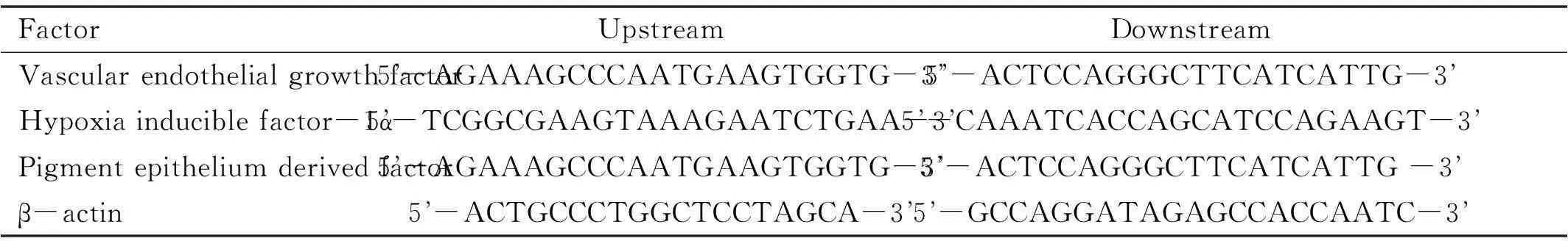
FactorUpstreamDownstreamVascularendothelialgrowthfactor5-AGAAAGCCCAATGAAGTGGTG-35-ACTCCAGGGCTTCATCATTG-3Hypoxiainduciblefactor-1α5-TCGGCGAAGTAAAGAATCTGAA-35-CAAATCACCAGCATCCAGAAGT-3Pigmentepitheliumderivedfactor5-AGAAAGCCCAATGAAGTGGTG-35-ACTCCAGGGCTTCATCATTG-3β-actin5-ACTGCCCTGGCTCCTAGCA-35-GCCAGGATAGAGCCACCAATC-3
The 5.0 software designer was used to design and synthesize the primer series (Shanghai Biological Engineering Company).
RESULTS
General ConditionsWeight were decreased and blood glucose were increased steady in the whole rats after modeling estimated, with diabetic duration extended, the blood glucose in each group keep high levels in the same time point. In another word, ozone and oxygen has no influence on blood glucose. (Figure 1A, B)
Immunohistochemical Determination of VEGFRetinal VEGF expression was identified by brown staining. VEGF expression in the control group was negative or weakly positive with a small amount of perivascular expression. VEGF expression was higher in the other groups, especially in the sub-limiting membrane, retinal ganglion cell (RGC) layer and the inner and outer plexiform layer. Group D showed a similar expression patterns to, but lower expression levels than group B and group C (Figure 2).
VEGF Expression in Each GroupReverse transcription-polymerase chain reaction (RT-PCR) results show that VEGF was detectable in all retina samples. The expression of VEGF mRNA in group A, B, C and D is 4.22±1.18, 11.60±3.42, 10.75±2.81 and 7.40±2.13 respectively, and there is statistic significance (F=23.923;P=0.000). VEGF expression in group B were nearly three times more than group A (P=0.000); expression of VEGF in group D were less than group B and C, but more than group A (P=0.000); there is no statistical significance between group B and C (Figure 3).
ELISA methods get the same results as RT-PCR and the VEGF level in group A, B, C and D, they are 4.29±1.11, 12.1±1.62, 10.65±1.58 and 4.44±1.76 respectively, there is statistical significance (F=62.249,P=0.000) and VEGF level in group B and C were more than group A and D (P=0.000); and there is no statistical significance between group A and D (P=0.997) as well as group B and C (P=0. 873) (Figure 4).
HIF-1α mRNA ExpressionHIF-1α mRNA expression by RT-PCR in group A was 2.3±0.3, which was significantly different from expression levels in groups B and C (P<0.05). Similar expression levels were found in group B and C (2.8±0.3 and 2.8±0.5, respectively;P>0.05). The lowest expression was found in group D (1.8±0.4), which was significantly different compared with group A (P<0.05) (Figure 5).

Figure 1Weight and blood glucose variation tendency between before and after modelingA: Weight; B: Blood glucose.
PEDF mRNA ExpressionPEDF mRNA expression in each group was follow: 0.22±0.12 in group A, 0.13±0.08 and 0.12±0.07 in groups B and C, and 0.19±0.09 in group D. Although PEDF mRNA expression was different in each group, the differences with no statistically significant (F=2.803;P>0.05) (Figure 6).
DISCUSSION
DR is considered to be an ischemic and hypoxic disorder that can lead to neovascularization and blindness. In the hypoxic state, it is thought that the retina produces growth factors that lead to vessel leakage and macular edema, angiogenesis causes fibrovascular tissue formation which can cause retinal detachment and finally loss of vision.
Retinal pathological changes that occur in STZ-induced rats vary with different tests. Some researchers found evidence of hypoxia in the diabetic mouse at a much earlier time point of 5mo using the oxygen-dependent probepimonidazole[9]. Others have found vasoconstriction and a substantial decrease in blood flow is as early as after 3-4wk of diabetics in both mice and rats. Thus, hypoxia plays a vital role in the process of DR, but the exact point at which hypoxia occurs in the histology is still undefined. Research has shown that capillary non-perfusion is representative of hypoxia in the tissue[10], and this may explain why the most clinical symptoms are posterior to the histopathology changes. Our study showed that VEGF occurred in each layer of the retinal in 1mo STZ-induced rat by immunohistochemical determination. There is no clinical symptom but the pathology changes occurring. It means that we should pay attention to the diabetic patients whose without obvious eye symptoms.

Figure 2VEGF expression byimmunohistochemistry, VEGF is indicated by the brown stainA: Group A with VEGF immunohistochemical staining, VEGF expression is hard to detected (DAB×400); B: Group B with VEGF expression has shown in GCL of retina (DAB×400); C: Group C with VEGF stained in the GCL and OPL (DAB×400); D: Group D with a low level of VEGF expressed in the GCL (DAB×400); GCL: Ganglion cell layer; OPL: Outer plexiform layer.

Figure 3VEGF expression in each group in retinalGB: Group blank; GM: STZ-induced model group; GO2: STZ-induced rats with treatment by O2; GO3: STZ-induced rats with treatment by ozone.

Figure 4HIF-1α expression in each groupGB: Group blank; GM: STZ-induced model group; GO2: STZ-induced rats with treatment by O2; GO3: STZ-induced rats with treatment by ozone.

Figure 5PEDF expression in each groupGB: Group blank; GM: STZ-induced model group; GO2: STZ-induced rats with treatment by O2; GO3: STZ-induced rats with treatment by ozone.
VEGF plays a key role in the DR process has been established[11-13]. VEGF is a protein that is a vascular endothelial cell mitogen and vascular permeability factor, and it is the most direct neovascularization factor, because VEGF mRNA expression increased during retinal hypoxia and remained elevated during the development of neovascularization. Animal studies revealed that VEGF synthesis occurs within the retina[14], which is consistent with the results of our study. Immuno-histochemical staining showed that VEGF mostly occurred in the inner layer of the retina. The VEGF content in the retina was significantly increased in groups B and C, which was different from the control STZ-induced rats. VEGF expression in ozone-treated rats (group D) was significantly lower than in the STZ-control group (group B) and in the oxygen treated group (group C). Meanwhile, the research show that even in the serum get the same results, that is VEGF can be detected in each group. But the model group and oxygen group was much more express than control group, just as other researches that VEGF no matter in retinal or in serum are involve in the process of the DR. To the contrary, the VEGF in retinal or in serum level decreased obviously compared to the model group or oxygen group, it means that ozone can reduce VEGF expression, whether it through antibiotic or improve oxygen supply still unknown, but it really worked.
VEGF is regulated by HIF-1α. Diabetics is a disorder that is associated with oxidative stress; research shown that HIF-1α is the primary hypoxia signaling protein in cells for regulating angiogenesis, and that it induces the transcription of several genes. Oxygen plays a key role in stabilizing HIF-1α and its function. When oxygen is normal, HIF-1α isoxidatively modified by prolyl 1 hydroxylase enzymes, but when cells are in hypoxic, the proline is not hydroxylated and HIF-1α escapes degradation. Thus, HIF-1α is a major determinant of VEGF cellular expression and secretion. Our study shows that HIF-1α expression in the model groups (groups B and C) was higher than the control group (group A). This is in agreement with the hypoxia results and is an initial change. However, after 1mo of ozone treatment, HIF-1α expression in the STZ+ozone group (group D) was even lower than that in the control group (group A). This may be because ozone consists of three oxygen atoms; the free oxygen can combine with another oxygen atom, and in this way improve the oxygen supply, which exceeds that in the oxygen treatment group. For the single methods to test HIF-1α, so it’s real but the exact mechanism require further study.
PEDF is another growth factor that is secreted by the retina, and it contributes to the angiogenic homeostasis in ocular tissues. PEDF was initially identified as a neurotrophic agent that is secreted by human fetal retinal pigment epithelium(RPE) cells[15]. However, in the eye, studies showed that endothelial quiescence and barrier function are achieved through a balance of VEGF and PEDF[16]. During the onset of active DR, VEGF and PEDF expression were reversed. Our study showed that low PEDF levels were expressed compared to that of VEGF and HIF-1α. Though there is no difference among each group of PEDF expression, however, the PEDF level in ozone group close to the blank group, and the group B and C still keep lower level of PEDF. As an angiogenic inhibitor, the function of PEDF is more universal because it can activate more tissues than other inhibitors, but the effects of angiogenic inhibition were dose-dependent, so the percentage of PEDF should be increased to the extent. low levels of PEDF performance in our study, which maybe ozone did not work by this way or the period too short for PEDF to have an effect on the retina.
Ozone, in theory, has oxidizing action that leads to the formation of hydrogen peroxide, which enters cells with various effects: in red blood cells, it shifts the hemoglobin dissociation curve to the right and facilitates release of oxygen[17-18]; and in leucocytes and endothelial cells, it induces the production of interleukins, interferon, transforming growth factor (TGF), nitrogen oxide and antacoids[19-20]. Previous studies have demonstrated that controlled ozone administration may promote an oxidative preconditioning or adaptation to oxidative stress, thus preventing the damage induced by reactive oxygen species (ROS)[21]. It has also been shown that ozone therapy can effectively reduce hypoxic and ischemic diseases, such as diabetic complications (for example, diabetic foot and diabetic nephropathy, and ischemia-reperfusion disease). There is been no previous research on using ozone to treat DR, but ozone may be beneficial for the DR hypoxia and ischemia.
In our study, we found that ozone can reduce the VEGF and HIF-1α expression, and increase PEDF expression to maintain the RPE barrier function. Although oxygen is effective for treating hypoxia, we found that a general oxygen concentration did not seem to work to treat DR. It is possible that effectively treating hypoxia diseases required a high percentage of oxygen.
Many physicians are worry about the side-effects of ozone and they refuse to use it. In fact, the side-effects can be avoided if an ozone generator is used correctly. The duration of our study was short, and a longer treatment and research duration may yield more information. However, our research still suggests that ozone can be used effectively to treat patients with DR. Further research is required to translate these results from the bench to the clinic.
REFERENCES
1 Sjølie AK, Stephenson J, Aldington S, Kohner E, Janka H, Stevens L, Fuller J. Retinopathy and vision loss in insulin-dependent diabetics in Europe. The EURODIAB IDDM Complications Study.Ophthalmology1997;104(2):252-260
2 Yokota T, Ma RC, Park JY,Isshiki K, Sotiropoulos KB, Rauniyar RK, Bornfeldt KE, King GL. Role of protein kinase C on the expression of platelet-derived growth factor and endothelin-1 in the retina of diabetic rats and cultured retinal capillary pericytes.Diabetics2003;52(3):838-845
3 Ablonczy Z, Prakasam A, Fant J, Fauq A, Crosson C, Sambamurti K. Pigment epithelium-derived factor maintains retinal pigment epithelium function by inhibiting vascular endothelial growth factor-R2 signaling through gamma-secretase.JBiolChem2009;284(44):30177-30186
4 Wei Zhang,Ning-Ning Liu, Jian-Hua Xu, Zhe-Li Liu. HIF-1αexpression and retinal cell apoptosis in rat retina ischemia-reperfusion injury.IntJOphthalmol2007;7(2):301-304
5 Wright WS,McElhatten RM, Messina JE, Harris NR. Hypoxia and the expression of HIF-1alpha and HIF-2alpha in the retina of streptozotocin-injected mice and rats.ExpEyeRes2010;90(3):405-412
6 Hernández F, Menéndez S, Wong R. Decrease of blood cholesterol and stimulation of antioxidative response in cardiopathy patients treated with endovenous ozone therapy.FreeRadicBiolMed1995;19(1):115-119
7 Morsy MD, Hassan WN, Zalat SI. Improvement of renal oxidative stress markers after ozone administration in diabetic nephropathy in rats.DiabetolMetabSyndr2010;2(1):29-34
8 Coppola L,Giunta R, Verrazzo G, Luongo C, Sammartino A, Vicario C, Giugliano D. Giugliano. Influence of ozone on haemoglobin oxygen affinity in type-2 diabetic patients with peripheral vascular disease:invitrostudies.DiabeticMetab1995;21(4):252-255.
9 de Gooyer TE, Stevenson KA, Humphries P, Simpson DA, Curtis TM, Gardiner TA, Stitt AW. Rod photoreceptor loss in Rho-/- mice reduces retinal hypoxia and hypoxia-regulated gene expression.InvestOphthalmolVisSci2006;47(12):5553-5560
10 Linsenmeier RA, Braun RD, McRipley MA, Padnick LB, Ahmed J, Hatchell DL, McLeod DS, Lutty GA. Retinal hypoxia in long-term diabetic cats.InvestOphthalmolVisSci1998;39(9):1647-1657
11 Cancarini A, Costagliola C, Dell’omo R, Romano M, Morescalchi F, Agnifili L, Ruggeri G, Semeraro F. Effect of intravitreal bevacizumab on serum, aqueous, and vitreous humor levels of erythropoietin in patients with proliferative diabetic retinopathy.MinervaEndocrinol2014;39(4):305-311
12 Semeraro F, Cancarini A, Morescalchi F, Romano MR, dell’Omo R, Ruggeri G, Agnifili L, Costagliola C. Serum and intraocular concentrations of erythropoietin and vascular endothelial growth factor in patients with type 2 diabetics and proliferative retinopathy.DiabeticsMetab2014;40(6):445-451
13 Wang J, Chen S, Jiang F, You C, Mao C, Yu J, Han J, Zhang Z, Yan H. Vitreous and plasma VEGF levels as predictive factors in the progression of proliferative diabetic retinopathy aftervitrectomy.PLoSONE2014;9(10):e110531
14 Mima A, Qi W, Hiraoka-Yamomoto J, Park K, Matsumoto M, Kitada M, Li Q, Mizutani K, Yu E, Shimada T, Lee J, Shoelson SE, Jobin C, Rask-Madsen C, King GL. Retinal not systemic oxidative and inflammatory stress correlated with VEGF expression in rodent models of insulin resistance and diabetes.InvestOphthalmolVisSci2012;53(13):8424-8432
15 Tombran-Tink J, Chader GG, Johnson LV. PEDF: a pigment epithelium-derived factor with potent neuronal differentiative activity.ExpEyeRes1991;53(3):411-414
16 Ohno-Matsui K, Morita I, Tombran-Tink J, Mrazek D, Onodera M, Uetama T, Hayano M, Murota SI, Mochizuki M. Novel mechanism for age-related macular degeneration: an equilibrium shift between the angiogenesis factors VEGF and PEDF.JCellPhysiol2001;189(3):323-333
17 Freeman BA,Mudd JB. Reaction of ozone with sulfhydryls of human erythrocytes.ArchBiochemBiophys1981;208(1):212-220
18 Van der Vliet A, O’Neil CA, Eiserich JP, Cross CE. Oxidative damage to extracellular fluids by ozone and possible protective effects of thiols.ArchBiochemBiophys1995;321(1):43-50
19 Bocci V, Luzzi E, Corradeschi F, Paulesu L, Rossi R, Cardaioli E, Di Simplicio P. Studies on the biological effects of ozone: 4. Cytokine production and glutathione levels in human erythrocytes.JBiolRegulHomeostAgents1993;7(4):133-138
20 de Groote D,Zangerle PF, Gevaert Y, Fassotte MF, Beguin Y, Noizat-Pirenne F, Pirenne J, Gathy R, Lopez M, Dehart I. Direct stimulation of cytokines (IL-1 beta, TNF-alpha, IL-6, IL-2, IFN-gamma and GM-CSF) in whole blood. I. Comparison with isolated PBMC stimulation.Cytokine1992;4(3):239-248
21 Cunha-Vaz J, Faria de Abreu JR, Campos AJ. Early breakdown of the blood-retinal barrier in diabetics.BrJOphthalmol1975;59(11):649-656
·Original article·
臭氧对STZ诱导的糖尿病大鼠视网膜表达VEGF, HIF-1α与PEDF的影响
谢婷玉, 张萃丽, 陈雪艺
摘要
目的:通过测定经臭氧灌肠治疗后的糖尿病大鼠视网膜及血清中血管内皮生长因子(VEGF)、缺氧诱导因子-1α(HIF-1α)与色素上皮衍生因子(PEDF)的表达差异,了解臭氧在早期糖尿病视网膜病变治疗的作用。
方法: 选取70只雄性SD大鼠中的10只做为正常对照组,予以正常饮食;余大鼠经适应性喂养后通过腹腔一次性注射链脲佐菌素(50mg/ml)制作糖尿病模型。将造模成功的大鼠模型随机分成模型对照组(B组)、氧气治疗组(C组)及臭氧治疗组(D组),其中臭氧治疗组给予臭氧灌肠治疗(2次/wk,共计1mo),氧气治疗组给予同等剂量及频次的氧气进行灌肠治疗,治疗1mo后取视网膜及血清,使用免疫组化法检测视网膜中VEGF的表达,使用ELISA及RT-PCR的方法检测视网膜及血清中VEGF、HIF-1α与PEDF的含量。
结果:免疫组化结果显示VEGF主要在内层视网膜表达,而臭氧治疗组VEGF表达接近于空白对照组;血清与视网膜中VEGFmRNA表达在四组间均有统计学差异(F=23.923;P=0.000),其中臭氧治疗组VEGF的表达接近于空白对照组,但差异仍有统计学意义(P<0.05);而模型对照组同氧气治疗组的VEGF表达亦无统计学差异(P>0.05);相对于空白对照组,臭氧治疗组HIF-1α表达下降(P<0.05), 而在模型对照组与氧气治疗组间无统计学差异(P>0.05)。PEDFmRNA在四组间的表达均有差异,但差异均无统计学意义(P>0.05)。
结论:臭氧灌肠治疗可以有效的降低血清及视网膜中VEGF 和HIF-1α 的表达,可以一定程度的抑制早期糖尿病视网膜病变的发展。
关键词:糖尿病视网膜病变;臭氧;血管内皮生长因子;色素上皮衍生因子;缺氧诱导因子-1α
DOI:10.3980/j.issn.1672-5123.2016.2.01
通讯作者:陈雪艺,广州中山眼科中心,硕士研究生,主任医师,教授,研究方向:玻璃体视网膜疾病. xtygood@126.com
作者简介:谢婷玉,新疆医科大学,硕士研究生,副主任医师,讲师,研究方向:玻璃体视网膜疾病的防治。
基金项目:(作者单位:830011新疆维吾尔自治区乌鲁木齐新疆医科大学第一附属医院眼科)