2014年诺贝尔化学奖(超分辨率荧光显微技术)
2015-10-28超分辨荧光显微镜显纳镜
超分辨荧光显微镜——显纳镜
袁景和,方晓红
(中国科学院化学研究所分子纳米结构与纳米技术院重点实验室 北京 100190)
几种超分辨率荧光显微技术的原理和近期进展
吕志坚,陆敬泽,吴雅琼,等
热点追踪
2014年诺贝尔化学奖(超分辨率荧光显微技术)
·编者按·
2014年10月8日,瑞典皇家科学院将2014年诺贝尔化学奖授予美国霍华德·休斯医学研究所的埃里克·白兹格(Eric Betzig)教授、美国斯坦福大学的威廉姆·莫纳尔(William E Moerner)教授和德国马普生物物理化学所的斯特凡·赫尔(Stefan W. Hell)教授,以表彰他们在超分辨率荧光显微技术领域取得的突出成就.他们的研究突破了光学显微镜分辨率不能高于0.2 μm的科学限制,将光学显微镜技术带到了纳米尺度,使科学家们可以观察到蛋白质的聚集,并可以在纳米尺度上跟踪细胞分裂.
所谓超分辨荧光显微镜是指超过衍射极限分辨率的荧光显微镜.17世纪,当科学家首次在光学显微镜下研究血红细胞、细菌、酵母细胞等生物活体时,微生物学由此诞生,光学显微镜成为生命科学研究中的重要工具之一.1873年,显微镜专家阿贝在其发表的论文中证明了阿贝方程,即显微镜的分辨率受到光的波长限制,人们无法利用光学显微镜观察到比光波长的一半,即0.2 μm还小的物体.直至埃里克·白兹格、威廉姆·莫纳尔和斯特凡·赫尔突破这一限制,利用荧光分子把光学显微镜带到了一个新的天地.2000年,斯特凡·赫尔发明了受激发射损耗(STED)显微技术.该技术使用一束激光激发荧光分子使其发光,另一束激光则将一块纳米大小的区域之外的大部分荧光抵消.显微镜对样本逐个进行扫描,所生成图像的分辨率高于阿贝分辨率的限制.埃里克·白兹格和威廉姆·莫纳尔的研究成果为第2种方法——单分子显微技术,这种方法的关键是能打开和关闭单个分子的荧光.科学家们对同一区域多次成像,每次只让几个零散的分子发出荧光,将得到的图像叠加在一起,就得到了一幅分辨率在纳米尺度的超稠密图像.2006年,埃里克·白兹格首次将单分子显微技术投入了实际运用.
在生命化学研究领域,越来越多的化学家将生理状态下的复杂生物体系,如活细胞/活体作为研究对象.如何在活细胞/活体中,对生物分子的结构、分子间(内)相互作用、生化反应等,实现分子水平的实时动态检测是科研人员所面临的首要问题.超分辨率荧光显微镜技术的发明,为人们突破生命化学研究的瓶颈,阐明生化反应和生命活动的分子机制,最终揭示生老病死的生命奥秘,带来了新的手段和新的机遇.
自2006年以来,超分辨光学显微技术发展迅速,各种新的超分辨成像方法不断涌现,并展现了激动人心的应用前景.作为新兴技术,超分辨光学显微镜的发展历史不长,它们在生命过程分子机制研究中的应用,还处于起步阶段,仍需要进一步发展适合于活细胞/活体动态变化研究的高时间分辨显纳镜.但是,2014年诺贝尔化学奖获得者已经为这项对人类最重要的技术的发展奠定了基础,极大地促进了分子水平上生化反应过程及生物大分子构效关系的研究,为生命化学的发展翻开了新的一页.
本期专题得到了席鹏研究员(北京大学工学院生物医学工程系)的大力支持.
·热点数据排行·
截至2015年1月20日,根据中国知网(CNKI)和Web of Science数据报告显示,有关超分辨率荧光显微技术研究与应用的期刊文献分别为21与1652条,本刊将相关数据按照:研究机构发文数、作者发文数、期刊发文数、被引用频次进行排行,结果如下.

研究机构发文数量排名(CNKI)

研究机构发文数量排名(WOS)
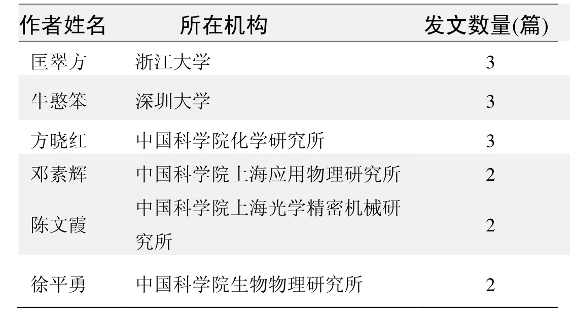
作者发文数量排名(CNKI)
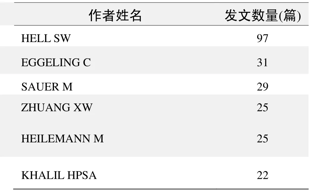
作者发文数量排名(WOS)
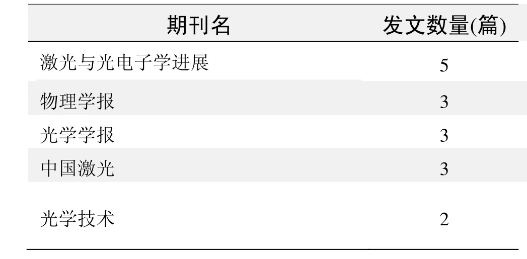
期刊发文数量排名(CNKI)
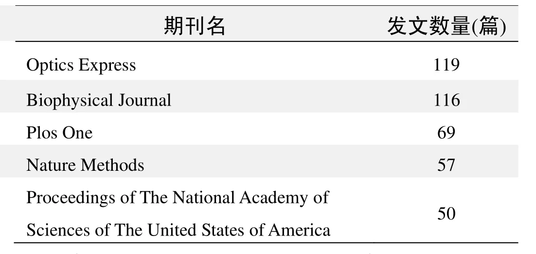
期刊发文数量排名(WOS)
根据中国知网(CNKI)数据报告,筛选出有关超分辨率荧光显微技术研究与应用的高被引论文,排行结果如下.
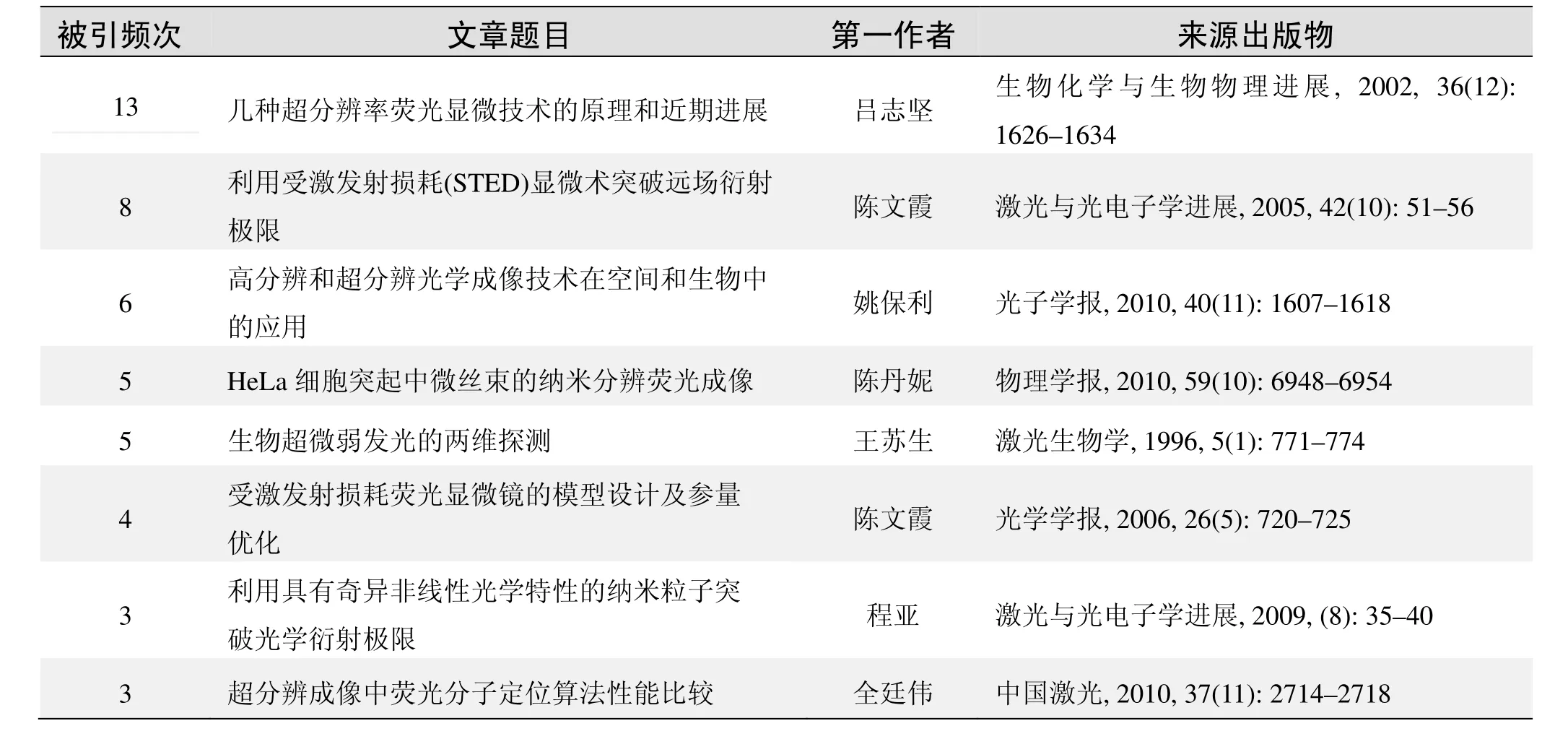
国内数据库高被引论文排行

(数据来源:中国知网,检索时间:2015.1.20)
根据Web of Science统计数据,有关超分辨率荧光显微技术研究与应用的高被引论文.*摘编自《化学通报》2014年77卷11期1029~1035页
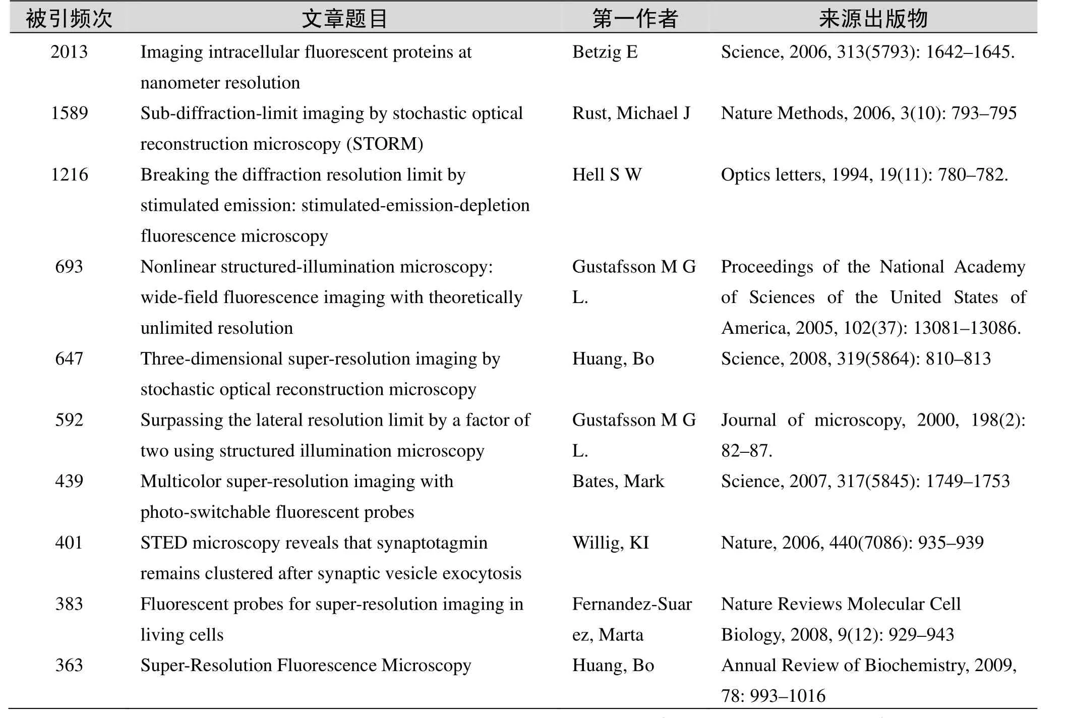
国外数据库高被引论文排行
超分辨荧光显微镜——显纳镜*
袁景和,方晓红
(中国科学院化学研究所分子纳米结构与纳米技术院重点实验室北京 100190)
化学是在分子水平研究物质组成、结构、性质及变化的科学.正如美国科学院院士、哈佛大学谢晓亮教授在《Science》评述中所指出的“活细胞已成为21世纪(科学研究中)的试管”,在生命化学研究领域,越来越多的化学家将生理状态下的复杂生物体系如活细胞或活体作为研究对象.研究人员首先面临的挑战问题是,如何在活细胞/活体中实现分子水平上生物分子的结构、分子间(内)相互作用、生化反应等的实时动态检测.2014年诺贝尔化学奖获得者Eric Betzig、Stefan W. Hell和William E. Moerner所发展的“超分辨率荧光显微镜”,为人们突破这一生命化学研究的瓶颈,阐明生化反应和生命活动的分子机制,最终揭示生老病死的生命奥秘,带来了新的手段和新的机遇.所谓超分辨荧光显微镜是指超过衍射极限分辨的荧光显微镜.光学显微镜由于其非侵入、动态表征等优势,广泛应用于生命科学研究.然而,物理学家Ernst. Abbe在1873年就指出,传统光学显微镜的分辨率有一极限值.因为一个无限小的点所发出的光,经显微光学系统成像后得到的是一个扩展的焦斑,即艾里斑.对于常用可见光波长的显微镜,分辨率不超过200 nm,也就是说无法分辨距离小于200 nm的两个点.这个Abbe衍射极限一直被人们认为是不可逾越的理论极限,阻碍了对生物体系的分子水平研究.
近年来随着超分辨荧光显微术的兴起,研究人员研制了多种突破衍射极限的超分辨光学显微镜,使得光学显微镜进入“显纳镜”(Nanoscopy)时代.这些超分辨显微镜主要分为两类:一类以Hell发明的受激辐射耗尽(Stimulated emission depletion,STED)显微镜为代表,通过调制光照明方式来实现超分辨,也可称为结构光照明超分辨显微镜;另一类是基于单分子定位的超分辨显微镜,典型代表是Betzig和Moerner等发展的光活化定位显微镜(Photoactivated localization microscopy,PALM),通过对具有光开关功能的荧光基团进行单分子成像和定位而实现.下面将简介这两类显微镜的原理、方法、应用进展,以及三位诺奖获得者的贡献.
1STED荧光显微镜
1.1原理
早在1994年Hell就提出了通过受激辐射耗尽的方式实现超分辨成像的设想.其基本原理为,激发光将荧光分子从基态S0激发到激发态S1,经过振动弛豫,荧光分子跃迁到S1的最低振动能级L2,当荧光分子从该振动能级跃迁回到基态S0时,能量以荧光的形式辐射出去,这就是自发荧光辐射过程,传统的共聚焦显微镜等即是通过检测这一自发辐射荧光信号来成像的.另一方面,处于激发态的荧光分子在外界辐射影响下,产生与外界辐射同频率、同位相和同偏振的辐射,即受激荧光辐射过程,如果这一过程竞争超过自发荧光辐射,就导致激发态分子进入非自发荧光辐射状态,不发荧光,称为受激辐射耗尽(STED).诺贝尔化学奖获得者Hell创造性地同时使用两束激光激发样品,其中一束为中间空心的面包圈形状STED光,两束光叠加后使得只有激发光焦斑中心区域的荧光分子可以自发辐射荧光,而被STED光束照射的区域,发生STED效应不再发荧光,这就使得有效的荧光激发半径大大降低,从而突破了衍射极限的限制.
1.2方法与应用
实现STED超分辨成像的关键在于获得面包圈形STED光,并且要求STED光波长位于荧光发射波长的红限,时间上比激发光有亚纳秒量级的延迟,强度上远高于激发光.2000年,Hell领导其研究组,利用两台同步的钛宝石飞秒脉冲激光器成功地构建了超分辨STED显微镜,成像48 nm直径荧光球,获得104 nm横向分辨率,第一次用实验实现了STED超分辨显微成像.后来经一系列设备改进,分辨率提高到29 nm.随后Hell等还发展了快速扫描STED荧光显微镜(80帧/s)、双色STED成像、多寿命多色STED成像等一系列技术.
早期的STED显微镜主要采用飞秒或者亚皮秒脉冲激光器激发,设备昂贵,调制难度大.后来Hell等发展了用连续激光激发的STED(CWSTED)显微镜,并实现了商品化.虽然成本降低,但为充分耗尽荧光分子,需使用比脉冲激光更高的激光功率,容易导致荧光样品的光漂白和热损伤.新的商品化超连续谱激光器,具有同时输出激发光和STED光、方便STED波长选择、避免同步失调等优势,将大大促进STED显微镜的普及.
应用方面,Hell实验室2006年在《Nature》上报道,STED显微镜分辨出直径仅40 nm的神经突触囊泡,并发现突触囊泡膜蛋白I(Synaptotagmin I)在突触囊泡胞吐后仍保持独立团簇状态.随后STED显微镜凭借其超高分辨能力很快在研究细胞膜微区结构、细胞器、神经突触、病毒等方面取得不少新的重要发现.如Hell及其合作者观测140 nm大小的HIV-1病毒颗粒上包膜蛋白的分布,发现这些糖蛋白发生聚集是病毒高效停靠宿主细胞及随后感染的必要条件.他们从对体外细胞成像进一步实现了对活老鼠大脑神经元的超高分辨率实时成像,展示了STED在活体成像中应用前景.
Hell发明STED显微镜,其意义不仅是分析技术上的进步,更重要的是观念上的突破.Hell提出了STED显微镜的分辨率公式.公式指出,只要STED光强足够大,显微镜的分辨率没有理论上的极限.由于有衍射极限理论的思维禁锢,在STED提出后的很长一段时间,并未得到大多数学者的支持.直到2006年Hell研究组实现了神经突触囊泡的超分辨成像,展示了令人信服的生物体系表征结果,才得到广泛认可,并启发和带动了一大批超分辨技术的迅速发展.值得一提的是,STED显微镜由于其技术难度,长期以来国际上只有少数实验室可实现.在国内,中科院化学所基于三态弛豫(T_Rex)概念研制成功了超连续脉冲激光激发SC-STED,获得了好于50 nm的超分辨荧光成像,并在此基础上研制了与AFM联用的仪器,在国际上首次实现了荧光和力学信号的同时同步成像,可实现更高精度的光学引导AFM测量,以及光学和力学性质的多参数测量.北京大学实现CWSTED显微镜,浙江大学使用方位角偏振光束,获得了更锐的STED光.
2PALM——基于单分子荧光成像的超分辨显微镜
2.1成像原理
与STED显微镜需要复杂的光学系统不同,PALM等超分辨显微镜最初是在对传统的全内反射显微镜做简单改造后实现的,通过对具有光开关性质的荧光分子进行单分子定位而重构出超分辨显微图像.因为尽管光学分辨极限为200 nm左右,如果这个衍射限范围内只有一个分子,那么通过对显微物镜点扩展函数(PSF)的拟合,其定位的精度与波长无关,可达到1 nm,远小于200 nm的衍射极限.因此,可以对一个样品采取多次成像的方法,每一次成像只激发少量的稀疏分布的单分子.利用一束激光使4个分子中的2个从非荧光态(暗态a)激活为荧光态(亮态b),对处于亮态的2个分子进行拟合定位获得纳米级超分辨定位(c,定位于十字交叉中心点);待这2个分子荧光猝灭后(d),再重复上述循环(e~f)可获得新激活分子的纳米级超分辨成像,这便是PALM的成像原理.由此可见,实现这类超分辨成像需要两个关键技术:一是单分子成像,二是使用具有光调控暗态—亮态开关性质的荧光分子.
2.2单分子成像
单分子荧光成像是PALM等超分辨成像的技术基础,Moerner是单分子成像领域的先驱.单个分子的成像检测,即获得1个分子的光学信号,对化学而言,可谓达到最高灵敏度,难度很大.1989年,Moerner等首次实现低温下单个分子的光谱检测,随后人们相继取得低温单分子荧光光谱、室温单分子荧光光谱、活细胞体系单分子荧光成像等一系列重要突破.1999年,《Science》以“单分子:化学研究的前沿”为题,出版了单分子研究的专刊,综述了单分子研究进展和未来发展方向,极大地推动了单分子检测技术的兴起和发展.
需要指出的是,单分子检测本身也是化学、生物、物理等交叉学科的前沿领域.单分子检测对于生物分子的研究具有特别重要的意义.因为传统的生物体系研究往往都是对大量分子在一段时间内的平均行为的描述,由于分子结构(如构象)的多样性、生化反应的非同步性和所处环境的(如细胞膜、质、核等不同微区)非均相性,使得生物分子通常具有显著的非均一性,在集群研究中单个分子的行为被平均化和掩盖,阻碍了对其结构和功能的深入认识.利用单分子成像,除了可实现超分辨单分子定位,还可研究生物单分子的物理化学性质、动态变化和生物学功能的分子机制,有望为生命科学和生命化学的研究带来重大突破.最近,《Nature Methods》点评了在过去十年中对生物学研究影响最深的十大技术,其中单分子技术和超高分辨率显微术共同上榜,今年的诺贝尔化学奖也是对单分子技术的肯定Moerner长期从事单分子成像研究,为该领域的发展做出了许多重要贡献,其中也包括一些超分辨成像新方法的发展,并一直推动单分子技术在生命科学中的应用.Moerner的获奖主要在于他对于PALM奠基技术-单分子成像的贡献.此外,他在1997年最早报道了具有光活化荧光开关效应的荧光蛋白并实现了对该蛋白的单分子检测.他发现绿色荧光蛋白(GFP)经488 nm激光激发荧光后会进入暗态,不再发荧光,但经405 nm激光照射后,能返回到原来可发射荧光的状态.这种光活化特性的荧光蛋白正是实现PALM超分辨成像所需要的荧光基团.可惜的是,Moerner当时并没有想到将该荧光蛋白用于超分辨成像.
2.3PALM
2006年,Eric Betzig和Lippincott-Schwartz等提出了光激活定位显微术(PALM)的概念,巧妙地利用一种GFP突变体作为光活化蛋白(PAFP)来标记靶蛋白,并在细胞中表达.用405 nm激光器低能量照射细胞表面,一次仅激活出稀疏分布的几个荧光分子,然后用561 nm激光激发得到荧光图像并精确定位这些荧光单分子.待长时间使用561 nm来漂白这些已经定位正确的荧光分子之后,再分别用405 nm和561 nm激光来激活、激发和漂白其他的荧光分子,多次成像后,将这些分子的荧光图像合成到一张图上,得到了比传统光学显微镜至少高10倍以上的分辨率.开发高性能的光活化蛋白是PALM技术发展的重要方向.一些常见的光活化蛋白存在光活化比率小、荧光信号弱等缺点,导致其成像质量受限.研究人员从荧光蛋白的突变体中筛选pH和光稳定性好、光活化速率快的各钟新型的光活化蛋白如PAmCherry1,在固定细胞中获得小于25 nm的空间分辨率.
2006年,Betzig等在《Science》发表首篇PALM论文,展示PALM显微镜可清楚分辨固定细胞粘着斑中的粘着斑蛋(Vinculin)、伪足上的肌动蛋白、质膜上逆转录蛋白Gag、线粒体等,随后又发展了活细胞双色共定位和单分子追踪PALM技术等,观察了多种亚细胞结构变化,如活细胞中粘附复合物(adhesioncomplexes,ACs)的形成过程以及在形成过程中单个桩蛋白(paxillin)的捕获和失去.PALM自提出以来受到了相关领域科研人员的广泛重视,并得到了长足发展.例如,对神经突触后密集区结构(PSD)高分辨成像,发现4种主要脚手架蛋白形成80 nm动态变化的聚集体,有的能选择性地富集AMPA受体而不是NMDA,提出PSD内部结构可能参与调节神经传递的强度和可塑性.
Betzig获得诺贝尔化学奖的工作经历颇有些传奇.他早年曾在建立近场光学显微镜SNOM方面做出过开创性的工作,1992年他在《Science》上发表利用SNOM实现单分子成像的成果,引起广泛关注.SNOM通过纳米光纤近场激发样品,可实现纳米尺度成像.Betzig意识到SNOM在生物体系研究的局限性,曾设想采用通过准确定位不同波长的荧光分子以绕开衍射极限,并在1995年发表了可以说是PALM技术雏型的理论文章,但当时无法在实验上实现.他曾一度失望地离开了学术界,到父亲的企业工作.不过他一直没有放弃突破衍射极限的梦想.当他了解到Lippincott-Schwartz实验室的光活化荧光蛋白后,重返实验室,终于梦想成真.
2.4随机光学重构显微镜(STORM)
与PALM同时诞生的,还有一种同样是基于单分子点扩展函数拟合定位实现超分辨的方法,即哈佛大学华裔女科学家Xiaowei Zhuang研究组开发的超分辨随机光学重构显微(stochastic optical reconstruction microscopy,STORM).STORM与PALM的基本原理一致,区别在于STORM使用的荧光开关基团是有机染料而不是荧光蛋白.Zhuang等发现可使用一对染料分子Cy3和Cy5作为荧光标记实现超分辨成像,因为不同波长光可以控制Cy5在荧光发射态和暗态之间切换,而Cy3分子可以加速该开关过程.
Zhuang及合作者在STORM方法改进和应用上做了大量的工作,如发展可同时记录2种甚至多种蛋白质空间相对位置的方法、三维STORM等.应用方面的成果包括:对神经元树突和轴突中肌动蛋白等骨架蛋白成像,发现肌动蛋白形成缠绕在神经轴突外周的环状结构,轴突中的钠离子通道呈现出与之关联的周期性分布,表明轴突质膜可能影响了轴突与其他细胞相互交流的方式;通过对端粒成像,发现了一种与端粒功能有关的t-loop构象,并提出了调控t-loop构象形成的新机制.
STORM和PALM一直是超分辨成像领域并驾齐驱、不分伯仲的两种技术.Zhuang在博士后期间就在单分子成像领域崭露头角,后来又以研究RNA构象变化、病毒感染等出色工作成为单分子成像领域代表性人物之一.她的课题组在使用荧光共振能量转移常用染料对进行单分子成像时,观察到其光开关效应,很快便独立地发展了超分辨成像新方法.STORM的工作与PALM于2006年同月发表,只是在投稿日期上晚了4个月.由于具有对细胞内源性生物分子成像的优势,目前STORM在活细胞等生物体系的应用似更领先一步,年仅40岁的Zhuang也因此当选为美国科学院院士.此次遗憾地与诺奖失之交臂,令人惋惜.不过,Zhuang及其发展的STORM对生物成像的重大贡献是不会被人忘记的.
北京大学、中科院化学所、生物物理所、物理所等是国内较早从事生物单分子成像和单分子研究的单位,在生物大分子相互作用、细胞信号转导、蛋白质折叠和分子马达等领域的研究都取得过一些重要的突破.生物物理所和北京大学分别在发展新的PALM成像光活化蛋白以及双分子荧光互补技术与PALM相结合等方面近期也有高水平的成果报道.不过总体来说,国内在单分子研究这一前沿交叉领域的研究力量还较薄弱,在单分子检测基础上从事PALM和STORM的研究队伍更小.可以说这一领域在国内的发展不免受到传统学科观念、评价体系、经费支持力度和人才知识结构等方面的影响.此次超分辨和单分子成像的工作获得诺奖,将对化学与生物前沿交叉领域的研究工作起到积极的推动作用.
3小结与展望
自2006年以来,超分辨光学显微镜迅速发展,并且在生物体系的应用中展现了激动人心的前景,各种新的超分辨成像方法不断涌现.但是作为新兴的技术,超分辨光学显微镜的发展历史不长,STED PALM/STORM分别于2000年、2006年研制成功.它们应用于生命过程分子机制的研究,还处于起步阶段,其技术本身仍有待进一步完善.例如,STED超分辨显微镜由于高强度STED光的使用,对荧光标记分子光稳定性的要求高,难以实现单分子水平检测,还可能对生物样品造成损伤.此外,STED显微镜的研制和维护通常需要具有一定光学专业技术知识的人员,影响了它的普及推广.PALM和STORM虽然较容易实现,但需要进行多次成像和拟合定位,时间分辨往往受到限制,对图像数据处理要求高.因此仍需要进一步发展适合于活细胞/活体动态变化研究的高时间分辨显纳镜.另一方面,超分辨光学显微镜与其他表征技术的联用也是今后的发展方向,特别是实现在对分子位置进行成像的同时,表征其化学组分和构象.超分辨显微技术的不断进步和广泛应用,将极大地促进分子水平上生化反应过程及生物大分子构效关系的研究,为生命化学的发展翻开新的一页.
·高被引论文摘要·
被引频次:13
几种超分辨率荧光显微技术的原理和近期进展
吕志坚,陆敬泽,吴雅琼,等
在生命科学领域,人们常常需要在细胞内精确定位特定的蛋白质以研究其位置与功能的关系.多年来,宽场/共聚焦荧光显微镜的分辨率受限于光的阿贝/瑞利极限,不能分辨出200 nm以下的结构.近年来,随着新的荧光探针和成像理论的出现,研究者开发了多种实现超出普通共聚焦显微镜分辨率的三维超分辨率成像方法.主要介绍这些方法的原理、近期进展和发展趋势.介绍了光源的点扩散函数(point spread function,PSF)的概念和传统分辨率的定义,阐述了提高xy平面分辨率的方法.通过介绍单分子荧光成像技术,引入了单分子成像定位精度的概念,介绍了基于单分子成像的超分辨率显微成像方法,包括光激活定位显微技术(photoactivated localization microscopy,PALM)和随机光学重构显微技术(stochastic optical reconstruction microscopy,STORM).介绍了两大类通过改造光源的点扩散函数来提高成像分辨率的方法,分别是受激发射损耗显微技术(stimulated emission depletion,STED)和饱和结构照明显微技术(saturated structure illumination microscopy,SSIM).比较了不同的z轴提取信息的方法,并阐述了这些方法与xy平面上的超分辨率显微成像技术相结合所得到的各种三维超分辨率显微成像技术的优劣.探讨了目前超分辨率显微成像的发展极限和方向.
超分辨率荧光显微技术;点扩散函数;PALM;STORM;STED;SSIM
来源出版物:生物化学与生物物理进展, 2002, 36(12): 1626–1634联系邮箱:陈良怡,chen_liangyi@yahoo.com
被引频次:8
利用受激发射损耗(STED)显微术突破远场衍射极限
陈文霞,肖繁荣,刘力,等
摘要:远场光学显微镜受衍射极限分辨率的限制,而近场光学显微镜南于缺乏层析能力,则无法实现超分辨的三维成像.研究了既可突破远场光学显微术的衍射极限分辨率又可实现三维成像的成像技术——受激发射损耗(STED),综述了STED的分辨率与STED光的光强、延迟时间、光斑空间分布等主要参数的关系,以及该技术的最新进展和应用前景.
关键词:分辨率;衍射极限;受激发射损耗;时间特性;空间特性
来源出版物:激光与光电子学进展, 2005, 42(10): 51–56
被引频次:6
高分辨和超分辨光学成像技术在空间和生物中的应用
姚保利,雷铭,薛彬,等
摘要:大到天文光学望远镜观察浩瀚的宇宙,小到光学显微镜探察细微的纳米世界,光学成像技术在人类探索和发现未知世界奥秘的活动中扮演着至关重要的角色.看得更远、看得更细、看得更清楚是人们不断追求的目标,传统光学理论已证明所有经典光学系统都是一个衍射受限系统,即光学系统空间分辨率的物理极限是由光的波长和系统的相对孔径(或数值孔径)决定的.能否突破这个极限?能否不断提高光学系统的成分辨率?围绕着这个问题,本文综述了近年来开展的各种光学高分辨和超分辨成像技术,及其在空间探测和生物领域中的应用.
关键词:高分辨;超分辨;光学成像;空间光学遥感;显微成像
来源出版物:光子学报, 2010, 40(11): 1607–1618联系邮箱:姚保利,yaobl@opt.ac.cn
被引频次:5
HeLa细胞突起中微丝束的纳米分辨荧光成像
陈丹妮,刘磊,于斌,等
摘要:在Matlab编程环境下模拟了单分子定位显微的纳米分辨成像,在传统实验参数条件下,对不同间隔分子带模型进行了模拟成像.模拟结果表明,单分子定位显微方法可以区分中心相隔20 nm的两个分子带.同时,也分析了不同像元大小对单分子定位精度的影响.此外,通过单分子定位显微方法在IX71倒置荧光显微镜上实现了纳米分辨,系统极限分辨率,即半高全宽为48 nm.在该系统上,获得了HeLa细胞突起中微丝束结构的纳米分辨图像.从重构获得的图像中可以看到微丝束的直径为75~200 nm.
关键词:纳米分辨;单分子定位;HeLa细胞;微丝束;细胞突起;
来源出版物:物理学报, 2010, 59(10): 6948–6953联系邮箱:牛憨笨,hhnin@5711edu. cn
被引频次:5
生物超微弱发光的两维探测
王苏生,陈天明,俞信,等
摘要:本文介绍了自行研制的二套系统及其应用.1.高灵敏荧光显微镜系统,该系统探测灵敏度达到10-6Ix量级,比普通CCD系统提高了104倍,系统用宽量程照度计对微弱光成像性能进行了标定,在给出细胞荧光图像的同时,可以给出每一象元的发光强度,并可给出视觉更易分辨的光强的三维显示和伪彩色图像.在该系统上得到了分红菌甲素在Hela细胞中的分布图像,Hela细胞加入竹红菌甲素后的光照损伤及抗氧化剂维生素E等对细胞的保护图象.2.光子计数成像系统,该系统灵敏度达10-8Ix量级,可探测到单个光子及其分布,在其上得到了绿豆芽,树叶,昆明鼠,人手及手指的超微弱发光的光子图象,并用统计理论进行了信号检验.
关键词:纳米分辨;单分子定位;HeLa细胞;微丝束;细胞突起;
来源出版物:激光生物学, 1996, 5(1): 771–774
被引频次:4
受激发射损耗荧光显微镜的模型设计及参量优化
陈文霞,肖繁荣,刘力,等
摘要:受激发射损耗荧光显微镜利用荧光饱和激发态荧光受激损耗的非线性关系,通过限制损耗区域,可突破远场光学显微术的衍射极限分辨力并实现三维成像.基于对粒子速率方程组的修正,建立了描述荧光团各能级粒子数概率时间特性的模型,并定义了时间平均损耗效率判据.采用高斯函数模拟两束入射激光脉冲通过对模型的数值计算,模拟了激发脉冲的SIED激光脉冲的光强、脉冲宽度以及两束光的延迟时间等参量与损耗效率之间的关系,并获得了各参量的最佳值,优化了损耗效率,为提高系统分辨力提供了有效的途径.
关键词:显微;损耗效率;粒子数概率;光强;脉冲宽度;延迟时间;
来源出版物:光学学报, 2006, 26(5): 720–725联系邮箱:陈文霞,wenxia@siom.ac.cn
被引频次:3
利用具有奇异非线性光学特性的纳米粒子突破光学衍射极限
程亚,陈建芳
摘要:受激发射损耗荧光显微镜利用荧光饱和激发态荧光受激损耗的非线性关系,通过限制损耗区域,可突破远场光学显微术的衍射极限分辨力并实现三维成像.基于对粒子速率方程组的修正,建立了描述荧光团各能级粒子数概率时间特性的模型,并定义了时间平均损耗效率判据.采用高斯函数模拟两束入射激光脉冲通过对模型的数值计算,模拟了激发脉冲的SIED激光脉冲的光强、脉冲宽度以及两束光的延迟时间等参量与损耗效率之间的关系,并获得了各参量的最佳值,优化了损耗效率,为提高系统分辨力提供了有效的途径.
关键词:远场光学成像;光学衍射极限;非线性光学
来源出版物:激光与光电子学进展, 2009, (8): 35–40
被引频次:3
超分辨成像中荧光分子定位算法性能比较
全廷伟,曾绍群,吕晓华
摘要:超分辨成像已成为活细胞结构和功能成像的关键工具,荧光分子定位是超分辨成像过程中不可缺少的步骤,从超分辨成像角度研究各种荧光分子定位算法性能具有重要的意义.选择5种典型的荧光分子定位算法:质心法、广义质心法、高斯拟合、解线性方程组和极大似然法,以定位精度和定位时间来评价所选择算法的性能.结果表明,1)高斯拟合、极大似然法和广义质心法能高精度对荧光分子定位,不受荧光分子所在子区域提取的影响;2)质心法和解线性方程组法能应用于图像在线分析,但定位精度较低,受子区域提取影响较大;3)当两个荧光分子位于一个衍射斑时,采用这5种算法的定位精度都会急剧下降.受激发射损耗荧光显微镜利用荧光饱和激发态荧光受激损耗的非线性关系,通过限制损耗区域,可突破远场光学显微术的衍射极限分辨力并实现三维成像.基于对粒子速率方程组的修正,建立了描述荧光团各能级粒子数概率时间特性的模型,并定义了时间平均损耗效率判据.采用高斯函数模拟两束入射激光脉冲通过对模型的数值计算,模拟了激发脉冲的SIED激光脉冲的光强、脉冲宽度以及两束光的延迟时间等参量与损耗效率之间的关系,并获得了各参量的最佳值,优化了损耗效率,为提高系统分辨力提供了有效的途径.
关键词:远场光学成像;光学衍射极限;非线性光学
来源出版物:中国激光, 2010,37(11): 2714-2718
被引频次:2
高分辨率光学显微术在生命科学中的应用
王娟,汤乐民
摘要:提高光学显微镜分辨率的研究主要集中在两个方面进行,一是利用经典方法提高各种条件下的空间分辨率,如用于厚样品研究的SPIM技术,用于快速测量的SHG技术以及用于活细胞研究的MPM技术等.二是将最新的非线性技术与高数值孔径测量技术(如STED和SSIM技术)相结合.生物科学研究离不开超高分辨率显微术的技术支撑,人们迫切需要更新显微术来适应时代发展的要求.近年来研究表明,光学显微镜的分辨率已经成功突破200 nm横向分辨率和400 nm轴向分辨率的衍射极限.高分辨率乃至超高分辨率光学显微术的发展不仅在于技术本身的进步,而且它将会极大促进生物样品的研究,为亚细胞级和分子水平的研究提供新的手段.
关键词:光学显微镜;高分辨率;非线性技术;纳米水平
来源出版物:南通大学学报(医学版), 2007, 27(1): 72–74
被引频次:2
可逆饱和光转移过程的荧光超分辨显微术
郝翔,匡翠方,李旸晖,等
摘要:基于可逆饱和光转移过程的荧光超分辨显微技术,从原理上打破了原有的光学远场衍射极限对光学系统极限分辨率的限制,在生物、化学、医学等多个学科拥有广泛的应用前景.回顾了近年来超分辨显微研究的历史,综述了目前常见的几种基于可逆饱和光转移过程的荧光超分辨显微方法,详细描述了各自的技术特点并对比了其优缺点,阐述了相关领域内最新的研究工作进展.
关键词:显微;超分辨;可逆饱和光转移过程;受激发射损耗显微镜;基态损耗显微镜;饱和图案激发显微镜;饱和结构光照明显微镜
来源出版物:激光与光电子学进展, 2012, 49(3): 030005联系邮箱:匡翠方,cfkuang@zju. edu. cn
被引频次:2
受激发射损耗显微技术中0/π圆形相位板参数优化
郝翔,匡翠方,王婷婷,等
摘要:采用光的矢量衍射理论,研究了0/π圆形相位板不同设计参数对于受激发射损耗(STED)显微镜中抑制光形成中空聚焦光斑聚焦质量的影响,并进行了数值模拟.相关计算结果表明,当采用不同偏振态、不同光强分布的入射光作为STED光,0/π圆形相位板内径的合理取值范围是不同的,而系统所用物镜的数值孔径对于内径的取值影响较小.在各种不同的参数组合下,使用线偏振光和圆偏振光作为入射光时,0/π圆形相位板内径取值范围基本保持一致;当入射光为径向偏振光时,内径取值则相对较大;而当使用切向偏振光时,任何取值均无法在系统焦平面附近得到满意的中空聚焦光斑.
关键词:受激发射损耗显微技术;相位板;衍射理论;数值孔径
来源出版物:光学学报, 2011,31(3): 03180001联系邮箱:匡翠方,cfkuang@zju. edu. cn
被引频次: 2013
来源出版物: Science, 2006, 313(5793): 1642-1645.
联系邮箱: Betzig E; betzige@janelia.hhmi.org
被引频次: 1589
Sub-diffraction-limit imaging by stochastic optical reconstruction microscopy (STORM)
Rust, Michael J; Bates, Mark; Zhuang Xiaowei
Abstract: We have developed a high-resolution fluorescence microscopy method based on high-accuracy localization of photoswitchable fluorophores. In each imaging cycle, only a fraction of the fluorophores were turned on, allowing their positions to be determined with nanometer accuracy. The fluorophore positions obtained from a series of imaging cycles were used to reconstruct the overall image. We demonstrated an imaging resolution of 20 nm. This technique can, in principle, reach molecular-scale resolution.
Keywords: Fluorescence Microscopy; Localization; resolution; superresolution
来源出版物: Nature Methods, 2006, 3(10): 793-795联系邮箱: Zhuang Xiaowei; zhuang@chemistry.harvard.edu
被引频次: 1216
Breaking the diffraction resolution limit by stimulated emission: stimulated-emission-depletion fluorescence microscopy
Hell S W; Wichmann J.
Abstract: We propose a new type of scanning fluorescence microscope Capable of resolving 35 nm in the far field. We overcome the diffraction resolution limit by employing stimulated emission to inhibit the fluorescence process in the outer regions of the excitation point-spread function. In contrast to near-field scanning optical microscopy, this method can produce three-dimensional images of translucent specimens.
来源出版物: Optics letters, 1994, 19(11): 780-782.
被引频次: 693
Nonlinear structured-illumination microscopy: wide-field fluorescence imaging with theoretically unlimited resolution
Gustafsson M G L.
Abstract: Contrary to the well known diffraction limit, the fluorescence microscope is in principle capable of unlimited resolution. The necessary elements are spatially structured illumination light and a nonlinear dependence of the fluorescence emission rate on the illumination intensity. As an example of this concept, this article experimentally demonstrates saturated structured-illumination microscopy, a recently proposed method in which the nonlinearity arises from saturation of the excited state. This method can be used in a simple, wide-field (nonscanning) microscope, uses only a single, inexpensive laser, and requires no unusual photophysical properties of the fluorophore. The practical resolving power is determined by the signal-to-noise ratio, which in turn is limited by photobleaching. Experimental results show that a 2D point resolution of <50 nm is possible on sufficiently bright and photostable samples.
Keywords: super resolution; moire; resolution extension; saturation
来源出版物: Proceedings of the National Academy of Sciences of the United States of America, 2005, 102(37): 13081-13086.
联系邮箱: Gustafsson M G L.; mats@msg.ucsf.edu
被引频次: 647
Three-dimensional super-resolution imaging by stochastic optical reconstruction microscopy
Huang, Bo; Wang, Wenqin; Bates, Mark; et al.
Abstract: Recent advances in far- field fluorescence microscopy have led to substantial improvements in image resolution, achieving a near-molecular resolution of 20 to 30 nanometers in the two lateral dimensions. Three- dimensional (3D) nanoscale-resolution imaging, however, remains a challenge. We demonstrated 3D stochastic optical reconstruction microscopy (STORM) by using optical astigmatism to determine both axial and lateral positions of individual fluorophores with nanometer accuracy. Iterative, stochastic activation of photoswitchable probes enables high- precision 3D localization of each probe, and thus the construction of a 3D image, without scanning the sample. Using this approach, we achieved an image resolution of 20 to 30 nanometers in the lateral dimensions and 50 to 60 nanometers in the axial dimension. This development allowed us to resolve the 3D morphology of nanoscopic cellular structures.
Keywords: Fluorescence nanoscopy; Axial resolution; localization; tracking; probes; endocytosis; precision; molecules; emitters; cells
来源出版物: Science, 2008, 319(5864): 810-813联系邮箱: Zhuang Xiaowei; zhuang@chemistry.harvard.edu
被引频次: 592
Surpassing the lateral resolution limit by a factor of two using structured illumination microscopy
Gustafsson M G L
Abstract: Lateral resolution that exceeds the classical diffraction limit by a factor of two is achieved by using spatially structured illumination in a wide-field fluorescence microscope. The sample is illuminated with a series of excitation light patterns, which cause normally inaccessible high-resolution information to be encoded into the observed image. The recorded images are linearly processed to extract the new information and produce a reconstruction with twice the normal resolution. Unlike confocal microscopy, the resolution improvement is achieved with no need to discard any of the emission light. The method produces images of strikingly increased clarity compared to both conventional and confocal microscopes.
Keywords: actin; cytoskeleton; fluorescence microscopy; interference; lateral resolution; moire microscopy; optical transfer function; patterned excitation; resolution; structured illumination; super-resolution; wide-field microscopy
来源出版物: Journal of Microscopy, 2000, 198(2): 82-87.
被引频次: 439
Multicolor super-resolution imaging with photo-switchable fluorescent probes
Bates, Mark; Huang, Bo; Dempsey, Graham T; et al.
Abstract: Recent advances in far-field optical nanoscopy have enabled fluorescence imaging with a spatial resolution of 20 to 50 nanometers. Multicolor super-resolution imaging, however, remains a challenging task. Here, we introduce a family of photo-switchable fluorescent probes and demonstrate multicolor stochastic optical reconstruction microscopy (STORM). Each probe consists of a photo-switchable "reporter" fluorophore that can be cycled between fluorescent and dark states, and an "activator" that facilitates photo-activation of the reporter. Combinatorial pairing of reporters and activators allows the creation of probes with many distinct colors. Iterative, color-specific activation of sparse subsets of these probes allows their localization with nanometer accuracy, enabling the construction of a super-resolution STORM image. Using this approach, we demonstrate multicolor imaging of DNA model samples and mammalian cells with 20-to30-nanometer resolution. This technique will facilitate direct visualization of molecular interactions at the nanometer scale.
Keywords: single molecules; energy-transfer; quantum dots; microscopy; localization; cells; resolution; nanoscopy
来源出版物: Science, 2007, 317(5845): 1749-1753联系邮箱: Zhuang Xiaowei; zhuang@chemistry.harvard.edu
被引频次: 401
STED microscopy reveals that synaptotagmin remains clustered after synaptic vesicle exocytosis
Willig, KI; Rizzoli, SO; Westphal, V; et al.
Abstract: Synaptic transmission is mediated by neurotransmitters that are stored in synaptic vesicles and released by exocytosis upon activation. The vesicle membrane is then retrieved by endocytosis, and synaptic vesicles are regenerated and re-filled with neurotransmitter (1). Although many aspects of vesicle recycling are understood, the fate of the vesicles after fusion is still unclear. Do their components diffuse on the plasma membrane, or do they remain together? This question has been difficult to answer because synaptic vesicles are too small (similar to 40 nm in diameter) and too densely packed to be resolved by available fluorescence microscopes. Here we use stimulated emission depletion (STED) (2) to reduce the focal spot area by about an order of magnitude below the diffraction limit, thereby resolving individual vesicles in the synapse. We show that synaptotagmin I, a protein resident in the vesicle membrane, remains clustered in isolated patches on the presynaptic membrane regardless of whether the nerve terminals are mildly active or intensely stimulated. This suggests that at least some vesicle constituents remain together during recycling. Our study also demonstrates that questions involving cellular structureswith dimensions of a few tens of nanometres can be resolved with conventional far-field optics and visible light.
Keywords: frog neuromuscular junction; hippocampal synapses; fluorescence microscopy; stimulated-emission; endocytosis; resolution; membrane; transmitter; release; domain
来源出版物: Science, 2006, 440(7086): 935-939联系邮箱: Jahn, R; rjahn@gwdg.de
被引频次: 383
Fluorescent probes for super-resolution imaging in living cells
Fernandez-Suarez, Marta; Ting, Alice Y.
Abstract: In 1873, Ernst Abbe discovered that features closer than similar to 200 nm cannot be resolved by lens-based light microscopy. In recent years, however, several new far-field super-resolution imaging techniques have broken this diffraction limit, producing, for example, video-rate movies of synaptic vesicles in living neurons with 62 nm spatial resolution. Current research is focused on further improving spatial resolution in an effort to reach the goal of video-rate imaging of live cells with molecular (1-5 nm) resolution. Here, we describe the contributions of fluorescent probes to far-field super-resolution imaging, focusing on fluorescent proteins and organic small-molecule fluorophores. We describe the features of existing super-resolution fluorophores and highlight areas of importance for future research and development.
Keywords: hotoactivated localization microscopy; optical reconstruction microscopy; diffraction resolution barrier; small-molecule probes; live cells; stimulated-emission; quantum dots; in-vivo; sted microscopy; near-field
来源出版物: Nature Reviews Molecular Cell Biology, 2008, 9(12): 929-943联系邮箱: Fernandez-Suarez, Marta; artafs@alum.mit.edu
被引频次: 363
Super-Resolution Fluorescence Microscopy
Huang, Bo; Bates, Mark; Zhuang Xiaowei
Abstract: Achieving a spatial resolution that is not limited by the diffraction of light, recent developments of super-resolution fluorescence microscopy techniques allow the observation of many biological structures not resolvable in conventional fluorescence microscopy. New advances in these techniques now give them the ability to image three-dimensional (3D) structures, measure interactions by multicolor colocalization, and record dynamic processes in living cells at the nanometer scale. It is anticipated that super-resolution fluorescence microscopy will become a widely used tool for cell and tissue imaging to provide previously unobserved details of biological structures and processes.
Keywords: diffraction limit; fluorescence imaging; fluorescent protein; live-cell imaging; photoswitchable dye; single molecule
来源出版物: Annual Review of Biochemistry, 2009, 78: 993-1016联系邮箱: Huang, Bo; bohuang@fas.harvard.edu
·推荐论文摘要·
Three dimensional multimodal sub-diffraction imaging with spinning-disk confocal microscopy using blinking/fluctuation probes
X. Chen; Z. Zeng; H. Wang; et al.
Abstract: Previous localization super-resolution techniques cannot offer three-dimensional imaging easily. Here we present a three-dimensional multimodal sub-diffraction imaging with spinning-disk (SD) confocal microscopy, 3D-MUSIC, which not only takes fully advantages of spinning-disk confocal microscopy, such as fast imaging speed, high signal-to-noise ratio, optical-sectioning capability, but also extends its spatial resolution limit along all three dimensions. Both axial and lateral resolution can be improved simultaneously by virtue of the blinking/fluctuation nature of the modified fluorescent probes, exemplified by the quantum dots (QDs). Further, dual-modality super-resolution image can be obtained, by super-resolution optical fluctuation imaging (SOFI), and bleaching/blinking assisted localization microscopy (BaLM). Therefore, fast super-resolution imaging can be achieved with SD-SOFI by only capturing 100 frames, yet a high-resolution imaging can be provided with SD-BaLM.
Keywords: multi-modality; super-resolution microscopy; three-dimensional; spinning-disk confocal
来源出版物:Nano Research, 2015, doi:10.1007/s12274–12015–10736–12278
联系邮箱:XI Peng; xipeng@pku.edu.cn
Fast Super-Resolution Imaging with Ultra-High Labeling Density Achieved by Joint Tagging Super-Resolution Optical Fluctuation Imaging
Z. Zeng; X. Chen; H. Wang; et al.
Abstract: Previous stochastic localization-based super-resolution techniques are largely limited by the labeling density and the fidelity to the morphology of specimen. We report on an optical super-resolution imaging scheme implementing joint tagging using multiple fluorescent blinking dyes associated with super-resolution optical fluctuation imaging (JT-SOFI), achieving ultra-high labeling density super-resolution imaging. To demonstrate the feasibility of JT-SOFI, quantum dots with different emission spectra were jointly labeled to the tubulin in COS7 cells, creating ultra-high density labeling. After analyzing and combining the fluorescence intermittency images emanating from spectrally resolved quantum dots, the microtubule networks are capable of being investigated with high fidelity and remarkably enhanced contrast at sub-diffraction resolution. The spectral separation also significantly decreased the frame number required for SOFI, enabling fast super-resolution microscopy through simultaneous data acquisition. As the joint-tagging scheme can decrease the labeling density in each spectral channel, thereby bring it closer to single-molecule state, we can faithfully reconstruct the continuous microtubule structure with high resolution through collection of only 100 frames per channel. The improved continuity of the microtubule structure is quantitatively validated with image skeletonization, thus demonstrating the advantage of JT-SOFI over other localization-based super-resolution methods.
来源出版物:Scientific Reports, 2015, 5 Article number: 8359
联系邮箱:XI Peng; xipeng@pku.edu.cn
Hundred-Thousand Light Holes Push Nanoscopy to go Parallel
Chen, Xuanze; Xi, Peng
Abstract: With the application of the elements of all major super-resolution techniques including stimulated emission depletion, structure illumination microscopy, and photo-activated localization microscopy, the incoherent crossed standing-wave microscopy achieves parallel super-resolution imaging.
Keywords: optical nanoscopy; STED; SIM; PALM
来源出版物: Microscopy Research and Technique, 2015, 78(1): 8–10
联系邮箱: Xi, Peng ; xipeng@pku.edu.cn
Sub-diffraction imaging of nitrogen-vacancy centers in diamond by stimulated emission depletion and structured illumination
Yang, Xusan; Tzeng, Yan-Kai; Zhu, Zhouyang; et al.
Abstract: Stimulated emission depletion (STED) and structured illumination (SIM) are two commonly used techniques for super-resolution imaging. However, the performance of these two techniques has never been quantitatively compared side-by-side. Taking advantage of the non-photobleaching characteristic of NV centres in fluorescent nanodiamond (FND), we performed a comparative study for the resolution of these two methods with 35 nm FNDs at the single particle level, as well as with FND grown in bulk diamond material. Results show that STED provides more structural details, whereas SIM provides a larger field of view with a higher imaging speed. SIMmay induce deconvolution smooth and orientational artifacts during its post-processing.
Keywords: fluorescent nanodiamonds; unlimited resolution; state depletion; STED microscopy; color-centers; superresolution; limit
来源出版物: RSC Advances, 2014, 4(22): 11305–11310
联系邮箱: Xi, Peng; xipeng@pku.edu.cn
Microscopy and its focal switch
Hell S W
Abstract: Until not very long ago, it was widely accepted that lens-based (far-field) optical microscopes cannot visualize details much finer than about half the wavelength of light. The advent of viable physical concepts for overcoming the limiting role of diffraction in the early 1990s set off a quest that has led to readily applicable and widely accessible fluorescence microscopes with nanoscale spatial resolution. Here I discuss the principles of these methods together with their differences in implementation and operation. Finally, I outline potential developments.
来源出版物: Nature Methods, 2009, 6(1): 24–32.
Nanoscopy with more than 100,000'doughnuts
Chmyrov A; Keller J, Grotjohann T; et al.
Abstract: We show that nanoscopy based on the principle called RESOLFT (reversible saturable optical fluorescence transitions) or nonlinear structured illumination can be effectively parallelized using two incoherently superimposed orthogonal standing light waves. The intensity minima of the resulting pattern act as 'doughnuts', providing isotropic resolution in the focal plane and making pattern rotation redundant. We super-resolved living cells in 120 mm × 100 mm-sized fields of view in <1 s using 116000 such doughnuts.
Keywords: structured-illumination microscopy; fluorescence microscopy; optical resolution; limit; protein; breaking; GFP
来源出版物: Nature Methods, 2013, 10(8): 737–740
联系邮箱: Hell S W; shell@gwdg.de
Toward fluorescence nanoscopy
Hell S W
Abstract: For more than a century, the resolution of focusing light microscopy has been limited by diffraction to 180 nm in the focal plane and to 500 nm along the optic axis. Recently, microscopes have been reported that provide three- to sevenfold improved axial resolution in live cells. Moreover, a family of concepts has emerged that overcomes the diffraction barrier altogether. Its first exponent, stimulated emission depletion microscopy, has so far displayed a resolution down to 28 nm. Relying on saturated optical transitions, these concepts are limited only by the attainable saturation level. As strong saturation should be feasible at low light intensities, nanoscale imaging with focused light may be closer than ever.
Keywords: emission depletion microscopy; diffraction resolution limit; stimulated-emission; axial resolution; light-microscopy; 4pi-confocal microscopy; 3-dimensional images; lateral resolution; opposing lenses; state-depletion
来源出版物: Nature Biotechnology, 2003, 21(11): 1347–1355.
联系邮箱: Hell S W; hell@nanoscopy.de
Lattice light-sheet microscopy: Imaging molecules to embryos at high spatiotemporal resolution
Chen B C; Legant W R; Wang K; et al.
Abstract: Although fluorescence microscopy provides a crucial window into the physiology of living specimens, many biological processes are too fragile, are too small, or occur too rapidly to see clearly with existing tools. We crafted ultrathin light sheets from two-dimensional optical lattices that allowed us to image three-dimensional (3D) dynamics for hundreds of volumes, often at subsecond intervals, at the diffraction limit and beyond. We applied this to systems spanning four orders of magnitude in space and time, including the diffusion of single transcription factor molecules in stem cell spheroids, the dynamic instability of mitotic microtubules, the immunological synapse, neutrophil motility in a 3D matrix, and embryogenesis in Caenorhabditis elegans and Drosophila melanogaster. The results provide a visceral reminder of the beauty and the complexity of living systems.
来源出版物: Science, 2014, 346(6208): 439
Nanoscopy in a living mouse brain
Berning S; Willig K I; Steffens H; et al.
Abstract: We demonstrated superresolution optical microscopy in a living higher animal. Stimulated emission depletion (STED) fluorescence nanoscopy reveals neurons in the cerebral cortex of a mouse with <70-nanometer resolution. Dendritic spines and their subtle changes can be observed at their relevant scales over extended periods of time.
Keywords: spines; GFP
来源出版物: Science, 2012, 335(6068): 551–551
联系邮箱: Willig K I; kwillig@gwdg.de
Video-rate far-field optical nanoscopy dissects synaptic vesicle movement
Westphal V; Rizzoli S O; Lauterbach M A; et al.
Abstract: We present video-rate (28 frames per second) far-field optical imaging with a focal spot size of 62 nanometers in living cells. Fluorescently labeled synaptic vesicles inside the axons of cultured neurons were recorded with stimulated emission depletion (STED) microscopy in a 2.5-micrometer by 1.8-micrometer field of view. By reducing the cross-sectional area of the focal spot by about a factor of 18 below the diffraction limit (260 nanometers), STED allowed us to map and describe the vesicle mobility within the highly confined space of synaptic boutons. Although restricted within boutons, the vesicle movement was substantially faster in nonbouton areas, consistent with the observation that a sizable vesicle pool continuously transits through the axons. Our study demonstrates the emerging ability of optical microscopy to investigate intracellular physiological processes on the nanoscale in real time.
Keywords: fluorescence microscopy; nerve-terminals; STED microscopy; resolution; boutons; synapses; dynamics; recovery; mobility; pools
来源出版物: Science, 2008, 320(5873): 246–249
联系邮箱: Hell S W; shell@gwdg.de
Imaging intracellular fluorescent proteins at nanometer resolution
Betzig E; Patterson G H; Sougrat R; et al.
We introduce a method for optically imaging intracellular proteins at nanometer spatial resolution. Numerous sparse subsets of photoactivatable fluorescent protein molecules were activated, localized (to similar to 2 to 25 nanometers), and then bleached. The aggregate position information from all subsets was then assembled into a superresolution image. We used this method - termed photoactivated localization microscopy - to image specific target proteins in thin sections of lysosomes and mitochondria; in fixed whole cells, we imaged vinculin at focal adhesions, actin within a lamellipodium, and the distribution of the retroviral protein Gag at the plasma membrane.
structured-illumination microscopy; diffraction limit; living cells; localization; spectroscopy; molecules
