红细胞分布宽度对判断急性胰腺炎病情和预后的意义
2015-07-18吕远军梁春娜刘健培
吕远军 梁春娜 刘健培
临床研究论著
红细胞分布宽度对判断急性胰腺炎病情和预后的意义
吕远军 梁春娜 刘健培
目的探讨红细胞分布宽度(RDW)对判断急性胰腺炎(AP)病情和预后的作用。方法63例AP患者被设为轻症AP组(35例)及重症AP组(28例),30名健康者被设为对照组。抽取患者就诊时和发病后72 h及对照组的血液检测RDW,对比轻症及重症AP患者以及AP患者与对照组RDW的差异,分析RDW与AP的病死率、局部并发症发生的关系。结果与对照组相比,AP患者就诊时的RDW较低[(14.1±2.1) %vs.(16.8±2.3) %,P<0.001];与轻症AP组相比,重症AP组就诊时的RDW更高[(14.9±1.9) %vs.(19.2±2.4) %,P<0.001)]。死亡重症AP患者就诊时的RDW比存活者高[(24.2±4.1) %vs. (18.6±2.3) %,P<0.001)];发生局部并发症的轻型AP患者就诊时的RDW比无局部并发症者高[(23.9±3.2) %vs. (18.2±2.3) %,P<0.001)]。结论RDW可能是判断AP病情的潜在指标,与AP局部并发症和病死率可能相关。
急性胰腺炎;红细胞分布宽度;预后
急性胰腺炎(AP)是临床常见的急腹症,具有病情多变、发展迅速的特点。重症急性胰腺炎(SAP)治疗困难、预后差,病死率可高达42%[1]。所以,区分SAP和轻症急性胰腺炎(MAP)对治疗及预后有重要意义。目前临床常用的淀粉酶、脂肪酶等诊断指标并不能预测胰腺炎的严重程度和预后。红细胞分布宽度(RDW) 是反映红细胞体积异质性的参数,升高表示其差异性增大。近年来,国内外学者发现RDW与心血管疾病、脑梗死、终末期肾功能不全和感染性休克等危重症的预后及临床转归密切相关,并认为RDW升高可以反映机体潜在的炎症状态[2-5]。但RDW在判断AP预后方面的临床价值,笔者见目前国内似无相关报道,故本研究拟检测AP患者血液RDW的改变,探讨RDW与AP严重程度、局部并发症的发生和患者死亡的相关性。
对象与方法
一、研究对象
以2009年2月1日至2012年10月30日在开平市中心医院就诊的AP患者为研究对象。病例入组标准:符合中华医学会消化病学分会2004 年《中国急性胰腺炎诊治指南(草案)》的诊断标准[6];起病24 h内就诊。排除标准:合并其他感染性疾病、代谢性疾病、恶性肿瘤和慢性器官功能不全。根据Ranson指标、急性生理学及慢性健康状况评分系统Ⅱ(APACHE-Ⅱ)及CT 分级结果进行AP的轻重分型:符合AP诊断标准并具有下列之一者定义为SAP:局部并发症(胰腺坏死、假性囊肿、胰腺脓肿);器官衰竭;Ranson指标≥3项;APACHE-Ⅱ评分≥8;CT分级为D、E;不符合上述标准定义为MAP。共入选患者63例,其中MAP组35例,年龄29~63岁;SAP组28 例,年龄23~72岁;选择30名同期来我院进行体检的健康者为对照组,年龄20~70岁。各组一般资料比较差异无统计学意义(P均>0.05),见表1。

表1 MAP组、SAP组及对照组一般资料比较
二、标本采集及检测
采集所有入选研究对象的静脉血,AP患者于就诊时和起病72 h后采集,采用Sysmex XS-800i全自动血液分析仪(日本森美康株式会社)自动检测血常规,RDW正常参考值为11.5%~15.0%。
三、统计学处理

结 果
一、AP患者就诊时RDW值与对照组比较结果
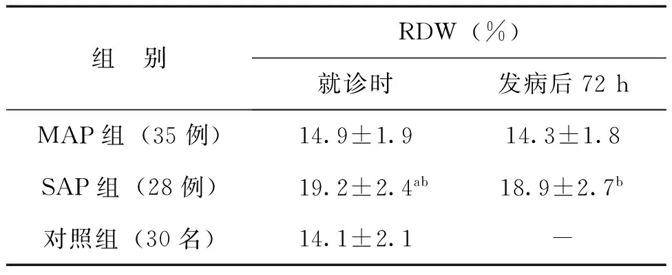
与对照组相比,AP患者就诊时的RDW较低[(14.1±2.1) %vs. (16.8±2.3) %,t=-5.44、P<0.001]。但MAP组就诊时的RDW值与对照组的RDW值比较差异无统计学意义(t=-1.61、P=0.110)。与MAP组相比,SAP组就诊时的RDW更高(t=7.94、P<0.001),见表2。
二、发病后72hAP患者RDW值变化情况
发病后72 h,MAP组的RDW值与就诊时的RDW值比较差异无统计学意义(t=-1.36、P=0.179)。SAP组发病后72 h的RDW值与就诊时RDW值比较差异也无统计学意义(t=-0.44、P=0.662)。与MAP组相比,SAP组发病后72 h的RDW更高(t=8.09、P<0.001),见表2。


组 别RDW(%)就诊时发病后72hMAP组(35例)14.9±1.914.3±1.8SAP组(28例)19.2±2.4ab18.9±2.7b对照组(30名)14.1±2.1-
注:与对照组比较,aP<0.05;与MAP组比较,bP<0.05
三、RDW与AP患者死亡及局部并发症发生的关系
MAP组无死亡病例;SAP组共死亡3例,死亡患者就诊时的RDW比存活患者高[(24.2±4.1) %vs. (18.6±2.3) %,t=3.69、P<0.001]。SAP组有1例出现胰腺坏死、2例出现胰腺假性囊肿、2例出现胰腺脓肿,发生局部并发症患者就诊时的RDW比无局部并发症患者高[(23.9±3.2) %vs. (18.2±2.3) %,t=4.70、P<0.001)],见图1。

图1 RDW与AP患者死亡及局部并发症发生的关系
A:存活患者与死亡患者RDW的比较情况;B:出现及无出现局部并发症患者RDW的比较情况;*表示与另一组数据比较P<0.05
讨 论
AP是临床常见的急症之一。MAP患者症状轻,预后良好,而SAP患者往往合并MODS和感染并发症,病死率可高达42%[1]。因此,寻找可靠的指标对AP的严重程度进行早期诊断和预测,及时将有限的医疗资源和有效措施应用于高危患者,对降低AP患者的病死率,改善其预后有重要意义。许多临床评分系统如Ranson指标和APACHE-Ⅱ等被应用于AP的评估,但这些评分标准均较复杂、操作不简便,故未能在临床上被广泛认可和推广[7-9]。单纯的实验室指标可行性更高,但目前反应炎症严重程度的指标如CRP、IL-6和IL-8等变化幅度大、特异性低[10]。而以往提出的诊断胰腺炎的血尿淀粉酶和脂肪酶已被证实与胰腺炎的严重程度不相关[11]。寻找更简便、有效的血清学指标用于判断AP的病情和预后显得尤为重要。
RDW是由血液分析仪测量获得反映周围红细胞体积异质性的参数,升高反映红细胞的体积变异增大,最初用于贫血的分类。随着对危重病研究的不断深入,RDW越来越受到国内外学者的重视。国内外研究结果显示,RDW与心脏急重症、卒中、外周动脉疾病、肾功能不全、重症感染等密切相关,可作为此类疾病风险评估中的一个独立、高效的预测指标[2-5]。AP,尤其是SAP是急诊科常见急重症,会进一步引发局部感染并发症、MODS和临床死亡。本研究发现SAP患者就诊时的RDW较对照组高,提示其可能可作为AP早期诊断的辅助指标。另外,患者发病后72 h与起病24 h内的RDW数值相近,与AP局部并发症和病死率相关,这也提示了起病72 h内的RDW对于AP病情程度和预后判断的潜在作用。再者,RDW是各级医院已经广泛应用的检验指标之一,检测技术成熟,且操作简便、经济可行,值得推广。
RDW与AP患者病情和预后密切相关的病理生理机制目前尚未明确。炎症机制是目前公认的理论之一:机体的炎症状态影响骨髓功能和铁代谢;炎症因子抑制红细胞成熟,刺激未成熟的、体积更大的网织红细胞进入循环,从而导致RDW增大[3]。氧化应激也可通过缩短外周血红细胞寿命、促进未成熟的红细胞进入外周血循环而增加RDW[12]。另外炎症也可改变红细胞膜表面糖蛋白和离子通道,导致红细胞形态发生改变[13]。故RDW反映了机体的炎症应激水平。这可能就是RDW与AP严重程度、病死率相关的原因。
综上所述,RDW可能与AP局部并发症和病死率相关,可能是判断AP病情和预后的一个简便有效的血清学指标。
[1]Wang X, Cui Z, Zhang J, Li H, Zhang D, Miao B, Cui Y, Zhao E, Li Z, Cui N. Early predictive factors of in hospital mortality in patients with severe acute pancreatitis. Pancreas, 2010, 39(1): 114-115.
[2]Makhoul BF, Khourieh A, Kaplan M, Bahouth F, Aronson D, Azzam ZS. Relation between changes in red cell distribution width and clinical outcomes in acute decompensated heart failure. Int J Cardiol, 2013, 167(4): 1412-1416.
[3]Ku NS, Kim HW, Oh HJ, Kim YC, Kim MH, Song JE, Oh DH, Ahn JY, Kim SB, Jeong SJ, Han SH, Kim CO, Song YG, Kim JM, Choi JY. Red blood cell distribution width is an independent predictor of mortality in patients with gram-negative bacteremia. Shock, 2012, 38(2): 123-127.
[4]Patel KV, Semba RD, Ferrucci L, Newman AB, Fried LP, Wallace RB, Bandinelli S, Phillips CS, Yu B, Connelly S, Shlipak MG, Chaves PH, Launer LJ, Ershler WB, Harris TB, Longo DL, Guralnik JM. Red cell distribution width and mortality in older adults: a meta-analysis. J Gerontol A Biol Sci Med Sci, 2010, 65(3): 258-265.
[5]Oh HJ, Park JT, Kim JK, Yoo DE, Kim SJ, Han SH, Kang SW, Choi KH, Yoo TH. Red blood cell distribution width is an independent predictor of mortality in acute kidney injury patients treated with continuous renal replacement therapy. Nephrol Dial Transplant, 2012, 27(2): 589-594.
[6]中华医学会消化病学分会胰腺疾病学组. 中国急性胰腺炎诊治指南(草案). 中华消化杂志, 2004,43(9): 236-238.
[7]Bollen TL, Singh VK, Maurer R, Repas K, van Es HW, Banks PA, Mortele KJ. A comparative evaluation of radiologic and clinical scoring systems in the early prediction of severity in acute pancreatitis. Am J Gastroenterol, 2012, 107(4): 612-619.
[8]Wang S, Feng X, Li S, Liu C, Xu B, Bai B, Yu P, Feng Q, Zhao Q. The ability of current scoring systems in differentiating transient and persistent organ failure in patients with acute pancreatitis. J Crit Care, 2014, 29(4): 693-697.
[9]李雅洁,黄智铭,陶利萍,洪万东. APACHE Ⅱ评分、Ranson评分及EPIC评分对急性胰腺炎预后评价的比较——附198例报告. 新医学, 2009,40(11): 716-718.
[10]Fisic E, Poropat G, Bilic-Zulle L, Licul V, Milic S, Stimac D. The role of IL-6, 8, and 10, sTNFr, CRP, and pancreatic elastase in the prediction of systemic complications in patients with acute pancreatitis. Gastroenterol Res Pract, 2013, 2013: 282645.
[11]Popa CC. Prognostic biological factors in severe acute pancreatitis. J Med Life, 2014, 7(4): 525-528.
[12]Zhao Z, Liu T, Li J, Yang W, Liu E, Li G. Elevated red cell distribution width level is associated with oxidative stress and inflammation in a canine model of rapid atrial pacing. Int J Cardiol, 2014, 174(1): 174-176.
[13]Song CS, Park DI, Yoon MY, Seok HS, Park JH, Kim HJ, Cho YK, Sohn CI, Jeon WK, Kim BI. Association between red cell distribution width and disease activity in patients with inflammatory bowel disease. Dig Dis Sci, 2012, 57(4): 1033-1038.
(本文编辑:洪悦民)
Clinicalsignificanceofredbloodcelldistributionwidthinthediagnosisandprognosisofacutepancreatitis
LyuYuanjun,LiangChunna,LiuJianpei.
DepartmentofEmergency,theCentralHospitalofKaipingCity,Kaiping529300,China
,LiuJianpei,E-mail:kamplau@126.com
ObjectiveTo investigate the clinical significance of red blood cell distribution width (RDW) in determining the disease severity and prognosis of patients with acute pancreatitis (AP).MethodsA total of 63 patients with AP were divided into a mild AP group (35 patients) and a severe AP group (28 patients); 30 healthy volunteers were selected as the control group. Patient blood samples collected at the time of admission and 72 hours after disease onset, as well as control subject blood samples, were submitted for RDW determination. The RDW values were compared between the mild AP and severe AP patients and between AP patients and the control subjects to analyze the relationship between RDW and the mortality and occurrence of local complications in patients with AP.ResultsCompared to the control group, the AP patients exhibited a lower RDW value at the time of admission (14.1%±2.1%vs. 16.8%±2.3%,P<0.001). Compared to the mild AP group, the severe AP group showed higher RDW values at the time of admission (14.9%±1.9%vs. 19.2%±2.4%,P<0.001). Among the patients with severe AP, those who died showed higher RDW values upon admission than those who survived (24.2%±4.1%vs. 18.6%±2.3%,P<0.001). In the mild AP group, the RDW values of the patients with local complications were higher than those without local complications (23.9%±3.2%vs. 18.2%±2.3%,P<0.001).ConclusionsRDW is a potential indicator of AP severity and might be related to the incidence of local complications and mortality in patients with AP.
Acute pancreatitis;Red blood cell distribution width;Prognosis

10.3969/g.issn.0253-9802.2015.08.005
广东省自然科学基金资助(S2013040016160)
529300 开平,广东省开平市中心医院急诊科(吕远军,梁春娜);510630 广州,中山大学附属第三医院胃肠外科(刘健培)
,刘健培, E-mail:kamplau@126.com
2015-03-01)
